Time to death in a prospective cohort of 252
patients treated for fracture of the proximal
femur in a major hospital in Portugal
Tempo até a morte após fratura do fêmur proximal:
uma coorte prospectiva de 252 doentes tratados
no segundo maior hospital em Portugal
Tiempo transcurrido hasta la muerte después de
una fractura de cadera: una cohorte prospectiva
de 252 pacientes del segundo mayor hospital
de Portugal
1 Instituto de Engenharia Biomédica, Universidade do Porto, Porto, Portugal. 2 Instituto de Investigação e Inovação em Saúde, Universidade do Porto, Porto, Portugal.
3 Escola Superior de Tecnologia da Saúde do Porto, Porto, Portugal. 4 Programa de Computação Científica, Fundação Oswaldo Cruz, Rio de Janeiro, Brasil.
5 Serviço de Ortopedia, Hospital São João, Porto, Portugal.
6 Instituto de Comunicação e Informação Científica e Tecnológica em Saúde, Fundação Oswaldo Cruz, Rio de Janeiro, Brasil. 7 Faculdade de Medicina, Universidade do Porto, Porto, Portugal.
8 Faculdade de Engenharia, Universidade do Estado do Rio de Janeiro, Rio de Janeiro, Brasil.
Correspondence S. M. F. Alves
Instituto de Engenharia Biomédica, Universidade do Porto.
Rua do Campo Alegre 823, 4150-180 Porto, Portugal. smfa@ineb.up.pt
Sónia Campos 1
Sandra Maria Ferreira Alves 1,2,3
Marilia Sá Carvalho 4
Nuno Neves 1,2,5
Abel Trigo-Cabral 5
Maria Fátima Pina 1,2,6,7,8
Abstract
The objectives were to analyze one-year sur-vival and mortality predictors in patients with fracture of the proximal femur (low/moderate trauma). A prospective cohort was formed by in-viting all patients hospitalized in the Orthopedic Ward of the second largest hospital in Portugal (May 2008-April 2009). Survival was assessed at 3, 6, 9, and 12 months after fracture and related to demographic factors, lifestyle, and clinical history, as well as to data from medical records (fracture type, surgery date, surgical treatment, and preoperative risk). Of the 340 patients hos-pitalized, 252 were included (78.9% women). Mortality at 3, 6, 9, and 12 months was 21.2%, 25%, 28.8%, and 34.6% for men and 7.8%, 13.5%, 19.2%, and 21.4% for women, respectively. Pre-dictors of death were male gender (HR = 2.54; 95%CI: 1.40-4.58), ASA score III/IV vs. I/II (HR = 1.95; 95%CI: 1.10-3.47), age (HR = 1.06; 95%CI: 1.03-1.10), and delay in days to surgery (HR = 1.07; 95%CI: 1.03-1.12). Factors related to death were mainly related to patients’ characteristics at admission.
Hip Fractures; Survival; Mortality
Resumo
Os objetivos foram analisar a sobrevivência após um ano e os fatores associados para doentes com fratura do fêmur proximal (baixo impacto). Foi constituída uma coorte com todos os doentes hospitalizados no serviço de ortopedia do segun-do maior hospital de Portugal (maio de 2008 a abril de 2009). A sobrevivência foi avaliada aos 3, 6, 9 e 12 meses após a fratura e relacionada com fatores demográficos, estilo de vida, história clínica e fatores médicos (tipo de fratura, data da cirurgia, tratamento e risco pré-operatório). Dos 340 doentes hospitalizados, 252 (78,9% mu-lheres) foram incluídos. Mortalidade aos 3, 6, 9 e 12 meses de seguimento foi 21,2%, 25%, 28,8%, 34,6% para homens e 7,8%, 13,5%, 19,2%, 21,4% para mulheres. Os fatores associados com a mortalidade foram: sexo masculino (HR = 2,54; IC95%: 1,40-4,58), escore da American Society of Anesthesiologists mais elevado, III/IV vs. I/II (HR = 1,95; IC95%: 1,10-3,47), idade (HR = 1,06; IC95%: 1,03-1,10) e dias de atraso na cirurgia (HR = 1,07; IC95%: 1,03-1,12). Fatores associados com a mortalidade estão na maioria relacionados com as características do doente na admissão.
Introduction
Proximal femur fractures (PFF) create a public health burden due to their negative impact on the well-being of patients and families as well as on health systems, due to the need for care dur-ing the acute and recovery phases 1,2. In Portugal, a retrospective study based on hospital admis-sions reported 77,083 PFF from 2000 to 2008, or an average of 8,500 such fractures per year 3.
Most patients with PFF are older women (> 75 years) with underlying bone fragility and increased fracture risk 4. However, mortality is higher in men, although on average they are younger than women at the time of fracture 5,6. Risk of death remains high in the first 3 to 6
months after the fracture in both men and wom-en, declining thereafter. However, mortality risk does not return to the same levels observed in the general population matched for age and gender but without PFF 7,8,9.
Early identification of patients at increased risk can help promote measures to reduce mor-tality 10,11. Factors such as age, gender, mobility prior to the fracture, fracture type, time to sur-gery, preexisting clinical conditions and their se-verity, medical complications following the frac-ture, and surgical treatment can influence the patient’s risk of death after a PFF 12,13,14,15.
As with incidence, survival after PFF dif-fers between countries 9. Predictors of mortality have received little research attention in Portu-gal. Available information on mortality after PFF in Portugal has relied on small patient samples from small hospitals 16,17,18.
We conducted a prospective cohort study with one year follow-up to analyze time to death in patients over 49 years of age with a PFF caused by low/moderate trauma and to determine the main predictors of mortality.
Methods
Participants
All patients admitted to the Orthopedic Ward of the São João Hospital (SJ Hospital) for treatment of an acute PFF caused by low/moderate trauma from May 1st, 2008, to April 30th, 2009, were in-vited to participate. The local ethics committee approved the study on April 11th, 2005 (protocol n. 4/2005), and participants provided informed written consent in compliance with the Helsinki Declaration.
The SJ Hospital, the second largest hospi-tal in Portugal, is located in Porto, a city of ap-proximately 240,000 inhabitants (2009) located
in Northwest Continental Portugal. Since all sus-pected cases of PFF undergo x-ray to confirm the diagnosis, the registry of imaging tests (PACS – Picture Archiving and Communication System) was searched daily to identify patients treated at the emergency service for PFF. Hospitalization was confirmed by consulting the hospital data-base or the nurses’ board in the Orthopedic Ward.
Data collection
A structured questionnaire adapted from the MEDOS Questionnaire19 was used to record the patients’ age, gender, marital status, and living arrangement and daily activities (housekeeping, grocery shopping, childcare, walking) before the fracture, history of previous fractures, comorbid-ities, and current medication. An open question for the description of the event that caused the fracture was also included. The same trained in-terviewer applied all the questionnaires during hospitalization. Patients were defined as cogni-tively impaired when they were unable to inform their age, date and place of birth, and place of residence. In such cases, whenever available the closest relative answered the questions (38% of respondents), otherwise the patient was exclud-ed from the study.
During the study period 340 patients were treated for PFF at SJ Hospital, but the final sam-ple consisted of 252 patients. Figure 1 shows the patient flow and exclusion criteria. Patients an-swered the questionnaire four days after admis-sion, on average.
To ensure accurate assessment of comor-bidities, we analyzed hospital discharge records, which compile the following data on all charges: gender, age, date of admission and dis-charge, cause of admission and main diagnosis (and up to 19 secondary causes and diagnoses) coded according to the International Classifica-tion of Diseases, version 9, Clinical ModificaClassifica-tion (ICD9-CM), procedures during hospitalization, and transfers to and from other hospitals. Based on the hospital discharge registry, we selected all cases of PFF (ICD9-CM codes 820.x) during the study period.
Figure 1
Flow chart of participants’ selection (n = 252) and exclusion criteria of patients admitted to the Orthopedic Ward of São João Hospital, Portugal (May 1st, 2008, to April 30th, 2009).
Follow-up consisted of a short telephone interview with questions on mobility, place of residence after the fracture, independence in activities of daily living, and occurrence of new fractures. The questionnaire was applied at 3, 6, 9, and 12 months after the index fracture by the same interviewer that applied the initial ques-tionnaire in the hospital. Survival time was mea-sured in days starting from the day of admission to day of death or end of study (for patients that were alive at the end of the study). For patients that died, the exact date of death was requested from the respondent (closest family member). Twenty-four patients could not be reached by telephone at any of the follow-up times, so sur-vival time was determined by the last contact: in some cases the discharge date (n = 7), in oth-ers the last appointment at any ward in the SJ Hospital (n = 16), or the last successful follow-up (n = 1).
Time (delay) to surgery was measured as the difference in calendar days between surgery date and admission date. For patients that suffered an additional fracture, survival time was measured from the second fracture.
Statistical analysis
Summary statistics on participants’ characteris-tics were computed (mean and standard devia-tion or SD). Chi-square or Fisher’s exact test was used to assess associations between categorical variables, and independent sample t-test to com-pare quantitative variables between two groups (after normality verification for groups less than 30). Statistical significance was set at p < 0.05.
No adjustment was conducted for history of previous fractures.
mellitus, respiratory disease, cerebrovascular disease, renal disease, and dementia). Medica-tion was analyzed using both a dichotomous variable (yes/no) as well as categories according to the Anatomical Therapeutic Chemical clas-sification system 21.
Proportional hazard assumption was verified using plot option, and residual analysis was con-ducted to assess the model’s quality. Interactions between variables were also tested.
The final multivariate Cox’s proportional haz-ards model included all independent variables (described previously) with significant p-value after age adjustment (p < 0.05). The results are presented using hazards ratios (HR) and respec-tive 95% confidence intervals (95%CI).
Results
Baseline
Table 1 shows the participants’ characteristics. Of the 252 patients included in the study, 78.9% were women, with mean age 80.3 years (SD 9.1) vs. 76.0 years (SD 11.5) in men (p = 0.015). Slightly more than half of the patients (52.8%) were over 80 years old (range 50-105). All patients were ad-mitted on the day of the fracture.
Only 7.5% of patients had no comorbidities. Fifty patients (19.8%) reported a history of previ-ous fractures (hip, wrist, or spine); 48 were wom-en. Six patients (one man) sustained a second fracture on the contralateral hip during the study period, one of which while hospitalized.
Table 1
Baseline characteristics of patients (n = 252) admitted to the Orthopedic Ward of São João Hospital, Portugal (May 1st, 2008, to April 30th, 2009).
Characteristics Women Men p-value
n % n %
Marital status
Widowed 114 57.3 13 24.5
Married 56 28.1 28 52.8
Single/Divorced 29 14.6 12 22.6 < 0.001
Living arrangement
With someone 132 66.3 37 69.8
Alone 51 25.6 13 24.5
Institution 16 8.0 3 5.7 0.815
Daily activities (hours per day)
≥ 4 63 31.7 22 41.5
< 4 136 68.3 31 58.5 0.178
Number of comorbidities
0-1 44 22.1 13 24.5
2-4 93 46.7 23 43.4
≥ 5 62 31.2 17 32.1 0.895
Comorbidities
Hypertension 111 55.8 20 37.7 0.019
Heart disease 64 32.2 16 30.2 0.784
Anemia 50 25.1 11 20.8 0.509
Diabetes mellitus 46 23.1 11 20.8 0.715
Respiratory disease 25 12.6 19 35.8 < 0.001
Thyroid disease 12 6.0 0 0.0
-Cerebrovascular disease 26 13.1 7 13.2 0.974
Renal disease 13 6.5 5 9.4 0.367
Dementia 38 19.1 9 17.0 0.725
Table 1 (continued)
Characteristics Women Men p-value
n % n %
Medication *
Yes 158 79.4 34 64.2
No 15 7.5 10 18.9 0.009
Type of medications used
Antihypertensive 84 42.2 20 37.7 0.713
Anxiolytics/Sedatives 63 31.7 8 15.1 0.021
Antithrombotic 44 22.1 14 26.4 0.393
Lipid modifying agents 38 19.1 4 7.5 0.054
Cardiac therapy 29 14.6 5 9.4 0.379
Antidepressants 27 13.6 2 3.8 0.054
Insulins and analogues 22 11.1 4 7.5 0.357
Blood glucose lowering drugs 16 8.1 4 7.5 0.619 Place of fall
Home 163 81.9 34 64.2
Outdoors 36 18.1 19 35.8 0.005
Type of fracture
Intracapsular 82 41.2 20 37.7
Extracapsular 117 58.8 33 62.3 0.647
Type of surgery **
Osteosynthesis 133 67.9 36 69.2
Arthroplasty 63 32.1 16 30.8 0.850
ASA score ***
I-II 102 52.6 24 47.1
III-IV 92 47.4 27 52.9 0.531
* Data not available for 35 patients; ** Patients not submitted to surgery n = 4; *** Data not available for 3 patients with surgery.
Treatment was surgical in 98.4% of the pa-tients. Mean time from admission to surgery was 3.5 days (SD 4.4) in women versus 3.2 days (SD 2.8) in men (p = 0.74). Approximately one- third (36.7%) of patients underwent surgery on the day of admission or the following day, 22% on the second day after admission, and 41% on sub-sequent days. Time from admission to surgery in patients with ASA I or II was shorter (2.6 days; SD 2.8) than in patients with ASA III or IV (4.2 days; SD 5.1) (p = 0.003). However, gender differ-ences were identified: in patients with ASA III or IV, 33.7% of women underwent surgery on the day of admission or the following day compared to 7.4% of men (p = 0.007).
In patients on antithrombotic medication prior to the fracture, waiting time for surgery was 4.8 days (SD 4.7) as opposed to other patients (3.1 days; SD 3.9) (p = 0.012). Men on antithrombotic medication prior to the fracture waited longer for surgery (5.2 days; SD 3.4) when compared to women on the same medication (4.7 days; SD
5.1) (p = 0.753), as well as compared to men not on prior antithrombotic medication (2.8 days; SD 2.5) (p = 0.035).
Among patients on antithrombotic medica-tion prior the fracture, 84.6% of men were ASA score III or IV, compared to 47.7% of women in the same ASA group (p = 0.018).
Mean length of hospital stay was similar in women and men, namely 14.9 days (SD 12.4) for women and 13.3 days (SD 12.3) for men (p = 0.421). Length of stay was greater for patients living alone prior to the fracture when compared to those living with someone else, i.e., 18.5 days (SD 15.7) and 13.4 days (SD 10.9) respectively (p = 0.021).
Follow-up
follow-up, namely 21.2%, 25%, 28.8%, and 34.6%, compared to 7.8%, 13.5%, 19.2%, and 21.4% in women, respectively.
Survival analysis
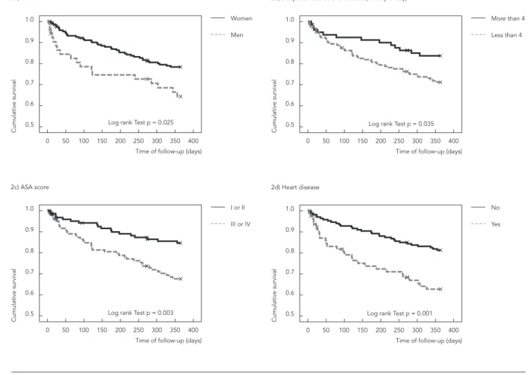
Overall one-year survival was 75% (95%CI: 70%-81%). In men it was 64% (52%-78%) and in women 82% (73%-84%). Figure 2 shows Kaplan-Meier curves for statistically significant variables according to the log-rank test. After adjustment for age, the variables gender, ASA score, presence of comorbidities, and number of comorbidities remained as significant predictors of mortality (Table 2). No interaction between variable was identified.
Table 3 shows the results of the multivari-ate Cox’s proportional hazards model. Accord-ingly, male gender (HR = 2.54; 95%CI: 1.40-4.58), III/V ASA score (HR = 1.95; 95%CI: 1.10-3.47), age (HR = 1.06; 95%CI: 1.03-1.10), and delay to sur-gery (HR = 1.07; 95%CI: 1.03-1.12) were indepen-dent and statistically significant predictors of survival after PFF.
Discussion
Mortality after PFF creates a major clinical and public health burden. One-fourth of patients in our study had died after one year. Increased mortality was associated with increasing age,
Figure 2
Kaplan-Meier curves with significant log-rank test for gender, daily activities before fracture, ASA score, heart disease, respiratory disease, dementia, anemia, and number of comorbidities. Cohort study of 252 patients admitted to the Orthopedic Ward of São João Hospital, Portugal (May 1st, 2008,
to April 30th, 2009).
Figure 2 (continued)
longer delay to surgery, male gender, and worse ASA score.
Overall one-year mortality in men (34.6%) was higher than reported in some European countries such as Ireland (30.1%), Norway (31%), and the Netherlands (33%). Meanwhile, in wom-en, one-year mortality (21.4%) was higher than in Ireland and Norway, but lower than in the Neth-erlands 22,23,24. In Portugal, as far as we know only one other prospective cohort (patients > 65 years old) studied survival after PFF 17, and overall mortality in women was similar (22.2%), while in men our study showed lower overall mortality (48.3%). However, the results should be analyzed with caution, since this other study included pa-tients more than 65 years of age.
Even after adjusting for age, ASA score, and delay to surgery, men showed 2.54 times high-er mortality risk (95%CI: 1.40-4.58) in women.
These results are similar to findings from a pro-spective cohort of 218 patients in Spain (2007), with a relative risk of 2.44 (95%CI: 1.01-5.93) after adjusting for age, type of fracture, living situation, functional status prior to the fracture, mental sta-tus, comorbidities, delirium during admission, and situation at discharge (patients discharged to the community versus to an institution) 25.
in men, and more men were on antithrombotic medication prior to the fracture, placing them at greater risk of respiratory complications and thromboembolic events.
Age increased the risk of mortality after a PFF, with a 6% increase in mortality for each year (HR = 1.06; 95%CI: 1.03-1.10) in our study. A similar prospective cohort study in Italy with 3,707 patients > 50 years of age found a similar risk (HR = 1.08; 95%CI: 1.06-1.09) 28.
A controversial factor affecting mortality af-ter PFF is time between admission and surgery 29,30,31. Although the optimal time for surgery
af-ter PFF in the elderly is not clear, most authors report that early surgical intervention (< 24, < 48, or < 72 hours) is associated with better prognosis and improved health status 15,31,32. Delay to sur-gery prolongs hospital stay and increases the risk of pulmonary embolism, deep venous thrombo-sis, heart failure, urinary infection, and pressure sores 33,34,35, thus delaying rehabilitation and in-creasing the risk of death.
Table 2
Age-adjusted hazard ratios (HR) for one-year mortality after proximal femur fracture. Cohort study of 252 patients admitted to the Orthopedic Ward of São João Hospital, Portugal (May 1st, 2008, to April 30th, 2009).
Variable Crude
HR (95%CI)
Age-adjusted HR (95%CI)
Gender
Female 1.00 1.00
Male 1.85 (1.06-3.21) 2.44 (1.38-4.31)
Daily activities before fracture (hours per day)
≥ 4 1.00 1.00
< 4 1.88 (1.02-3.48) 1.54 (0.82-2.90)
ASA score
ASA I-II 1.00 1.00
ASA III-IV 2.38 (1.36-4.17) 2.27 (1.29-3.98)
Comorbidities
Without heart disease 1.00 1.00
With heart disease 2.31 (1.39-3.86) 2.06 (1.23-3.44)
Without respiratory disease 1.00 1.00
With respiratory disease 2.03 (1.14-3.61) 2.11 (1.19-3.76)
Without dementia 1.00 1.00
With dementia 1.83 (1.03-3.25) 1.82 (1.02-3.22)
Without anemia 1.00 1.00
With anemia 1.91 (1.12-3.26) 1.72 (1.01-2.96) Number of comorbidities
0-1 1.00 1.00
2-4 1.70 (0.73-3.95) 1.66 (0.71-3.84)
≥ 5 3.32 (1.45-7.60) 3.08 (1.34-7.06)
Delay to surgery 1.08 (1.03-1.12) 1.08 (1.03-1.12)
95%CI: 95% confidence interval.
Table 3
Final Cox’s proportional hazards analysis. Predictors of one-year survival after proximal femur fracture. Cohort study of 252 patients admitted to the Orthopedic Ward of São João Hospital, Portugal (May 1st, 2008, to April 30th, 2009).
Variable Crude
HR (95%CI)
Adjusted * HR (95%CI)
Gender
Female 1.00 1.00
Male 1.85 (1.06-3.21) 2.54 (1.40-4.58) ASA score
ASA I/II 1.00 1.00
ASA III/IV 2.38 (1.36-4.17) 1.95 (1.10-3.47) Age 1.05 (1.02-1.08) 1.06 (1.03-1.10) Delay to surgery 1.08 (1.03-1.12) 1.07 (1.03-1.12)
95%CI: 95% confidence interval; HR: hazard ratio.
In our study, each day of delay to surgery was associated with a 7% increase in risk of death. Time to surgery can help explain gender differ-ences in survival. In our study, men were less likely than women to undergo surgery on the day of admission or on the following day. Sur-gery is often postponed to stabilize patients’ clinical condition and optimize their hemody-namic status, since most patients admitted after a PFF are clinically unstable, dehydrated, ane-mic, and malnourished 36. In addition, patients on antithrombotic medication prior to the frac-ture (highly common in the elderly for prevent-ing thrombosis and atrial fibrillation) normally have their surgery delayed to prevent excessive intraoperative bleeding 37,38. However, accord-ing to our findaccord-ings a simple analysis of the use of antithrombotic medication does not appear to result in differences in time to surgery, since men on antithrombotic medication prior to sur-gery waited an average of 0.5 days longer than women. On the other hand, when analyzing ASA score and time to surgery, only 7% of men with ASA III or IV underwent surgery on the day of ad-mission or the following day, compared to 34% of women with the same ASA score. This indicates that even if there is no difference in ASA between men and women, the comorbidities that contrib-ute to higher ASA scores in men require longer time for stabilization, thus resulting in longer time to surgery.
In our final Cox’s proportional hazards analy-sis, ASA score, which takes severity of comorbidi-ties into consideration, was found to be useful in predicting mortality in patients with a PFF, cor-roborating other studies 10,39.
The study presents some limitations. Partici-pants’ mental status was not tested objectively.
However, we evaluated their orientation in time and space based on their answers on age, date and place of birth, and place of residence. Con-trary to other studies 40,41, we did not find an as-sociation between mortality and daily activities prior to hip fracture; however, this may have re-sulted from limitations in the way the variable was collected, by categories: < 4 hours versus ≥ 4 hours of daily activity, not taking into consid-eration previous limitation in walking or use of walking aids.
The study’s strong points are related to the de-sign, as a prospective cohort study conducted in the second largest hospital in Portugal. Data were collected directly from patients and from objec-tive and standardized records, thereby increasing data quality. In addition, unlike other studies, the same interviewer applied all the questionnaires at baseline and follow-up, also enhancing data quality. Finally, diagnosis and treatment of PFF were confirmed by consulting imaging records, thereby avoiding misclassification of fractures, while type of surgery and comorbidities were as-sessed from patients’ clinical charts, thus avoid-ing recall bias.
Resumen
Los objetivos del estudio fueron analizar la superviven-cia tras un año y los factores asosuperviven-ciados para enfermos con fractura de la cadera (bajo impacto). Fue constitui-da una cohorte con todos los enfermos hospitalizados en el servicio de ortopedia del segundo mayor hospital de Portugal (mayo/2008 – abril/2009). La supervivencia fue evaluada a los 3, 6, 9 y 12 meses tras la fractura y re-lacionada con factores demográficos, estilo de vida, his-toria clínica y factores médicos (tipo de fractura, fecha de la cirugía, tratamiento y riesgo preoperatorio). De los 340 enfermos hospitalizados, 252 (78,9% mujeres) fueron incluidos. La mortalidad a los 3, 6, 8 y 12 meses de seguimiento fue de un 21,2%, 20%, 28,8%, 34,6% en hombres y un 7,8%, 13,5%, 19,2%, 21,4% en mujeres. Los factores asociados con la mortalidad fueron: sexo mas-culino (HR = 2,54; IC95%: 1,40-4,58), ASA puntuación más elevada, III/IV vs. I/II (HR = 1,95; IC95%: 1,10-3,47), edad (HR = 1,06; IC95%: 1,03-1,10) y días de retraso en la cirugía (HR = 1,07; IC95%: 1,03-1,12). Los factores es-tán en su mayoría relacionados con las características del enfermo en la admisión.
Fracturas de Cadera; Supervivencia; Mortalidad
Contributors
S. Campos gathered the data from the interviews at ad-mission and follow-up, conducted the analysis, discus-sion of the results, and wrote the draft. S. M. F. Alves participated in the analysis, discussion of the results, and writing of the final manuscript. M. S. Carvalho par-ticipated in the statistical analysis, discussion of the results, and final version of the manuscript. N. Neves participated in the interpretation and discussion of the results and final version of the manuscript. A. Trigo-Ca-bral participated in the discussion and interpretation of the results and writing of the final manuscript. M. F. Pi-na was responsible for the study design and conception, interpretation and discussion of the results, and writing of the final manuscript.
Acknowledgments
This study was financed by Portuguese funds through the FCT (Fundação para a Ciência e Tecnologia) in the framework of the Pest-C/SAU/LA0002/2011. Authors would also liked to thank PTDC/SAU-EPI/113424/2009 grant supported by FCT.
The authors wish to thank PhD students Brenda Gas-parini, Davi da Silveira Barroso Alves, and Leidjaira Ju-vanhol Lopes for their helpful comments on the article. M.S.C. was supported by CNPq (309692/2013-0) and FAPERJ (E-26/103.204/2011).
References
1. Cooper C. The crippling consequences of fractures and their impact on quality of life. Am J Med 1997; 103(2A):12S-7S.
2. Nurmi I, Narinen A, Luthje P, Tanninen S. Cost analysis of hip fracture treatment among the el-derly for the public health services: a 1-year pro-spective study in 106 consecutive patients. Arch Orthop Trauma Surg 2003; 123:551-4.
3. Alves SM, Castiglione D, Oliveira CM, de Sousa B, Pina MF. Age-period-cohort effects in the in-cidence of hip fractures: political and economic events are coincident with changes in risk. Osteo-poros Int 2013; 25:711-20.
4. Cummings SR, Melton LJ. Epidemiology and out-comes of osteoporotic fractures. Lancet 2002; 359:1761-7.
5. Kannegaard PN, van der Mark S, Eiken P, Abraha-msen B. Excess mortality in men compared with women following a hip fracture. National analysis of comedications, comorbidity and survival. Age Ageing 2010; 39:203-9.
6. Trombetti A, Herrmann F, Hoffmeyer P, Schurch MA, Bonjour JP, Rizzoli R. Survival and potential years of life lost after hip fracture in men and age-matched women. Osteoporos Int 2002; 13:731-7. 7. Kanis JA, Oden A, Johnell O, De Laet C, Jonsson B,
Oglesby AK. The components of excess mortality after hip fracture. Bone 2003; 32:468-73.
8. Haentjens P, Magaziner J, Colon-Emeric CS, Vanderschueren D, Milisen K, Velkeniers B, et al. Meta-analysis: excess mortality after hip fracture among older women and men. Ann Intern Med 2010; 152:380-90.
9. Abrahamsen B, van Staa T, Ariely R, Olson M, Coo-per C. Excess mortality following hip fracture: a systematic epidemiological review. Osteoporos Int 2009; 20:1633-50.
10. Norring-Agerskov D, Laulund AS, Lauritzen JB, Duus BR, van der Mark S, Mosfeldt M, et al. Meta-analysis of risk factors for mortality in patients with hip fracture. Dan Med J 2013; 60:A4675. 11. Gunasekera N, Boulton C, Morris C, Moran C. Hip
fracture audit: the Nottingham experience. Osteo-poros Int 2010; 21 Suppl 4:S647-53.
12. Roche JJ, Wenn RT, Sahota O, Moran CG. Effect of comorbidities and postoperative complications on mortality after hip fracture in elderly people: prospective observational cohort study. BMJ 2005; 331:1374.
13. Haentjens P, Autier P, Barette M, Venken K, Vander-schueren D, Boonen S, et al. Survival and function-al outcome according to hip fracture type: a one-year prospective cohort study in elderly women with an intertrochanteric or femoral neck fracture. Bone 2007; 41:958-64.
14. Tosteson AN, Gottlieb DJ, Radley DC, Fisher ES, Melton 3rd LJ. Excess mortality following hip frac-ture: the role of underlying health status. Osteopo-ros Int 2007; 18:1463-72.
16. Salvador MJ, Ferreira A, Gomes C, Moniz T, Judas F. Fracturas da extremidade superior do fêmur: mor-bilidade e mortalidade. Acta Reumatol Port 2002; 27:91-100.
17. da Costa JA, Ribeiro A, Bogas M, Costa L, Varino C, Lucas R, et al. Mortality and functional im-pairment after hip fracture: a prospective study in a Portuguese population. Acta Reumatol Port; 34:618-26.
18. Cruz M. Why do we close our eyes while the world is falling? A study on proximal femur osteoporotic fractures in a Portuguese population. Acta Reuma-tol Port 2009; 34(2B):370-7.
19. Dequeker J, Ranstam J, Valsson J, Sigurgevisson B, Allander E. The Mediterranean Osteoporosis (ME-DOS) Study questionnaire. Clin Rheumatol 1991; 10:54-72.
20. Owens WD, Felts JA, Spitznagel Jr. EL. ASA physical status classifications: a study of consistency of rat-ings. Anesthesiology 1978; 49:239-43.
21. World Health Organization. Anatomical therapeu-tic chemical (ATC) classification index with de-fined daily doses (DDDs). Geneva: World Health Organization; 2003.
22. Boereboom FT, Raymakers JA, Duursma SA. Mor-tality and causes of death after hip fractures in The Netherlands. Neth J Med 1992; 41:4-10.
23. Elliott J, Beringer T, Kee F, Marsh D, Willis C, Ste-venson M. Predicting survival after treatment for fracture of the proximal femur and the effect of de-lays to surgery. J Clin Epidemiol 2003; 56:788-95. 24. Forsen L, Sogaard AJ, Meyer HE, Edna T, Kopjar B.
Survival after hip fracture: short- and long-term excess mortality according to age and gender. Os-teoporos Int 1999; 10:73-8.
25. Alegre-Lopez J, Cordero-Guevara J, Alonso-Valdivielso JL, Fernandez-Melon J. Factors associ-ated with mortality and functional disability after hip fracture: an inception cohort study. Osteopo-ros Int 2005; 16:729-36.
26. Wehren LE, Hawkes WG, Orwig DL, Hebel JR, Zim-merman SI, Magaziner J. Gender differences in mortality after hip fracture: the role of infection. J Bone Miner Res 2003; 18:2231-7.
27. Panula J, Pihlajamaki H, Mattila VM, Jaatinen P, Vahlberg T, Aarnio P, et al. Mortality and cause of death in hip fracture in patients aged 65 or older: a population-based study. BMC Musculoskelet Dis-ord 2011; 12:105.
28. Maggi S, Siviero P, Wetle T, Besdine RW, Saugo M, Crepaldi G, et al. A multicenter survey on profile of care for hip fracture: predictors of mortality and disability. Osteoporos Int 2010; 21:223-31.
29. Orosz GM, Magaziner J, Hannan EL, Morrison RS, Koval K, Gilbert M, et al. Association of timing of surgery for hip fracture and patient outcomes. JA-MA 2004; 291:1738-43.
30. Leung F, Lau TW, Kwan K, Chow SP, Kung AW. Does timing of surgery matter in fragility hip fractures? Osteoporos Int 2010; 21 Suppl 4:S529-34.
31. Khan SK, Kalra S, Khanna A, Thiruvengada MM, Parker MJ. Timing of surgery for hip fractures: a systematic review of 52 published studies involv-ing 291,413 patients. Injury 2009; 40:692-7. 32. Moja L, Piatti A, Pecoraro V, Ricci C, Virgili G,
Salanti G, et al. Timing matters in hip fracture surgery: patients operated within 48 hours have better outcomes. A meta-analysis and meta-re-gression of over 190,000 patients. PLoS One 2012; 7:e46175.
33. Siegmeth AW, Gurusamy K, Parker MJ. Delay to surgery prolongs hospital stay in patients with fractures of the proximal femur. J Bone Joint Surg Br 2005; 87:1123-6.
34. McLaughlin MA, Orosz GM, Magaziner J, Hannan EL, McGinn T, Morrison RS, et al. Preoperative sta-tus and risk of complications in patients with hip fracture. J Gen Intern Med 2006; 21:219-25. 35. Lefaivre KA, Macadam SA, Davidson DJ, Gandhi R,
Chan H, Broekhuyse HM. Length of stay, mortality, morbidity and delay to surgery in hip fractures. J Bone Joint Surg Br 2009; 91:922-7.
36. Kumar D, Mbako AN, Riddick A, Patil S, Williams P. On admission haemoglobin in patients with hip fracture. Injury 2011; 42:167-70.
37. Al-Rashid M, Parker MJ. Anticoagulation manage-ment in hip fracture patients on warfarin. Injury 2005; 36:1311-5.
38. Ranhoff AH, Martinsen MI, Holvik K, Solheim LF. Use of warfarin is associated with delay in surgery for hip fracture in older patients. Hosp Pract (1995) 2011; 39:37-40.
39. Donegan DJ, Gay AN, Baldwin K, Morales EE, Es-terhai Jr. JL, Mehta S. Use of medical comorbidi-ties to predict complications after hip fracture sur-gery in the elderly. J Bone Joint Surg Am 2010; 92: 807-13.
40. Aharonoff GB, Koval KJ, Skovron ML, Zuckerman JD. Hip fractures in the elderly: predictors of one year mortality. J Orthop Trauma 1997; 11:162-5. 41. Meyer HE, Tverdal A, Falch JA, Pedersen JI. Factors
associated with mortality after hip fracture. Osteo-poros Int 2000; 11:228-32.
Submitted on 19/May/2014