Shrimp Serine Proteinase Homologues
Pm
MasSPH-1 and -2 Play a Role in the
Activation of the Prophenoloxidase System
Miti Jearaphunt1, Piti Amparyup1,2, Pakkakul Sangsuriya1,2, Walaiporn Charoensapsri2,3, Saengchan Senapin2,3, Anchalee Tassanakajon1*
1Center of Excellence for Molecular Biology and Genomics of Shrimp, Department of Biochemistry, Faculty of Science, Chulalongkorn University, Bangkok, Thailand,2National Center for Genetic Engineering and Biotechnology (BIOTEC), National Science and Technology Development Agency (NSTDA), Klong 1, Klong Luang, Pathumthani, Thailand,3Center of Excellence for Shrimp Molecular Biology and Biotechnology (Centex Shrimp), Mahidol University, Bangkok, Thailand
Abstract
Melanization mediated by the prophenoloxidase (proPO) activating system is a rapid immune response used by invertebrates against intruding pathogens. Several masquerade-like and serine proteinase homologues (SPHs) have been demonstrated to play an essential role in proPO activation in insects and crustaceans. In a previous study, we characterized the mas-querade-like SPH,PmMasSPH1, in the black tiger shrimpPenaeus monodonas a multifunc-tional immune protein based on its recognition and antimicrobial activity against the Gram-negative bacteriaVibrio harveyi. In the present study, we identify a novel SPH, known as PmMasSPH2, composed of an N-terminal clip domain and a C-terminal SP-like domain that share high similarity to those of other insect and crustacean SPHs. We demonstrate that gene silencing ofPmMasSPH1 andPmMasSPH2 significantly reduces PO activity, resulting in a high number ofV.harveyiin the hemolymph. Interestingly, knockdown ofPmMasSPH1 suppressed not only its gene transcript but also other immune-related genes in the proPO system (e.g.,PmPPAE2) and antimicrobial peptides (e.g.,PenmonPEN3,PenmonPEN5, crustinPm1 and Crus-likePm). ThePmMasSPH1 andPmMasSPH2 also show binding activi-ty to peptidoglycan (PGN) of Gram-positive bacteria. Using a yeast two-hybrid analysis and co-immunoprecipitation, we demonstrate thatPmMasSPH1 specifically interacted with the final proteinase of the proPO cascade,PmPPAE2. Furthermore, the presence of both PmMasSPH1 andPmPPAE2 enhances PGN-induced PO activityin vitro. Taken together, these results suggest the importance ofPmMasSPHs in the activation of the shrimp proPO system.
OPEN ACCESS
Citation:Jearaphunt M, Amparyup P, Sangsuriya P, Charoensapsri W, Senapin S, Tassanakajon A (2015) Shrimp Serine Proteinase HomologuesPm MasSPH-1 and -2 Play a Role in the Activation of the Prophenoloxidase System. PLoS ONE 10(3): e0121073. doi:10.1371/journal.pone.0121073
Academic Editor:Irene Söderhäll, Uppsala University, SWEDEN
Received:December 2, 2014
Accepted:January 29, 2015
Published:March 24, 2015
Copyright:© 2015 Jearaphunt et al. This is an open access article distributed under the terms of the
Creative Commons Attribution License, which permits unrestricted use, distribution, and reproduction in any medium, provided the original author and source are credited.
Data Availability Statement:All relevant data are within the paper.
Introduction
Invertebrates primarily rely on innate immunity mediated by cellular and humoral responses to combat invading pathogens. A cellular immune response involves different types of blood cells that recognize conserved pathogen associated molecular patterns (PAMPs) and leads to activation of nodule formation, encapsulation, and phagocytosis of the microorganisms [1–3]. In contrast, the humoral defense involves production and secretion of several immune proteins such as antimicrobial peptides (AMPs), as well as proteinases and their inhibitors that associat-ed with the clotting and melanization cascade in hemolymph [1–3]. Melanization is one of the most rapid and effective humoral responses that is driven by a key enzyme called phenoloxi-dase (PO) and is tightly regulated by the proPO activation cascade [4–6]. Upon recognition of microbial PAMPs, such as lipopolysaccharide (LPS) from Gram-negative bacteria, peptidogly-can (PGN) from Gram-positive bacteria orβ-1,3-glucans from fungi, by pattern- or pathogen-recognition receptors (PRPs) [7], several serine proteinases generate the active PO required for melanin synthesis [4,8,9].
Several reports in insects have demonstrated the essential function of proPO in surviving microbial infection [10–13]. In crustaceans, it has also been demonstrated that the freshwater crayfishPacifastacus leniusculusis highly susceptible toAeromonas hydrophilainfection after suppression of proPO [14]. The absence of proPO in the kuruma shrimp,Marsupenaeus japo-nicus, significantly increases mortality and bacterial load in the hemolymph [15]. In the shrimp
Penaeus monodon, co-silencing ofPmproPO1 and 2 also had increased mortality after the bac-terialV.harveyiand the fungusFusarium solaniinfections [16,17]. In addition, the melani-zation reaction products from shrimp hemocyte also hadin vitroantimicrobial activities against Gram-negative bacteria (V.harveyiandV.parahaemolyticus), Gram-positive bacteria (Bacillus subtilis) and fungus (F.solani) [17].
Because the highly toxic intermediates generated from the melanization process are harmful to pathogens as well as the host tissue itself, this process must be tightly regulated and localized [18]. Thus, several serine proteinases (SPs) and serine proteinase homologs (SPHs) are in-volved in controlling the proteolysis steps. SPHs are SP-like enzymes that contain one or more clip-domain(s) at the N-terminus and an SP-like domain at the C-terminus, but the catalytic triad serine has been substituted with glycine, and therefore, SPHs lack proteolysis activity. Sev-eral SPHs have been identified in insects and crustaceans and their roles in the proPO cascade have been characterized. In insects, theAnopheles gambiaeSPH, CLIPA8, is required for mela-nization because knockdown of CLIPA8 will completely abolish melanin synthesis on the sur-face of the fungusBeauveria bassiana[12]. TheManduca sextaSPHs (SPH1 and SPH2) are also required for generating the active PO and enhancing PO activity via a collaborative func-tion with the proPO activating proteinase-1 (PAP-1) and PAP-3 [19,20]. Moreover, a non-clip domain-containing SPH-3 ofM.sextais required for PO activation, nodule formation, and gene transcription of antimicrobial effectors [21]. The proPO-activating factor-II (PPAF-II) of
Holotrichia diomphaliahas been characterized as the co-activator of PPAF-I, a clip-domain SP that cleaves proPO into PO, and the clip domain of PPAF-II also plays a role in proPO activa-tion by interacting with PO through its central cleft [22,23]. InTenebrio molitor, SPH1 but not SPH2 is a proPO activating cofactor that functions together with active PO to regulate melanin production by forming an active melanization complex that efficiently produces melanin on the surface of bacteria and has strong antibacterial activity [24,25]. In addition to proPO activa-tion, SPHs also possess other functions related to embryonic development such as in Drosophi-la[26] and are also involved in PRP recognition. The crayfishP.leniusculusmasquerade-like proteinPl-MasI functions as a PRP that binds LPS,β-1,3-glucan, Gram-negative bacteria and yeast and is involved in the clearance of microorganisms [27], whilstPlSPH1 andPlSPH2 may
exchanges are from the Japan Science and Technology Agency/Japan International Cooperation Agency, Science and Technology Research Partnership for Sustainable Development (JST/JICA, SATREPS) and the JSPS core-University Program. MJ is supported by the program Strategic Scholarships for Frontier Research Network for the Joint Ph.D. Program: The Office of the Higher Education Commission (28/2551) and the 90th Anniversary of Chulalongkorn University Fund (Ratchadaphiseksomphot Endowment Fund). The funders had no role in study design, data collection and analysis, decision to publish, or preparation of the manuscript.
act as the PRP to the Gram-positive bacteria cell wall component PGN [28]. In the mud crab
Scylla paramamosain,Sp-SPH can also bind various microbial cell wall components such as PGN, LPS andβ-1,3-glucans [29].
Our research group has identified and characterized several important proteins in the shrimp proPO system [16,30–33] including theP.monodonmasquerade-like SPH called
PmMasSPH1 that acts as a multifunctional immune protein [34,35]. Transcription of
PmMasSPH1 has been shown to be induced in response toV.harveyiinfection [34]. Further-more, the C-terminal SP-like domain ofPmMasSPH1 can act as a cell adhesive molecule and binds toV.harveyi, LPS and virus [35,36]. The N-terminal region ofPmMasSPH1, containing clip and glycine-rich domains, also has antimicrobial activity against Gram-positive bacteria [35]. In the present study, a newly identifiedPmMasSPH2 is described and bothPmMasSPH1 andPmMasSPH2 are further investigated for their roles in activation of the proPO system using RNA interference (RNAi) and protein-protein interaction approach. Moreover, the PRP properties forPmMasSPHs in recognizing and binding to the Gram-positive bacterial cell wall component PGN are also examined.
Materials and Methods
Sample preparation
Specific pathogen-free (SPF)Penaeus monodon(~4 g, fresh weight) were purchased from the Shrimp Genetic Improvement Center, BIOTEC, Thailand. Shrimp were reared in an aerated tank with seawater at 20 ppt salinity for 7 days before the experiments began. The hemolymph was collected and total RNA was extracted using the TRI Reagent (Molecular Research Center) following the manufacturer’s protocol. First-strand cDNA was synthesized using the ImProm-II Reverse Transcriptase System kit (Promega) with 1.5μg of total RNA and 0.5μg of oligo (dT)15primer. For the gene expression analysis at different developmental stages, RNA
extrac-tion and cDNA synthesis as described above was done on three individual shrimp from four stages including nauplius 3 (N3), protozoea 2 (Z2), mysis 2 (M2) and post-larvae 15 (PL15).
Sequence analysis
The sequence similarity search was performed using the BLAST algorithm (http://www.ncbi. nlm.nih.gov/BLAST/). The putative signal peptide cleavage site and the structural protein do-mains were predicted using the SignalP 3.0 server (http://www.cbs.dtu.dk/services/SignalP/) and the simple modular architecture research tool (SMART) (http://smart.embl-heidelberg.de/), respectively. Multiple amino acid sequence alignments were carried out using the ClustalW2 program (http://www.ebi.ac.uk/Tools/clustalw2/). The phylogenetic tree was created in Molecu-lar Evolutionary Genetics Analysis (MEGA) version 5.2.
Double-stranded RNAs (dsRNAs) preparation and gene silencing
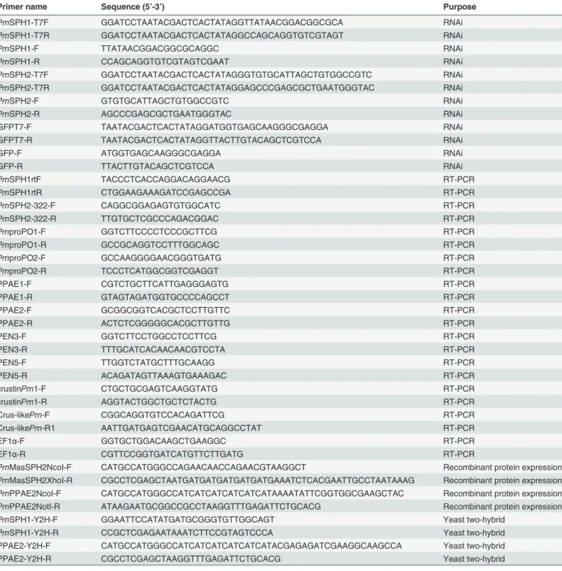
dsRNA was generated and purified as described previously [16] using gene specific primers (PmSPH1-T7F,PmSPH1-T7R,PmSPH1-F andPmSPH1-R forPmMasSPH1,PmSPH2-T7F,
Table 1. Primers used in the experiments.
Primer name Sequence (5’-3’) Purpose
PmSPH1-T7F GGATCCTAATACGACTCACTATAGGTTATAACGGACGGCGCA RNAi
PmSPH1-T7R GGATCCTAATACGACTCACTATAGGCCAGCAGGTGTCGTAGT RNAi
PmSPH1-F TTATAACGGACGGCGCAGGC RNAi
PmSPH1-R CCAGCAGGTGTCGTAGTCGAAT RNAi
PmSPH2-T7F GGATCCTAATACGACTCACTATAGGGTGTGCATTAGCTGTGGCCGTC RNAi
PmSPH2-T7R GGATCCTAATACGACTCACTATAGGAGCCCGAGCGCTGAATGGGTAC RNAi
PmSPH2-F GTGTGCATTAGCTGTGGCCGTC RNAi
PmSPH2-R AGCCCGAGCGCTGAATGGGTAC RNAi
GFPT7-F TAATACGACTCACTATAGGATGGTGAGCAAGGGCGAGGA RNAi
GFPT7-R TAATACGACTCACTATAGGTTACTTGTACAGCTCGTCCA RNAi
GFP-F ATGGTGAGCAAGGGCGAGGA RNAi
GFP-R TTACTTGTACAGCTCGTCCA RNAi
PmSPH1rtF TACCCTCACCAGGACAGGAACG RT-PCR
PmSPH1rtR CTGGAAGAAAGATCCGAGCCGA RT-PCR
PmSPH2-322-F CAGGCGGAGAGTGTGGCATC RT-PCR
PmSPH2-322-R TTGTGCTCGCCCAGACGGAC RT-PCR
PmproPO1-F GGTCTTCCCCTCCCGCTTCG RT-PCR
PmproPO1-R GCCGCAGGTCCTTTGGCAGC RT-PCR
PmproPO2-F GCCAAGGGGAACGGGTGATG RT-PCR
PmproPO2-R TCCCTCATGGCGGTCGAGGT RT-PCR
PPAE1-F CGTCTGCTTCATTGAGGGAGTG RT-PCR
PPAE1-R GTAGTAGATGGTGCCCCAGCCT RT-PCR
PPAE2-F GCGGCGGTCACGCTCCTTGTTC RT-PCR
PPAE2-R ACTCTCGGGGGCACGCTTGTTG RT-PCR
PEN3-F GGTCTTCCTGGCCTCCTTCG RT-PCR
PEN3-R TTTGCATCACAACAACGTCCTA RT-PCR
PEN5-F TTGGTCTATGCTTTGCAAGG RT-PCR
PEN5-R ACAGATAGTTAAAGTGAAAGAC RT-PCR
crustinPm1-F CTGCTGCGAGTCAAGGTATG RT-PCR
crustinPm1-R AGGTACTGGCTGCTCTACTG RT-PCR
Crus-likePm-F CGGCAGGTGTCCACAGATTCG RT-PCR
Crus-likePm-R1 AATTGATGAGTCGAACATGCAGGCCTAT RT-PCR
EF1α-F GGTGCTGGACAAGCTGAAGGC RT-PCR
EF1α-R CGTTCCGGTGATCATGTTCTTGATG RT-PCR
PmMasSPH2NcoI-F CATGCCATGGGCCAGAACAACCAGAACGTAAGGCT Recombinant protein expression
PmMasSPH2XhoI-R CGCCTCGAGCTAATGATGATGATGATGATGAAATCTCACGAATTGCCTAATAAAG Recombinant protein expression
PmPPAE2NcoI-F CATGCCATGGGCCATCATCATCATCATCATAAAATATTCGGTGGCGAAGCTAC Recombinant protein expression
PmPPAE2NotI-R ATAAGAATGCGGCCGCCTAAGGTTTGAGATTCTGCACG Recombinant protein expression
PmSPH1-Y2H-F GGAATTCCATATGATGCGGGTGTTGGCAGT Yeast two-hybrid
PmSPH1-Y2H-R CCGCTCGAGAATAAATCTTCCGTAGTCCCA Yeast two-hybrid
PPAE2-Y2H-F CATGCCATGGGCCATCATCATCATCATCATACGAGAGATCGAAGGCAAGCCA Yeast two-hybrid
PPAE2-Y2H-R CGCCTCGAGCTAAGGTTTGAGATTCTGCACG Yeast two-hybrid
(LPS) (Sigma-Aldrich) and 20μg of laminarin (β-1,3-glucan; Sigma-Aldrich). Shrimp were reared for 48 h after second injection prior to further analysis.
Hemolymph phenoloxidase (PO) activity assay
The total PO activity in shrimp hemolymph was measured as described previously [16]. 48 h after the second dsRNA injection, hemolymph was collected without using any anticoagulant. Total protein concentration was quantified using the Bradford protein assay kit (Bio-Rad). Subsequently, 2 mg total protein was analyzed for PO activity using L-3,4-dihydroxyphenylala-nine (L-DOPA) as the substrate. PO activity was defined asΔA490per mg total protein/min.
The results were analyzed from independent three experiments using a one-way analysis of variance (ANOVA).
Determining bacterial load in
Pm
MasSPHs silenced shrimp
To investigate the involvement ofPmMasSPHs in bacterial clearance, shrimp were injected with dsRNA specific toPmMasSPH1,PmMasSPH2 and control GFP as described above. For the second injection, shrimp were givenV.harveyi639 at a concentration of 2×10⁵colony forming units (CFUs). 6 h after bacteria challenge, the hemolymph was collected from each shrimp and serially diluted on LB agar plates and incubated at 30°C overnight. The number of colonies formed on the plate was calculated as CFU/ml.
Gene expression analysis in
Pm
MasSPHs silenced shrimp
To test whether gene silencing ofPmMasSPHs could affect the expression of other immune-re-lated gene transcripts, gene expression analysis was carried out inPmMasSPHs silenced shrimp. The hemolymph was collected 48 h after dsRNA injection and then RNA was extracted and cDNA was made as described previously. Gene expression levels of several proPO-related proteins (PmproPO1,PmproPO2,PmPPAE1 andPmPPAE2) and antimicrobial proteins ( Pen-monPEN3,PenmonPEN5, crustinPm1 and Crus-likePm) were determined by semi-quantita-tive RT-PCR using the gene specific primers found inTable 1. The PCR conditions were as follows: 94°C 1 min, followed by 25 cycle of 94°C for 30 s, 55°C for 30 s and 72°C for 1 min, and finally 72°C for 5 min for the final step. PCR products were visualized by agarose gel elec-trophoresis and band intensity was analyzed using a Gel-Pro Analyzer. The expression levels of each gene are shown as relative expression normalized to EF1-α. The statistical significance of the triplicate experiments was determined by one-way ANOVA.
Expression and purification of recombinant proteins
The C-terminal SP-like domain ofPmMasSPH2 and the SP domain ofPmPPAE2 were ampli-fied usingPfuDNA polymerase (Promega) with the primer pairsPmMasSPH2NcoI-F /
PmMasSPH2XhoI-R andPmPPAE2NcoI-F /PmPPAE2NotI-R, respectively (Table 1). The PCR products were purified and cloned into the expression vector pET28b (Novagen). The His-tagged recombinant plasmids of the C-terminal SP-like domain ofPmMasSPH1 [35],
with 20 mM Tris-HCl (pH 8.0). The protein concentration was then determined using a Brad-ford protein assay kit (Bio-Rad).
Quantitative determination of
Pm
MasSPHs binding activity to PGN
An ELISA assay was performed to investigate the binding activities of bothPmMasSPH1 and
PmMasSPH2 to the bacterial cell wall component PGN. A 96-well plate was coated with 2μg PGN fromBacillus subtilis(InvivoGen) per well in 50μl water and then incubated at 37°C overnight. After fixing at 60°C for 2 h and blocking with 5% BSA in TBS for 1 h, the PGN-coat-ed plate was washPGN-coat-ed with TBS three times. 100μl of recombinant protein at various concentra-tions was added in each well and incubated at 4°C overnight. After washing, 100μl of a mouse anti-His antibody (1:5000) was added in each well and incubated at room temperature for 3 h. After washing, the alkaline phosphatase-conjugated goat anti-mouse antibody (1:10000) was added and incubated for 1 h. The plate was then washed twice with TBST and twice with water before adding the substrate (AP Substrate Kit; Bio-Rad). The results were analyzed by Scatch-ard plot analysis. The dissociation constant (Kd) and the maximum binding (Amax) was
calcu-lated from the Y-intercept and slope of linear equation as 1/A =Kd/Amax[L]+1/Amax(A is
defined as the absorbance at 405 nm and [L] is the protein concentration).
Protein-protein interaction assay
To determine if there is an interaction betweenPmMasSPH1 andPmPPAE2, we performed a yeast two-hybrid analysis from Matchmaker GAL4 Two-Hybrid System (Clontech). Full-length cDNA encodingPmMasSPH1 was cloned and fused in-frame with the activation do-main (AD) of the pGADT7 vector by PCR amplification usingPmSPH1-Y2H-F and
PmSPH1-Y2H-R primers (Table 1). Full-length cDNA encodingPmPPAE2 was fused in-frame with the GAL4 DNA binding domain (BD) of the pGBKT7 vector using PPAE2-Y2H-F and PPAE2-Y2H-R primers (Table 1). The recombinant plasmids ofPmMasSPH1/AD and
PmPPAE2/BD were co-transformed into theSaccharomyces cerevisiaestrain AH109 using the lithium acetate/dimethyl sulfoxide method [37]. Transformed yeast cells were selected on mini-mal media plates lacking leucine and tryptophan (-L/-W). Positive interactions were indicated by growth on high stringency medium lacking adenine, histidine, leucine and tryptophan (-A/-H/-L/-W) and by a blue color change from X-α-gal (Apollo Scientific) present in the media. The empty vector pGADT7 was also co-transformation as negative control and an interaction between murine p53 bait fusion (pVA3) and SV40 prey fusion (pTD1) served as a positive con-trol (Clontech).
In addition to the yeast two-hybrid assay, co-immunoprecipitation (co-IP) was also per-formed between the rPmMasSPHs and rPmPPAEs that were produced inE.colias described above. The procedure was conducted according to Isono and Schwechheimer [38] with a slight modification. Briefly, to test if rPmMasSPH1 and rPmPPAE2 interact, 40μg of purified anti-SP-like domain ofPmMasSPH1 antibody was mixed with 40μl of protein A agarose (Sigma) in 20 mM Tris-HCl (pH 8.0) and incubated at 4°C for 3 h. 50μM of rPmMasSPH1 and 50μM of thePmPPAE2 were incubated together at 4°C for 3 h and then loaded onto the antibody-tagged resin and further incubated at 4°C for 3 h. After washing 3 times with freshly prepared wash buffer (50 mM Tris-HCl (pH 7.5), 100 mM NaCl, 10% (v/v) glycerol and 0.05% (v/v) Triton X-100), the protein complex was eluted with 2X SDS-PAGE sample loading buffer. The protein complex was then separated by SDS-PAGE, transferred to a nitrocellulose membrane and de-tected with anti-His antibody. The co-IP assay betweenPmMasSPH2 andPmPPAEs was also performed using the purified anti-SP-like domain ofPmMasSPH2 antibody as described for
Effect of
Pm
MasSPH1 and
Pm
PPAE2 on the PGN-triggered
hemolymph PO activity
in vitro
To identify whetherPmMasSPH1 andPmPPAE2 are necessary for activation of proPO after infection by Gram-positive bacteria, PO activity was measured in the presence of PGN and the recombinant proteins. Fresh shrimp hemolymph (HL) was collected and the total protein con-centration measured by the Bradford assay (Bio-Rad). Total reaction included 0.8 mg HL with the addition of 0.25μg PGN and 1μM of each recombinant protein (PmPPAE2 and
PmMasSPH1 or BSA as the protein control). The CAC buffer (10 mM CAC containing 10 mM CaCl2) was added to a final volume of 174μl. 26μl of 3 mg/ml L-DOPA was added and the
ΔA490measured at 5 min. Each experimental treatment was performed in triplicate. Data were
analyzed using a one-way analysis of variance (ANOVA) followed by Duncan’s test and a
p<0.05 was considered statistically significance.
Results
Sequence analysis of
Pm
MasSPH1 and
Pm
MasSPH2
Based on the shrimpP.monodonEST database (http://pmonodon.biotec.or.th/) [39], three full-length cDNAs of clip SPHs (PmMasSPH1,PmMasSPH2 andPmMasSPH3) have been iso-lated [6]. Among these, a masquerade-like SPH1 (PmMasSPH1) of shrimpP.monodonhas been cloned and its role in shrimp immunity has been characterized [34,35]. In this study,
PmMasSPH1 and the novel SPHPmMasSPH2, were investigated to further elucidate their function in proPO activation. A full-length sequence ofPmMasSPH2 was obtained with an open reading frame of 1164 bp encoding 387 amino acid residues. ThePmMasSPH2 cDNA se-quence was deposited in the GenBank database under accession number FJ620686. Apart from a signal peptide of 20 amino acids at the N-terminus, the mature protein (367 amino acids) has the calculated molecular mass of 39.37 kDa and the predicted pIof 7.52. The conserved clip-domain and serine proteinase (SP)-like clip-domain with the catalytic triad (His184, Asp234and
Gly338) were also found inPmMasSPH2,PmMasSPH1 and other SPHs (Fig. 1). BLAST
analy-sis ofPmMasSPH1 andPmMasSPH2 showed high homology to the SPs of crustaceans and in-sects. High similarity and conserved function involved in proPO cascade have also been reported for the crayfishPacifastacus leniusculus PlSPH1 andPlSPH2, crabScylla paramamo-sain Sp-SPH and beetleHolotrichia diomphalia HdPPAF-II [28,29,40], and thus, we aligned these amino acid sequences with bothPmMasSPH1 andPmMasSPH2 (Fig. 1). Interestingly,
PmMasSPH1 andPmMasSPH2 have only 34.6% sequence similarity but high homology was observed across other species. For instance,PmMasSPH1 has 55.5% similarity toPlSPH2, and
PmMasSPH2 andPlSPH1 share 68.2% similarity. Sequence comparison of only the SP-like do-main showed consistency in similarity among selected species. The SP-like dodo-mains of
PmMasSPH1 andPmMasSPH2,PmMasSPH1 andPlSPH2, andPmMasSPH2 andPlSPH1 share 51.6%, 73.6% and 78.9% similarity, respectively. A phylogenetic analysis of the SP-like domain of insect and crustacean PPAFs was also carried out to examine the evolutionary rela-tionships. A phylogenetic tree constructed using the Neighbor-Joining method revealed that
PmMasSPH1 andPmMasSPH2 were grouped in a clade of crustacean PPAFs (Fig. 2).
Gene expression in various developmental stages of shrimp larva
We next examined the expression profile ofPmMasSPH1 andPmMasSPH2 transcripts to bet-ter understand regulation of immune genes during shrimp development. Gene expression pat-terns ofPmMasSPH1 andPmMasSPH2 were obtained and compared to the internal EF1-α control by RT-PCR analysis at four shrimp larval developmental stages including nauplius 3 (N3), protozoea 2 (Z2), mysis 2 (M2) and post-larvae 15 (PL15). We found that both
PmMasSPH1 andPmMasSPH2 were expressed at all the developmental stages we tested but with different expression patterns. Expression levels of thePmMasSPH1 transcripts were stable at all stages, while thePmMasSPH2 transcripts were lower at the early stages and then gradual-ly increased as development progressed (Fig. 3).
Knockdown of
Pm
MasSPHs decreases hemolymph PO activity
Because several proteins together participate in the proPO cascade to defend against invading pathogens, we evaluated the involvement ofPmMasSPH1 andPmMasSPH2 in proPO activa-tion by RNAi-mediated gene silencing. Shrimp were injected twice with dsRNA against
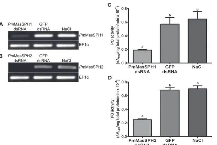
PmMasSPH1 orPmMasSPH2 with GFP dsRNA or NaCl solution used as control groups. 48 h after the second injection, shrimp hemolymph was collected and gene transcription analyzed. Our results show that shrimp receivingPmMasSPH1 orPmMasSPH2 dsRNA efficiently sup-pressedPmMasSPH1 orPmMasSPH2 transcript expression (Fig. 4A and B). Injection of GFP dsRNA or NaCl solution had no effect onPmMasSPH1 orPmMasSPH2 mRNA
transcript levels.
To examine whetherPmMasSPH1 andPmMasSPH2 are involved in the activation of proPO, the total hemolymph PO activity ofPmMasSPH1 orPmMasSPH2 silenced shrimp was measured using L-DOPA as a substrate. The hemolymph PO activity ofPmMasSPH1 and
PmMasSPH2 knockdown shrimp decreased significantly by 66.5% and 63.7%, respectively. No significant change in PO activity was observed in the control groups (p<0.05) (Fig. 4C and
D). These results indicate thatPmMasSPH1 andPmMasSPH2 are involved in proPO activa-tion in shrimp.
Pm
MasSPH silencing increase bacterial load in shrimp hemolymph
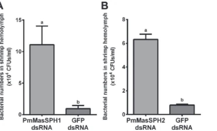
The above results clearly demonstrate that bothPmMasSPH1 andPmMasSPH2 have potential roles in proPO activation. Therefore, we investigated whetherPmMasSPH1 andPmMasSPH2 have immune activity against shrimp pathogenic bacteria. ThePmMasSPH1 orPmMasSPH2 silenced shrimp were challenged with pathogenicV.harveyiat 2 × 10⁵CFU/shrimp. As a con-trol, shrimp injected with GFP dsRNA were also challenged withV.harveyi. 6 h after bacterial challenge, hemolymph from individual shrimp was collected and the CFU ofV.harveyiwas de-termined. Our results show that the number of viable bacterial CFUs in thePmMasSPH1 and
PmMasSPH2 silenced shrimp increased 11.5 and 7.9-fold, respectively, compared to the con-trol shrimp injected with GFP dsRNA (Fig. 5A and B). It is worth noting that thePmMasSPH1 silenced shrimp have a higher bacterial hemolymph load than thePmMasSPH2 silenced Fig 1. A multiple amino acid sequence alignment ofPmMasSPH1 andPmMasSPH2 with other arthropod SPHs.The amino acid sequence ofPenaeus monodon PmMasSPH1 (ABE03741),PmMasSPH2 (ACP19560),Pacifastacus leniusculus PlSPH1 (AAX55746),PlSPH2 (ACB41379),Scylla paramamosain Sp -SPH (ADG83846) andHolotrichia diomphalia HdPPAFII (CAC12665) were collectively compared. The predicted signal peptides are in bold and underlined. The dash line indicates the glycine-rich domain of
PmMasSPH1. The conserved clip-domains are indicated below the line. The light-grey highlight indicates the cysteine residues in the clip-domains. The black box shows the conserved serine proteinase-like domain with the grey highlight indicating the catalytic triad (His, Asp, and Gly residues) in the domain.
Fig 2. The phylogenetic relationship between serine proteinase domains fromPmMasSPH1 andPmMasSPH2 and other serine proteinases.The deduced amino acid sequences of SP-domains from various clip-SPH species were used to generate a phylogenetic tree by the neighbor-joining method. Percent bootstrap values (1000 replicates) are shown at each branch point.PmMasSPH1 (ABE03741);PmMasSPH2 (ACP19560);PmSP,P.monodon
serine proteinase (ABW87872);PmSPL,P.monodonserine proteinase-like protein (ABD62888);PmSPL3 (ABO33174);PmMas,P.monodonmas-like protein (AAT42131);FcPPAF,Fenneropenaeus chinensisprophenoloxidase activating factor (AFW98986);FcMas (AFW98983);LvPPAF,Litopenaeus vannameiPPAF (AFW98993);LvSP (AY368151);LvMas (AFW98990);PlSPH1,P.leniusculusSPH1 (AAX55746);PlSPH2 (ACB41379);MsSPH3,
Manduca sextaSPH3 (AF413067);HdPPAFII,Holotrichia diomphaliaPPAFII (CAC12665);MjSPH,Marsupenaeus japonicusSPH (AB161692);NvSPH21,
Nasonia vitripennisSPH21 (NP_001155060);CqSP,Culexquin quefasciatusSP (XP_001868413);AaSP,Aedes aegyptiSP (XP_001655705);PtPPAF,
Portunus trituberculatusPPAF (ACN87221);CsPAF,Callinectes sapidusPAF (AAS60227);EsPPAF,Eriocheir sinensisPPAF (ACU65942);Es Trypsin-likeSP (ACT78700);LsST,Lucilia sericatasalivary trypsin (AEX33291);CpPPAF,Cancer pagurusPPAF (CCE46009);PpSP,Papilio polytesSP (BAM19108);HaSPL1,Helicoverpa armigeraSPL1 (ACI32835);TmPPAF,Tenebrio molitorPPAF (CAC12696);StSPL,Scylla tranquebaricaSPL (ADN44616);LoPPAF1,Lonomia obliquaPPAF1 (AAV91458);PxSP,Papilio xuthusSP (BAM17901);PrMSPH,Pieris rapaeMSPH (ACZ68116);TcSP4,
Tribolium castaneumSP4 (EEZ99183);DmMas,Drosophila melanogasterMas (AAC46512);Sp-SPH,S.paramamosainSPH (ADG83846).
Fig 3. Expression ofPmMasSPH1 andPmMasSPH2 transcripts at various stages of shrimp larval development.Expression profiles ofPmMasSPH1 andPmMasSPH2 were examined at four larval stages including nauplius 3 (N3), protozoea 2 (Z2), mysis 2 (M2) and post-larvae 15 (PL15) by semi-quantitative RT-PCR. The elongation factor 1-α(EF1-α) served as an internal control. Each lane represents the result of individual shrimp (n = 3).
doi:10.1371/journal.pone.0121073.g003
Fig 4. Hemolymph PO activity ofPmMasSPH1 andPmMasSPH2 silenced shrimp.The efficiency of gene silencing ofPmMasSPH1 andPmMasSPH2 was determined by semi-quantitative RT-PCR. Transcription levels ofPmMasSPH1 (A) andPmMasSPH2 (B) were examined in shrimp injected with the corresponding dsRNA. Shrimp injected with GFP dsRNA or NaCl served as control groups. Expression of EF1-αwas used as an internal control. Each band represents the pooled cDNA of triplicate samples of each treatment group. Hemolymph PO activity of thePmMasSPH1 (C) andPmMasSPH2 (D) silenced shrimp was also examined. Shrimp injected with GFP dsRNA or NaCl served as control groups. The total hemolymph PO activity was measured asΔA490/mg
total protein/min. The data are shown as the mean±standard deviation (error bars) from three independent experiments. Significantly different means (p<0.05)
are indicated by the lower case letters (a, b) above each bar.
shrimp. These results suggest that bothPmMasSPH1 andPmMasSPH2 are involved in the pro-cess of hemolymph bacterial clearance in the shrimpP.monodon.
Pm
MasSPH1 silencing alters expression of other immune genes
Several studies in insect immunity have shown evidence that a single gene does not strictly reg-ulate one pathway but can play a role in several pathways. Previous studies in the lepidopteran
Manduca sextashowed that SPH-3 is not only required for proPO activation but is also impor-tant for transcription of other immune effector genes [21]. We therefore, examined the effect ofPmMasSPHs silencing on the expression of other immune genes by semi-quantitative RT-PCR. The hemocyte cDNAs fromPmMasSPH1 orPmMasSPH2 silenced shrimp was ex-amined for transcript expression of other proPO-related genes (PmproPO1,PmproPO2,
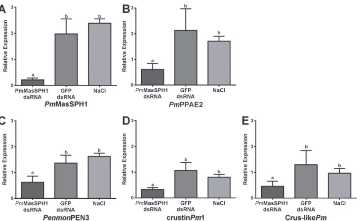
PmPPAE1 andPmPPAE2) and antimicrobial peptide genes (PenmonPEN3,PenmonPEN5, CrustinPm1 and Crus-likePm), with EF1-αas an internal control. Additionally, expression of these other immune-related genes was examined in the control groups injected with GFP dsRNA and NaCl. As shown inFig. 6, shrimp injected withPmMasSPH1 dsRNA have de-creased expression ofPmMasSPH1 (89%),PmPPAE2 (71%) (Fig. 6B),PenmonPEN3 (54%) (Fig. 6C), CrustinPm1 (69%) (Fig. 6D) and Crus-likePm(65%) (Fig. 6E) when compared to GFP dsRNA control shrimp. This suppression of immune genes byPmMasSPH1 silencing was not caused by an off-target effect of RNAi because sequence comparison between the region corresponding toPmMasSPH1 dsRNA and the other immune-related genes have no signifi-cant similarity (data not shown).PmMasSPH2 gene silencing was also performed in the same manner asPmMasSPH1. We found that only thePmMasSPH2 transcripts (98%) were sup-pressed in this model, while expression of other immune-related genes did not change (data not shown). Taken together,PmMasSPH1 appeared to be required for proPO activation and is involved in the synthesis of shrimp antimicrobial peptides.
In vitro
binding of
Pm
MasSPH1 and
Pm
MasSPH2 to PGN
It has been shown thatPmMasSPH1 might act as a PRP because it can bind to the Gram-nega-tive bacteriaV.harveyi, bacterial cell wall component LPS, and virus [35,36]. However, it is un-known ifPmMasSPHs binds to Gram-positive bacteria. In this study,PmMasSPH1 and Fig 5. Increase in bacterial load in hemolymph fromPmMasSPHs silenced shrimp.Shrimp were injected twice with dsRNA specific toPmMasSPH1 (A) orPmMasSPH2 (B). At the second dsRNA injection, shrimp were also injected withV.harveyi(2×10⁵CFU/shrimp). Control groups given injections of GFP dsRNA
or NaCl. The number of viable bacteria in the knockdown shrimp hemolymph is shown as the bacterial CFUs 6 h after challenge. The data are shown as the mean±standard deviation derived from three independent
experiments. Means with lower case letters above each bar indicate significant differences (p<0.05).
PmMasSPH2 were examined for a role in recognition of peptidoglycan (PGN), a Gram-positive bacterial cell wall component. An ELISA assay was used to quantitatively measure the binding activity between the recombinant proteins of maturePmMasSPH1 or maturePmMasSPH2 (with varying concentrations of 0–12.5μg/ml) with 2μg of ligand PGN fromBacillus subtilis. GFP served as a recombinant control protein. As shown inFig. 7, the rPmMasSPH1 and rPmMasSPH2 directly bound PGN in a concentration-dependent manner. The dissociation constant (Kd) of rPmMasSPH1-PGN and rPmMasSPH2-PGN were calculated as 6.51x10⁻⁹M and 5.79x10⁻⁹M, respectively. These results clearly indicate thatPmMasSPH1 and
PmMasSPH2 have a functional role as PRPs for PGN.
Pm
MasSPH1 interacts with
Pm
PPAE2
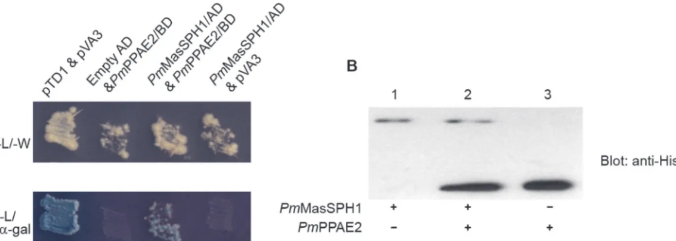
Because it has been demonstrated that some insect SPHs function as co-factors of the terminal PPAEs of proPO activation [11], it is important to investigate whetherPmMasSPHs interact withPmPPAEs. BecausePmPPAE2 changed expression whenPmMasSPH1 was silenced, we examined if there is a protein-protein interaction betweenPmMasSPH1 andPmPPAE2 using yeast two-hybrid and co-immunoprecipitation (co-IP) assays. In the yeast two-hybrid analysis, bothPmMasSPH1/AD andPmPPAE2/BD plasmids were co-transformed into the yeast AH109 strain and plated on selective media. Our results show thatPmMasSPH1 specifically binds toPmPPAE2 as indicated by blue colony formation on the selective media-A/-H/-L/-W/ X-α-gal (Fig. 8A, lanePmMasSPH1/AD andPmPPAE2/BD). In addition, no self-activation Fig 6. The effect ofPmMasSPH1 gene silencing on expression of other immune genes.Transcription levels ofPmMasSPH1 (A),PmPPAE2 (B),
PenmonPEN3 (C), crustinPm1 (D), and Crus-likePm(E) were analyzed inPmMasSPH1 silenced shrimp and control groups (GFP dsRNA or NaCl treatment). Each transcript is expressed as the relative expression normalized to the EF1-αtranscript. Each bar is displayed as the mean±standard
deviation (error bar). The lower case letters represent significant differences between treatments.
occurred whenPmPPAE2/BD was co-transformed with an empty AD plasmid (Fig. 8A, lane empty AD andPmPPAE2/BD). Co-transformation ofPmMasSPH1/AD with the control pVA3 plasmid was also performed to confirm thatPmMasSPH1 did not interact with the BD plasmid. The yeast two-hybrid assay suggests a genuine interaction betweenPmMasSPH1 and
PmPPAE2.
To further confirm the protein-protein interaction, a co-IP assay with rPmMasSPH1 and rPmPPAE2 was carried out using anti-SPH1-conjugated protein A agarose beads to precipitate the protein complex. As shown inFig. 8Blane 2, bothPmMasSPH1 andPmPPAE2 were ob-served in the elute protein fraction indicating an interaction between the proteins. In addition, other pairwise protein interactions (rPmMasSPH1 and rPmPPAE1, rPmMasSPH2 and Fig 7. Ability of rPmMasSPHs to bind a Gram-positive bacterial cell wall component.The binding ability of rPmMasSPH1 or rPmMasSPH2 (at varying concentrations 0–12.5μg/ml) toB.subtilisPGN was quantified by ELISA assay. rGFP was included as a recombinant protein control. The data at each point was fitted to the trend line and is shown as the mean±standard deviation from three individual experiments.
doi:10.1371/journal.pone.0121073.g007
Fig 8. Interaction ofPmMasSPH1 withPmPPAE2.The protein-protein interaction betweenPmMasSPH1 andPmPPAE2 were analyzed by yeast two-hybrid and co-IP assays. (A) The yeast two-two-hybrid results indicate the presence of BD and AD plasmids in transformed cells growing on an-L/-W plate. A putative interaction betweenPmMasSPH1/AD andPmPPAE2/BD as indicated by growth and blue color on the selective media-A/-H/-L/-W/X-α-gal. The positive control consisted of an interaction between murine p53 bait fusion (pVA3) and SV40 prey fusion (pTD1). The negative controls consisted of yeast cells containingPmPPAE2/BD with an empty AD vector or pVA3 vector andPmMasSPH1/AD. (B) The interaction betweenPmMasSPH1 andPmPPAE2 was confirmed by co-IP assay. The immunoprecipitated complex between rPmMasSPH1 and rPmPPAE2 was detected using the monoclonal anti-His antibody (lane 2). The rPmMasSPH1 or rPmPPAE2 alone (lanes 1 and 3, respectively) was used as a protein indicator.
rPmPPAE1, and rPmMasSPH2 and rPmPPAE2) were also examined by co-IP assay. However, no other positive interactions were found (data not shown). Altogether, the yeast two-hybrid and co-IP analyses indicate a direct interaction betweenPmMasSPH1 andPmPPAE2.
Enhancement of PGN-triggered hemolymph PO activity by
Pm
MasSPH1 and
Pm
PPAE2
in vitro
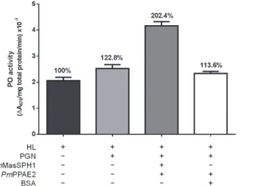
BecausePmMasSPH1 can recognize PGN and bind toPmPPAE2, we examined the role of these proteins in proPO activation. rPmMasSPH1, rPmPPAE2 and PGN were mixed with shrimp hemolymph and then PO activity was measured using L-DOPA as a substrate. In the presence of PGN, shrimp hemolymph PO activity was induced by approximately 23% com-pared with the non-activated hemolymph (HL) (Fig. 9). Interestingly, high PO activity from PGN was significantly enhanced by 102% when rPmMasSPH1 and rPmPPAE2 was graduated added to the hemolymph (Fig. 9). In addition, incubation of HL with PGN together with
PmPPAE2 and BSA (instead of rPmMasSPH1) resulted in no significant change in PO activity (14% compared to the control HL) (Fig. 9). These results suggest that hemolymph PO activity is slightly activated by PGN andPmMasSPH1 may be a co-factor toPmPPAE2 inP.monodon
proPO activation.
Discussion
In invertebrates, including insects and crustaceans, the proPO-activating system is essential for the melanization process that rapidly responds to the intruding pathogens [4–6,11]. The activa-tion of zymogen proPO to active PO, a critical step required for melanin synthesis, depends on the cleavage of terminal clip-serine proteinase and sometimes occurs with the aid of non-cata-lytic serine proteinase homologues (SPHs) cofactors [4–6,9]. Several studies have shown the re-quirement of SPHs for proPO activation by enhancing the cleavage of proPO to active PO by PPAEs [20,23,24]. For example, in insects, proPO activation ofHolotrichia diomphalianeeds PPAF-I to cleave proPO (79 kDa) into PO (76 kDa), and non-catalytic PPAF-II to bind the Fig 9. Enhancement of PGN-triggered hemolymph PO activity by rPmMasSPH1 and rPmPPAE2in
vitro.The total PO activity of shrimp hemolymph (HL) alone or mixed withB.subtilisPGN, rPmMasSPH1 and
rPmPPAE2 was determined. BSA served as a protein control. Each bar represents the PO activity as the mean±standard deviation from three replicates. The percentages of each treatment are relative to the PO activity of HL alone.
cleaved proPO and generate the catalytically active PO [23]. The process inManduca sexta re-quires both PPAE1 (PAP-1) and SPH to simultaneously activate the inactive proPO [20].
In the shrimpP.monodon,PmMasSPH1 has been previously identified and characterized as a multifunctional immune protein because it possesses opsonic abilities, antimicrobial activi-ty against gram-positive bacteria, and binds to LPS,V.harveyiand virus [34–36]. Nevertheless, the function ofPmMasSPH1 as it relates to proPO activation and otherPmMasSPHs remains unknown. Therefore, in the present study, we examinedPmMasSPH1 and the newly identified
PmMasSPH2 for their potential roles in proPO activation. The pattern recognition properties ofPmMasSPHs were also investigated because knowledge on the recognition of PGN by
PmMasSPHs has not yet been reported.
The novelPmMasSPH2 consists of an N-terminal clip-domain and a C-terminal SP-like do-main with a catalytic triad containing a Gly residue (instead of a Ser residue) similar to what has been previously identified inPmMasSPH1 and other arthropod SPHs. A multiple sequence alignment showed thatPmMasSPH2 has a similar primary structure asPlSPH1,PlSPH2,
HdPPAFII andSp-SPH, containing a one clip-domain at the N-terminus and an SP-like do-main at the C-terminus, unlikePmMasSPH1, which contains a glycine-rich domain at the C-terminus. The highest similarity was found betweenPmMasSPH1 andPlSPH2 and between
PmMasSPH2 andPlSPH1, suggesting that a similar role forPlSPHs in proPO activation. In the developmental expression profile ofPmMasSPHs, we found that both transcripts are expressed in all larval stages tested, similar to other proPO-related genes such asPmPPAE1 andPmproPO2 [31], althoughPmMasSPH2 had a slightly low level of gene expression at the N3 stage. This is in contrast toPmPPAE2 andPmproPO1 transcripts that are expressed at the middle phase of larval development (e.g., mysis and post-larvae stages) [31,41]. It has previous-ly been suggested thatPmPPAE1 andPmproPO2 together might function at an early stage of larval development whilePmPPAE2 andPmproPO1 may play a role at later developmental stages [31]. However, it was shown inP.monodonthat there is no PO activity at the N4 stage, suggesting incomplete proPO activation [41]. In addition, the absence of proPO expression in the middle phase of embryo development has also been reported in the crayfishPacifastacus leniusculus[42]. Thus, it is possible that the presence ofPmMasSPHs at early larval develop-mental stages ofP.monodonmight function in immune defense reactions other than in proPO activation. This hypothesis is also supported by previous work that shows thatPmMasSPH1 has multiple immune functions including opsonic capabilities, antimicrobial activity and rec-ognition of bacterial pathogens [35].
To evaluate the relevance ofPmMasSPHs in the proPO-activating system,in vivosilencing ofPmMasSPH1 andPmMasSPH2 using dsRNA was carried out and total shrimp PO activity was then examined. InPmMasSPH1 andPmMasSPH2 silenced shrimp, the hemolymph PO activity was significantly decreased compared to the control GFP dsRNA by approximately 67% and 64%, respectively. In comparison to the other shrimp proPO-related genes, this de-crease inPmMasSPHs is quite high and resemblesPmproPO1 andPmproPO2 silencing in which the PO activity was reduced by 75% and 73%, respectively [16]. In addition, knockdown ofPmPPAE1 andPmPPAE2 inP.monodonreduced the hemolymph PO activity only by 37% and 41%, respectively [31]. All evidence suggests thatPmMasSPHs,PmPPAEs andPmproPOs together participate in proPO activation, especiallyPmMasSPHs that are required factors in the activation cascade because their gene silencing resulted in a significant reduction of PO ac-tivity. Similarly, inP.leniusculus, knockdown ofPlproPO,PlSPH1 andPlSPH2 also disrupt PO activity, indicating their essential roles in proPO activation [14,28].
activity was examinedin vivo. The number of viableV.harveyiwas significantly increased in the hemolymph ofPmMasSPHs silenced shrimp thereby suggested the importance of
PmMasSPH1 andPmMasSPH2 in bacterial clearance. It was noted that there are differences in the rate ofV.harveyiclearance betweenPmMasSPH1 andPmMasSPH2. In comparison to the control GFP dsRNA, the bacterial number inPmMasSPH1 silenced shrimp was higher than in
PmMasSPH2 silenced shrimp (11.5-fold versus 7.9-fold, respectively) and may suggest a differ-ent way to activate proPO and eliminate the bacteria. In addition toPmMasSPHs, knockdown ofPmPPAEs andPmproPOs may also increase bacterial loads and induce high mortality in shrimp [16,30,31]. Likewise, inP.leniusculus, silencing ofPlproPO decreases PO activity and increases hemolymph bacterial number [14]. These relationships may explain the significant role ofPmMasSPHs in promoting the PO activity and subsequently limiting the number of bacteria in shrimp hemolymph.
A previous report in shrimp demonstrated that suppression of proPO genes (PmproPO1 andPmproPO2) alters the expression levels of other proPO-related genes and antimicrobial peptide (AMP) genes [15,32]. In the present study, the effect ofPmMasSPHs gene silencing on immune gene expression was therefore investigated. We found that transcription levels of the proPO-related gene (PmPPAE2) and AMP genes (PenmonPEN3,PenmonPEN5, CrustinPm1 and Crus-likePm) were significantly decreased by suppression ofPmMasSPH1 but not
PmMasSPH2 transcripts. This suggests that proPO andPmMasSPH1 could contribute to the crosstalk between the proPO-activating system and the AMP synthesis pathway in shrimp. This phenomenon is often found in invertebrate immunity because several studies have re-ported that a single regulator plays a role in more than one pathway. For example, in the meal-wormTenebrio molitor, Tm-SPE is a terminal serine proteinase that cleaves pro-spӓtzle in the
Toll pathway to produce AMPs and also activates proPO and the cofactor SPH1 in the melani-zation process [25]. This is consistent with a report inDrosophilashowing that the melani-zation reaction depends on activation of the Toll pathway and removal of the serine protease inhibitor Serpin27A [43]. InM.sexta, the hemolymph proteinase HP6 was also shown to acti-vate PPAE1 (proPAP1) in the proPO system and HP8 in the Toll-like pathway [44].
Because it has not yet been reported for the peptidoglycan recognition proteins (PGRPs) in crustaceans, there have been attempts to elucidate the mechanism of proPO activation by pep-tidoglycan in these animals. In the crayfishP.leniusculus, the involvement ofPlSPHs in PGN-induced proPO activation has been demonstrated [28]. In the mud crabScylla paramamosain,
Sp-SPH is also able to activate the PO activity triggered by PGN [29]. Nevertheless, these cray-fish and crab SPHs have no PGN binding motif typically found in PGRPs [45]. Therefore, in the present study, we investigated whetherPmMasSPHs can recognize PGN similar to our pre-vious study showing thatPmMasSPH1 could bind to the bacterial cell wall component, LPS [35]. Our ELISA assay clearly shows the ability of bothPmMasSPH1 andPmMasSPH2 to bind PGN. The slightly differentKdvalue ofPmMasSPH1 (6.51x10⁻⁹M) andPmMasSPH2
(5.79x10⁻⁹M) to PGN suggests that the binding ability ofPmMasSPH1 is stronger than that of
PmMasSPH2. Thus, we conclude thatPmMasSPH1 recognizes PGN but a different shrimp PRP,PmLGBP, binds to LPS andβ-1,3-glucan [32] and subsequently leads to proPO activa-tion. Our results also suggest that aside from the PGRPs, there is another mechanism for PGN-induced immunity in crustaceans probably via other immune related proteins that do not be-long to the PGRP family. This was also observed inP.monodonQM and C-type lectin proteins that did not contain a PGN binding motif but could interact with PGN and induce melani-zation activityin vitro[46]. However, the mechanism on how non-PGRP proteins recognize PGN and activate the proPO system requires further investigation.
(NF-κB)-dependent AMP synthesis [47]. In addition, the requirement of PGRP has been shown for the activation of Relish, an NF-κB transcription factor family [48]. Because
PmMasSPH1 have the ability to bind PGN and suppression of its transcription affects AMP gene expression, this might suggest that in addition to proPO activation,PmMasSPH1 may play a role in the Toll-induced AMP synthesis pathway. InM.sexta, SPH-3 is not only respon-sible for the proPO system but is also required for the expression of antimicrobial effector genes [21]. In addition, the silencing ofPmMasSPH2 did not affect transcription of other genes and therefore,PmMasSPH2 may play a role only in proPO activation.
To clarify the role ofPmMasSPH1 in proPO activation, protein-protein interaction assays were carried out because suppression ofPmMasSPH1 alteredPmPPAE2 transcript levels. We demonstrated a specific interaction betweenPmMasSPH1 andPmPPAE2, suggesting that
PmMasSPH1 anchors and acts as a cofactor toPmPPAE2 to activatePmproPO1 to active PO. This is supported by anin vitroassay showing that the presence of bothPmMasSPH1 and
PmPPAE2 can promote hemolymph PO activity triggered by PGN. The ability to bind micro-organisms and activate proPO might suggest an effective immune response method. When bound to a pathogen,PmMasSPH1 may recruit other components such asPmPPAE2 and
PmproPO1 to generate active PO at the site of pathogen infection. It has been shown inM.
sextathat after hemolymph proteinase HP21 activates proPAP2 and 3 [49], these active pro-teinases, along with SPHs, then produce active PO at the site of infection [19]. Although the crucial role ofPmMasSPH1 in proPO-related immunity is extensive described in this study, the role ofPmMasSPH2 is still unclear and requires further investigation. In summary, this study is the first to demonstrate that shrimp SPHs function as pattern recognition proteins for Gram-positive bacteria, act as co-activators of proPO and are involved in AMP synthesis.
Author Contributions
Conceived and designed the experiments: MJ PA PS WC SS AT. Performed the experiments: MJ PA PS WC. Analyzed the data: MJ PA PS WC SS AT. Contributed reagents/materials/anal-ysis tools: PA SS AT. Wrote the paper: MJ PA PS WC AT.
References
1. Lavine MD, Strand MR. Insect hemocytes and their role in immunity. Insect Biochem Mol Biol. 2002; 32: 1295–1309. PMID:12225920
2. Iwanaga S, Lee BL. Recent advances in the innate immunity of invertebrate animals. J Biochem Mol Biol. 2005; 38: 128–150. PMID:15826490
3. Jiravanichpaisal P, Lee BL, Söderhäll K. Cell-mediated immunity in arthropods: hematopoiesis, coagu-lation, melanization and opsonization. Immunobiology. 2006; 211: 213–236. PMID:16697916 4. Cerenius L, Söderhäll K. The prophenoloxidase-activating system in invertebrates. Immunol Rev.
2004; 198: 116–126. PMID:15199959
5. Kanost MR, Gorman MJ. Phenoloxidase in insect immunity. In: Beckage NE, editor. Insect Immunolo-gy. San Diego: Academic Press; 2008. pp. 69–96.
6. Amparyup P, Charoensapsri W, Tassanakajon A. Prophenoloxidase system and its role in shrimp im-mune responses against major pathogens. Fish Shellfish Immunol. 2013; 34: 990–1001. doi:10.1016/j.
fsi.2012.08.019PMID:22960099
7. Janeway CA Jr, Medzhitov R. Innate immune recognition. Annu Rev Immunol. 2002; 20: 197–216. PMID:11861602
8. Cerenius L, Kawabata S, Lee BL, Nonaka M, Söderhäll K. Proteolytic cascades and their involvement in invertebrate immunity. Trends Biochem Sci. 2010; 35: 575–583. doi:10.1016/j.tibs.2010.04.006 PMID:20541942
10. Ayres JS, Schneider DS. A Signaling protease required for melanization inDrosophilaaffects resis-tance and tolerance of infections. PLoS Biol. 2008; 6: e305. doi:10.1371/journal.pbio.0060305
11. Cerenius L, Lee BL, Söderhäll K. The proPO-system: pros and cons for its role in invertebrate immunity. Trends Immunol. 2008; 29: 263–271. doi:10.1016/j.it.2008.02.009PMID:18457993
12. Yassine H, Kamareddine L, Osta MA. The Mosquito melanization response is implicated in defense against the entomopathogenic fungusBeauveria bassiana. PLoS Pathog. 2012; 8: e1003029. doi:10. 1371/journal.ppat.1003029PMID:23166497
13. Binggeli O, Neyen C, Poidevin M, Lemaitre B. Prophenoloxidase activation is required for survival to mi-crobial infections inDrosophila. PLoS Pathog. 2014; 10: e1004067. doi:10.1371/journal.ppat.1004067 PMID:24788090
14. Liu H, Jiravanichpaisal P, Cerenius L, Lee BL, Söderhäll I, Söderhäll K. Phenoloxidase is an important component of the defense againstAeromonas hydrophilainfection in a crustacean,Pacifastacus leniusculus. J Biol Chem. 2007; 282: 33593–33598. PMID:17855335
15. Fagutao FF, Koyama T, Kaizu A, Saito-Taki T, Kondo H, Aoki T, et al. Increased bacterial load in shrimp hemolymph in the absence of prophenoloxidase. FEBS J. 2009; 276: 5298–5306. doi:
10.1111/j.1742-4658.2009.07225.xPMID:19682072
16. Amparyup P, Charoensapsri W, Tassanakajon A. Two prophenoloxidases are important for the survival ofVibrio harveyichallenged shrimpPenaeus monodon. Dev Comp Immunol. 2009; 33: 247–256. doi:
10.1016/j.dci.2008.09.003PMID:18834900
17. Charoensapsri W, Amparyup P, Suriyachan C, Tassanakajon A. Melanization reaction products of shrimp display antimicrobial properties against their major bacterial and fungal pathogens. Dev Comp Immunol. 2014; 47: 150–159. doi:10.1016/j.dci.2014.07.010PMID:25043262
18. Nappi AJ, Christensen BM. Melanogenesis and associated cytotoxic reactions: applications to insect innate immunity. Insect Biochem Mol Biol. 2005; 35: 443–459. PMID:15804578
19. Yu XQ, Jiang H, Wang Y, Kanost MR. Nonproteolytic serine proteinase homologs are involved in pro-phenoloxidase activation in the tobacco hornworm,Manduca sexta. Insect Biochem Mol Biol. 2003; 33: 197–208. PMID:12535678
20. Gupta S, Wang Y, Jiang H.Manduca sextaprophenoloxidase (proPO) activation requires proPO-acti-vating proteinase (PAP) and serine proteinase homologs (SPHs) simultaneously. Insect Biochem Mol Biol. 2005; 35: 241–248. PMID:15705503
21. Felföldi G, Eleftherianos I, ffrench-Constant RH, Venekei I. A Serine proteinase homologue, SPH-3, plays a central role in insect immunity. J Immunol. 2011; 186: 4828–4834. doi:10.4049/jimmunol.
1003246PMID:21398604
22. Kim MS, Baek MJ, Lee MH, Park JW, Lee SY, Söderhäll K, et al. A new easter-type serine protease cleaves a masquerade-like protein during prophenoloxidase activation inHolotrichia diomphalialarvae. J Biol Chem. 2002; 277: 39999–40004. PMID:12185078
23. Piao S, Song YL, Kim JH, Park SY, Park JW, Lee BL, et al. Crystal structure of a clip-domain serine pro-tease and functional roles of the clip domains. EMBO J. 2005; 24: 4404–4414. PMID:16362048 24. Lee KY, Zhang R, Kim MS, Park JW, Park HY, Kawabata S, et al. A zymogen form of masquerade-like
serine proteinase homologue is cleaved during pro-phenoloxidase activation by Ca2+in coleopteran
andTenebrio molitorlarvae. Eur J Biochem. 2002; 269: 4375–4383. PMID:12199717
25. Kan H, Kim CH, Kwon HM, Park JW, Roh KB, Lee H, et al. Molecular control of phenoloxidase-induced melanin synthesis in an insect. J Biol Chem. 2008; 283: 25316–25323. doi:10.1074/jbc.M804364200 PMID:18628205
26. Murugasu-Oei B, Rodrigues V, Yang X, Chia W. Masquerade: a novel secreted serine protease-like molecule is required for somatic muscle attachment in theDrosophilaembryo. Genes Dev. 1995; 9: 139–154. PMID:7851790
27. Lee SY, Söderhäll K. Characterization of a pattern recognition protein, a masquerade-like protein, in the freshwater crayfishPacifastacus leniusculus. J Immunol. 2001; 166: 7319–7326. PMID:11390482 28. Liu H, Wu C, Matsuda Y, Kawabata S, Lee BL, Söderhäll K, et al. Peptidoglycan activation of the
proPO-system without a peptidoglycan receptor protein (PGRP)? Dev Comp Immunol. 2011; 35: 51– 61. doi:10.1016/j.dci.2010.08.005PMID:20713082
29. Zhang QX, Liu HP, Chen RY, Shen KL, Wang KJ. Identification of a serine proteinase homolog (Sp-SPH) involved in immune defense in the mud crabScylla paramamosain. PLoS One. 2013; 8: e63787. doi:10.1371/journal.pone.0063787PMID:23724001
31. Charoensapsri W, Amparyup P, Hirono I, Aoki T, Tassanakajon A.PmPPAE2, a new class of crusta-cean prophenoloxidase (proPO)-activating enzyme and its role in PO activation. Dev Comp Immunol. 2011; 35: 115–124. doi:10.1016/j.dci.2010.09.002PMID:20837056
32. Amparyup P, Sutthangkul J, Charoensapsri W, Tassanakajon A. Pattern recognition protein binds to li-popolysaccharide andβ-1,3-glucan and activates shrimp prophenoloxidase system. J Biol Chem. 2012; 287: 10060–10069. doi:10.1074/jbc.M111.294744PMID:22235126
33. Amparyup P, Promrungreang K, Charoensapsri W, Sutthangkul J, Tassanakajon A. A serine protein-asePmClipSP2 contributes to prophenoloxidase system and plays a protective role in shrimp defense by scavenging lipopolysaccharide. Dev Comp Immunol. 2013; 41: 597–607. doi:10.1016/j.dci.2013.
06.013PMID:23817140
34. Amparyup P, Jitvaropas R, Pulsook N, Tassanakajon A. Molecular cloning, characterization and ex-pression of a masquerade-like serine proteinase homologue from black tiger shrimpPenaeus mono-don. Fish Shellfish Immunol. 2007; 22: 535–546. PMID:17174571
35. Jitvaropas R, Amparyup P, Gross PS, Tassanakajon A. Functional characterization of a masquerade-like serine proteinase homologue from the black tiger shrimpPenaeus monodon. Comp Biochem Phy-siol B Biochem Mol Biol. 2009; 153: 236–243. doi:10.1016/j.cbpb.2009.03.007PMID:19328243 36. Sriphaijit T, Flegel TW, Senapin S. Characterization of a shrimp serine protease homolog, a binding
protein of yellow head virus. Dev Comp Immunol. 2007; 31: 1145–1158. PMID:17451807
37. Gietz RD, Schiestl RH, Willems AR, Woods RA. Studies on the transformation of intact yeast cells by the LiAc/SS-DNA/PEG procedure. Yeast. 1995; 11: 355–360. PMID:7785336
38. Isono E, Schwechheimer C. Co-immunoprecipitation and protein blots. Methods Mol Biol. 2010; 655: 377–387. doi:10.1007/978-1-60761-765-5_25PMID:20734274
39. Tassanakajon A, Klinbunga S, Paunglarp N, Rimphanitchayakit V, Udomkit A, Jitrapakdee S, et al.
Penaeus monodongene discovery project: the generation of an EST collection and establishment of a database. Gene. 2006; 384: 104–112. PMID:16945489
40. Kwon TH, Kim MS, Choi HW, Joo CH, Cho MY, Lee BL. A masquerade-like serine proteinase homo-logue is necessary for phenoloxidase activity in the coleopteran insect,Holotrichia diomphalialarvae. Eur J Biochem. 2000; 267: 6188–6196. PMID:11012672
41. Jiravanichpaisal P, Puanglarp N, Petkon S, Donnuea S, Söderhäll I, Söderhäll K. Expression of im-mune-related genes in larval stages of the giant tiger shrimp,Penaeus monodon. Fish Shellfish Immu-nol. 2007; 23: 815–824. PMID:17490892
42. Zhang Y, Söderhäll I, Söderhäll K, Jiravanichpaisal P. Expression of immune-related genes in one phase of embryonic development of freshwater crayfish,Pacifastacus leniusculus. Fish Shellfish Immu-nol. 2010; 28: 649–653. doi:10.1016/j.fsi.2009.12.021PMID:20060476
43. Ligoxygakis P, Pelte N, Ji C, Leclerc V, Duvic B, Belvin M, et al. A serpin mutant links Toll activation to melanization in the host defence ofDrosophila. EMBO J. 2002; 21: 6330–6337. PMID:12456640 44. An C, Ishibashi J, Ragan EJ, Jiang H, Kanost MR. Functions ofManduca sextahemolymph proteinases
HP6 and HP8 in two innate immune pathways. J Biol Chem. 2009; 284: 19716–19726. doi:10.1074/
jbc.M109.007112PMID:19487692
45. Dziarski R, Gupta D. Mammalian PGRPs: novel antibacterial proteins. Cell Microbiol. 2006; 8: 1059– 1069. PMID:16819960
46. Udompetcharaporn A, Junkunlo K, Senapin S, Roytrakul S, Flegel TW, Sritunyalucksana K. Identifica-tion and characterizaIdentifica-tion of a QM protein as a possible peptidoglycan recogniIdentifica-tion protein (PGRP) from the giant tiger shrimpPenaeus monodon. Dev Comp Immunol. 2014; 46: 146–154. doi:10.1016/j.dci.
2014.04.003PMID:24736204
47. Royet J, Gupta D, Dziarski R. Peptidoglycan recognition proteins: modulators of the microbiome and in-flammation. Nat Rev Immunol. 2011; 11: 837–851. doi:10.1038/nri3089PMID:22076558
48. Choe KM, Werner T, Stöven S, Hultmark D, Anderson KV. Requirement for a peptidoglycan recognition protein (PGRP) in relish activation and antibacterial immune responses inDrosophila. Science. 2002; 296: 359–362. PMID:11872802