Phosphoryl Transfer Active Sites
Jean C. Summerton1, Gregory M. Martin1, Jeffrey D. Evanseck2, Michael S. Chapman1*
1Department of Biochemistry and Molecular Biology, School of Medicine, Oregon Health and Science University, Portland, Oregon, United States of America,2Center for Computational Sciences and the Department of Chemistry and Biochemistry, Duquesne University, Pittsburgh, Pennsylvania, United States of America
Abstract
Phosphoryl transfer reactions figure prominently in energy metabolism, signaling, transport and motility. Prior detailed studies of selected systems have highlighted mechanistic features that distinguish different phosphoryl transfer enzymes. Here, a top-down approach is developed for comparing statistically the active site configurations between populations of diverse structures in the Protein Data Bank, and it reveals patterns of hydrogen bonding that transcend enzyme families. Through analysis of large samples of structures, insights are drawn at a level of detail exceeding the experimental precision of an individual structure. In phosphagen kinases, for example, hydrogen bonds with the O3bof the nucleotide substrate are
revealed as analogous to those in unrelated G proteins. In G proteins and other enzymes, interactions with O3bhave been
understood in terms of electrostatic favoring of the transition state. Ground state quantum mechanical calculations on model compounds show that the active site interactions highlighted in our database analysis can affect substrate phosphate charge and bond length, in ways that are consistent with prior experimental observations, by modulating hyperconjugative orbital interactions that weaken the scissile bond. Testing experimentally the inference about the importance of O3binteractions in phosphagen kinases, mutation of arginine kinase Arg280decreases kcat, as predicted, with little impact upon KM.
Citation:Summerton JC, Martin GM, Evanseck JD, Chapman MS (2014) Common Hydrogen Bond Interactions in Diverse Phosphoryl Transfer Active Sites. PLoS ONE 9(9): e108310. doi:10.1371/journal.pone.0108310
Editor:Pratul K. Agarwal, Oak Ridge National Laboratory, United States of America
ReceivedJune 2, 2014;AcceptedAugust 27, 2014;PublishedSeptember 19, 2014
Copyright:ß2014 Summerton et al. This is an open-access article distributed under the terms of the Creative Commons Attribution License, which permits unrestricted use, distribution, and reproduction in any medium, provided the original author and source are credited.
Data Availability:The authors confirm that all data underlying the findings are fully available without restriction. All relevant data are within the Supporting Information files.
Funding:This work was supported by National Institutes of Health (NIH) GM077643 (MSC) and pre-doctoral fellowships from the American Heart Association (09PRE2020112, JCS) and Vertex, Inc. (JCS), as well as the Center for Computational Sciences at Duquesne and the National Science Foundation for Computer Resources (CHE-1126465 and CHE-0736146, JDE). The funders had no role in study design, data collection and analysis, decision to publish, or preparation of the manuscript. Furthermore the authors state that they have received funding from a commercial source, Vertex, Inc., and this does not alter the authors’ adherence to PLOS ONE policies on sharing data and materials.
Competing Interests:The authors have received funding from a commercial source, Vertex, Inc., and this does not alter the authors’ adherence to PLOS ONE policies on sharing data and materials.
* Email: [email protected]
Introduction
Enzymes that catalyze the transfer of a phosphate from ATP are widespread in biology. Free energy liberated in hydrolysis of phosphoandyride bonds, such as those in ATP, is essential for cellular energy metabolism, motility and the generation of transmembrane potentials. Much has been learned about enzyme-catalyzed phosphoryl transfer through the detailed study of individual enzymes. The most frequently cited mechanisms include; a) precise positioning of substrates for phosphate transfer; b) base-assisted activation of the nucleophile; c) activation of the electrophile; and finally d) electrostatic stabilization of the transition state [1]. Additional mechanisms have been proposed for specific enzymes, including: proton relay systems [2], substrate-assisted catalysis [3] and strain of theb andcphosphate groups [1]. Phosphoryl transfer enzymes are a broad class within which several mechanisms may have evolved. However, one is struck by the diversity in mechanistic proposals and the lack of consensus on key characteristics of active sites that might implicate common elements of mechanism that might bridge across diverse enzyme families.
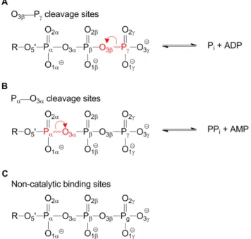
The Protein Data Bank (PDB) provides an opportunity for a top-down analysis of common active site configurations, including hydrogen bonding interactions with water. In this work, phosphoryl transferases are used to test the postulate that comparative analysis can complement traditional reductionist investigations of individual systems, providing additional enzymo-logical insights. Three classes of structures are compared here: those that cleave an O3b—Pcbond (Figure 1a); those that cleave a
Pa—O3abond (Figure 1b), and a ‘‘control’’ group of those that
analysis of an active site mutant affecting interactions with the bridgingb-oxygen in the reaction of arginine kinase, an enzyme in which such interactions had not previously been implicated. Finally, quantum mechanical calculations reveal a possible stereochemical rationale for the observed interactions with both bridging and non-bridging b-oxygens, adding to a number of plausible proposals that seek to explain the bond selectivity and mechanism of phosphoryl transfer.
Materials and Methods
Database analysis
Structure sets. Coordinates for protein-nucleotide complex-es (Table S1) were downloaded from the PDB, including those containing ATP, UTP, TTP, GTP, CTP or their analogs. Two sets of data were compiled, one containing structures up to 2.0 A˚ resolution, where solvent molecules were defined with confidence, and a larger group (inclusive of the first) up to 2.7 A˚ resolution from which solvent water was excluded, due to insufficient confidence in their positions at this resolution. Of the 1866 protein-NTP complexes to 2.7 A˚ , as of October 3rd, 2012, 305 were non-redundant and satisfied inclusion criteria (Table S2) that, for example, excluded structures in conformations known to be non-catalytic. Of the 305 vetted structures, 134 were at resolutions higher than 2.0 A˚ . Molprobity’s Reduce [7] was used to flip asparagine, glutamine and histidine side chains for optimal hydrogen bonding.
Structures were grouped by the site of bond cleavage in the ligand: 1) at the O3b—Pc bond (Figure 1a); 2) at the Pa—O3a
(Figure 1b); and 3) those in which the ligand binding site is non-catalytic (Figure 1c). The groups had 155, 100, and 50 PDB structures in the 2.7 A˚ set, respectively and 72, 45 and 17 structures in the 2.0 A˚ set. An underlying premise was that
enzymes might share catalytic features regardless of base-type (GTP, ATP,etc.). The set of proteins with non-catalytic binding sites serves as a control to distinguish potentially catalytic interactions from those primarily involved in ligand-binding. Comparisons between the sets of enzymes with Pa—O3a and
O3b—Pc cleavage sites also help to distinguish binding and
catalytic interactions, due to spatial separation of binding and catalytic sub-sites.
Identification of interactions. Coordinates were expanded according to the crystallographic symmetry and riding hydrogen atoms were added. These hydrogens had not been seen directly by x-ray crystallography, so their positions were inferred from the heavy atom coordinates using Reduce from the MolProbity program [7]. Assessed interactions included hydrogen bonds and coordination of metal ions, all analyzed using an in-house Python program.
Heavy atom distance and angle criteria were based on previous work [8]: ONNND distance#3.5 A˚ , P—ONNND angle$90u, where D
is the hydrogen bond donor, P is the ligand phosphorus atom, and O is the oxygen atom of the ligand. An additional criterion was added: ONNND—R angle$90uwhere R is the antecedent atom of a
hydrogen bond donor. These criteria depend only on the heavy atoms directly observable and can therefore be applied directly to medium resolution crystal structures. Assessments were also made using additional criteria possible once riding hydrogens were added: ONNNH#2.7 A˚ , P—ONNNH$80u and ONNNH—D$80u.
In-spection revealed that a small number of atom pairs satisfied the criteria without interacting directly, because they were brought into proximity by coordinating with a common metal ion. These were excluded using criteria shown in Figure S1. Metal interactions were subject only to a distance cut-off of 2.8 A˚ derived from analysis of metal coordination in the Cambridge Structural Database.
Classification and comparison of interactions. Ligand oxygens were organized into six groups: Non-bridgingc-,b- and
a-oxygens, and bridgingb-,a- and O59-oxygens. Distributions of enzyme-ligand interactions for each of the groups were compared using pair-wise two-tailed t-tests in RStudio using pooled standard deviations. When comparing the numbers of interactions with different atoms for the different structure sets, the Benjamini & Hochberg correction for multiple comparisons was applied. For each comparison, the null hypothesis was tested that the two sample means could be drawn from the same population, at a significance level of a= 0.05. Results are fully tabulated in the supplemental material (Table S3 and Table S4).
Quantum mechanical analysis of interactions
The Gaussian 09 program [9] was used for all electronic structures calculated with the B3LYP/6-311++G(d,p) level of theory [10]. Bulk solvent was modeled implicitly with the polarizable continuum model (PCM) using a solvent dielectric of 78.36 for water [11]. This level of theory had previously been shown to be appropriate for capturing solvation effects [12]. Natural bond orbital (NBO) analysis [13] was performed with the GenNBO5.0 program [14] using the B3LYP/6-311++ G(d,p)-optimized structures and HF/cc-PVTZ model chemistry [15]. The NBO program facilitates a Lewis-like description of electron density by transforming nonorthogonal atomic orbitals from a HF wave function into natural bond orbitals. Second order perturba-tion E(2) energy, given by Equaperturba-tion 1, estimates the magnitude of electron delocalization and hyperconjugation.
Figure 1. Enzyme structures can be categorized according to the fate of the bound nucleoside triphosphate (NTP). A) Phosphoryl transfer in which the O3b_Pcbond is cleaved. B) Reactions
in which the Pa_O3abond is cleaved and C) Structures where the bound NTP does not undergo a chemical reaction. Red lettering indicates the atoms in the scissile bond and red arrows depict the transfer of electrons in going from reactants to products.
E(2)~qi Fi,j 2
Ej{Ei
ð1Þ
qiis the donor orbital occupancy, and Ej– Eiis the energy gap between donor orbital i and acceptor orbital, j. Fi,jis the Fock matrix element which describes donor and acceptor orbital overlap. In this work, the donor will be a lone pair orbital of the
c-phosphate oxygens and the acceptor orbital iss*(O3b—Pc), the
antibonding orbital of the O3b—Pcbond in methyl triphosphate.
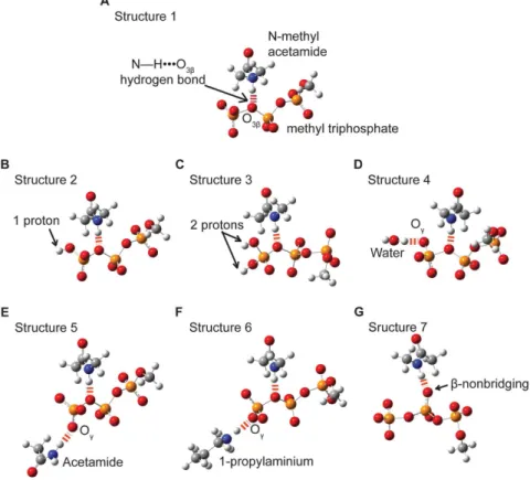
Methyl triphosphate has been used previously, to model interac-tions in ATP (Figure 2) [16].
The influence of a N—HNNNO3bhydrogen bond on the O3b—Pc
bond length and hyperconjugation was evaluated with Structures 1 through 6 (Figure 2a–f). (Similar results were obtained with the addition of Mg++coordinated tob- andc-oxygens.) Structure 7
was used to evaluate the effects of a hydrogen bond to a nonbridging b-oxygen (Figure 2g). N-methylacetamide was used to model a peptide backbone (neutral) hydrogen bond donor. The N—HNNNObhydrogen bond was weakened by increasing its length
from 2.5 A˚ to 3.5 A˚ in 0.1 A˚ increments for Structures 1 through 3 and in 0.2 A˚ increments for Structures 4 through 7. These changes were made by translating the N-methylacetamide, fixing the O3band N atoms at a specific hydrogen bond distance, then
optimizing other aspects of the structure with the N—HNNNOb
angle fixed at 170u. Structures 2 through 6 were used to probe the
electronic effects of either protontion or hydrogen bonding at the
c-oxygens of methyl triphosphate (Figure 2b–f). For protonated structures the O3b—Pc—Oc—H dihedral angle was fixed at 180u.
Acetamide was used as a model for an asparagine side chain in Structure 5, and 1-propylaminium was used as a model for a lysine side chain in Structure 6. All hydrogen bonds toc-oxygens, were fixed at a DNNNOcdistance of 2.8 A˚ , where D is the hydrogen bond
donor heavy atom. The DNNNOc—Pc—O3band the (D)HNNNOc—
Pc—O3b dihedral angles were each fixed at 180u. Changes in
O3b—Pcbond lengths and in E(2) energies were calculated relative
to each molecule without a hydrogen bond between N-methylacetamide and Ob.
Mutagenesis and Kinetics
Mutants of Limulus polyphemus arginine kinase (AK) were made using the QuikChange mutagenesis kit (Stratagene, Inc.). Expression, purification and kinetic analysis of mutants R280L and R280K followed previously published methods [17], though only R280K had sufficient activity for full analysis. Initial velocities were measured using a 666 grid of substrate concentrations: ATP
at 0.2, 0.4, 0.8, 1.2, 1.6, and 2.0 mM, and arginine at 0.2, 0.4, 0.8, 1.6, 3.2, and 7.0 mM. Duplicate measurements were made with separate protein preparations. Steady state parameters Kia, Km and Vmaxfor a random order sequential bimolecular-bimolecular (bi-bi) reaction mechanism were fitted to the data by nonlinear least squares using SigmaPlot.
Figure 2. Models used to test the dependence of hyperconjugation and O3b_Pcbond length on enzyme-ligand interactions.
N-methylacetamide (A) was used to model a (neutral) protein backbone amide hydrogen bond to O3b, using methyl triphosphate to model an NTP
nucleotide. Structures 2 (B) and 3 (C) were used to investigate the effects of protonation at thec-oxygens. Additional active site hydrogen bonds, represented in Structures 4 through 6 (D–F), were used to assess the secondary effects of different types of Ochydrogen bonds. Acetamide (E) 1-propylaminium (F) were used to model asparagine and lysine side chains respectively. Structure 7 (G) was used to investigate the impact of hydrogen bonding at a nonbridging b-oxygen on hyperconjugation and O3b_Pc bond length. Hydrogen bonds are shown with dashed red lines.
Results & Discussion
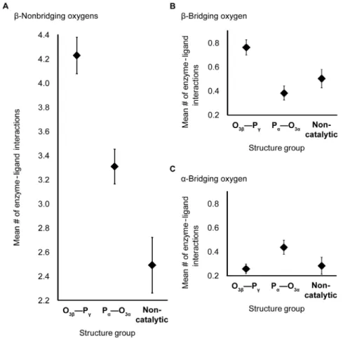
Database analysis of enzyme-nucleotide interactions Nucleotide interactions included hydrogen bonds with the enzyme, water or other substrates, as well as interactions with metal ions. The numbers of interactions were compared between phosphoryl transfer enzymes (Figure 1a) and two comparison groups (Figure 1b, c) to distinguish potentially catalytic from non-catalytic associations. T-tests reveal two overall trends (Table S3 and Table S4), that interactions with both non-bridgingb-oxygens and with the bridging oxygen of a scissile phosphoanhydride bond are more prevalent in phosphoanhydride bond-cleaving sites than in non-catalytic nucleotide-binding sites.
For non-bridgingb-oxygens, the mean numbers of interactions in both O3b—Pc–cleaving sites (m= 4.2, s= 0.2) and Pa—O3a–
cleaving sites (m= 3.3, s= 0.1) are significantly greater than in non-catalytic sites (m= 2.5, s= 0.2; p,0.05; 2.7 A˚ ; Figure 3a, Table S3a). The database analysis extends the finding in Ras, by Fourier Transform Infrared (FTIR) spectroscopy, of stronger interactions with b-oxygens than a- or c-oxgyens in [18], indicating that strong Ob interactions are widespread among
enzymes. Enzymes that cleave the O3b—Pcbond favor neutral
H-bond partners with the non-bridgingb-oxygens, whereas positively charged hydrogen bond donors are common in non-catalytic NTP binding sites. Backbone amide Ob interactions have been
implicated with Walker A motif P-loops [1,19], so the pre-eminence of neutral hydrogen bonding in the database indicates
that analogous interactions are favored in a more general array of phosphoryl transfer enzymes.
Although not every protein interacts with the bridging O3b,
interactions are about twice as common in O3b—Pccleaving sites
(mean number of 0.8,s= 0.1) as in either Pa—O3acleaving sites
(m= 0.4, s= 0.1) or non-catalytic NTP-binding sites (m= 0.5,
s= 0.1; p,0.05; 2.7 A˚ ; Figure 3b, Table S3a). (The statistics quoted are for the larger data set, but similar results are also obtained with the smaller 2.0 A˚ set that included solvent waters, Table S3b and Table S4b). Hydrogen bonds with the O3bmost
commonly involve neutral donors, particularly from backbone nitrogens (Table S5). Other donors include water and positively charged amino acids, especially arginine.
Interactions with bridging O3bhave previously been cited for G
proteins and several kinases, including 6-phosphofructo-2-kinase/ fructose-2,6-bisphosphatase and pyruvate kinase [2,5,20]. Thus, the structural survey captures results expected from detailed prior analyses and shows that analogous interactions actually occur in a diverse array of phosphoryl transfer enzymes including the MutL DNA mismatch repair protein [21], the DEAD-box RNA helicase [22], and Phosphoenolpyruvate carboxykinase [23]. The donor is often, but need not be, within some type of conserved motif, for example at position 4 of the Walker A motif (p-loop; Table S5). O3b interactions have previously been rationalized in terms of
stabilizing development of negative charge on the leaving group in proposed dissociative transfer mechanisms [2,5,20], though Warshel has noted that associative mechanisms would likely also
Figure 3. Mean number of enzyme-ligand interactions.Interactions are shown with nonbridgingb- (A), bridgingb- (B) and bridginga-oxygens (C) in the O3b_Pccleaving-, Pa_O3acleaving- and non-catalytic-NTP-binding sites. The resolution cutoff of structures used is 2.7 A˚, and water is not included. Bars show standard errors.
benefit from electrostatic stabilization of increased electron density on the O3b [24,25]. Our survey demonstrates O3b interactions
occur in diverse enzyme families, such as the phosphagen kinases, N-Acetyl-L-Glutamate kinase and Adenosine kinase, in which the mechanisms have been reported to be significantly associative [26,27,28,29]. Thus, the interaction may transcend the associa-tive/dissociative mechanistic categorization. The expanded set of enzymes exhibiting O3b interactions represent a wide array of
protein folds and differ in other aspects of active site configuration. Indeed, the proposed primary catalytic mechanisms of these enzymes are quite diverse, so it was not obvious that such a broad array of enzymes would share analogous interactions. The much greater incidence of O3binteractions among enzymes cleaving at
O3b—Pc over Pa—O3a, and over proteins with non-catalytic
nucleotide binding sites, suggests that most of these interactions have evolved to have a role in the chemistry and not just binding. Interactions with O3ain enzymes that cleave the Pa—O3abond
are more frequent than in the comparison groups, but more marginally than the O3binteractions. Only the difference between
Pa—O3a cleaving sites (m= 0.4, s= 0.1) and O3b—Pc cleaving
sites (m= 0.3, s,0.1) reaches statistical significance (p,0.05, Table S3a).
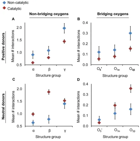
A preference was found for positively charged hydrogen bond donors in non-catalytic binding sites relative to the catalytic sites (O3b—Pc and the Pa—O3a combined group; Figure 4a, b).
Differences manifested particularly at the non-bridgingc-oxygens (catalytic:m= 2.0,s= 0.2; non-catalytic:m= 1.4,s= 0.1; p,0.05; 2.7 A˚ ; Table S6)). The number of neutral hydrogen bond donors in catalytic sites exceeded the average in non-catalytic sites (Figure 4c, d) particularly for the non-bridging (catalytic:m= 0.8,
s= 0.2; non-catalytic: m= 1.9, s= 0.1) and bridgingb- oxygens (catalytic: 0.2, s= 0.1; non-catalytic: m= 0.4, s,0.1; p,0.05; 2.7 A˚ ; Table S6). There are several possible explanations for the observed differences in disposition of charged interactions, including the possibility that non-catalytic binding sites have evolved without pressure for efficient dissociation of reaction products.
Quantum mechanical modeling of nucleotide interactions
Several mechanistic rationales have been offered for the O3b
interactions in G proteins and related enzymes [2,4,5,6,20,25,28,30,31,32]. This section examines if such inter-actions could (also) affect nucleotide stereoelectronics in light of its recently appreciated importance in the modulation of phosphoan-hydride bond energies [12,16]. The total of hyperconjugative effects has been estimated at,300 kJ/mol, with hydrogen bonds to c-oxygens each lessening this by ,20 kJ/mol [16], so modulation of active site interactions could plausibly impact reactions with activation barriers like the 34 kJ/mol of arginine kinase [17]. Our approach is Natural Bond Order analysis [13] which can inform us of the ground state disposition, but is not applicable to the transition structure, so the approach allows only limited inferences about rate enhancement.
For G proteins, the O3b interactions have been rationalized
through electrostatics [5]. Hydrogen bonding or cation interac-tions would favor charge accumulation on O3bin the progression
towards a dissociative transition state, or it could favor the transient accumulation of electron density in an associative mechanism [24,25]. It has also been noted that favoring charge accumulation on O3bwould also tend to increase the susceptibility
of Pc (and Pb) to nucleophilic attack [28]. A dissociative
mechanism has been cited as consistent with FTIR experiments on Ras-GAP [33] that indicated reduction ofb- and increase inc
-phosphoryl group bond orders. It is thought that electrostatic stabilization of negative charge at theb-oxygens draws negative charge (by induction) out of the O3b—Pc bond onto the O3b
oxygen, which is then partially transferred to the non-bridgingb -oxygens through resonance [33,34,35]. A small molecule quantum study of a series of organic phosphates indicated, however, that hyperconjugation was likely to be a greater factor than induction in scissile bond lengths [12]. Hyperconjugation is a stereoelec-tronic effect, where electron density is transferred from an electron rich donor orbital to the antibonding orbital of a neighboring bond [36]. An example is depicted in Figure 5, where electron density is transferred from ac-oxygen lone pair orbital in methyl triphosphate into the antibonding orbital of the O3b—Pc bond.
Rationalization of the mechanistic impact of O3binteractions has
already been controversial [5,24,25,28]. The goal here was not to arbiter this discussion, but to examine the plausibility of O3b
interactions having impact mediated through hyperconjugation, possibly in addition to a mixture of other effects.
Past quantum mechanical modeling of methyl triphosphate [12,16] has shown that enhanced hyperconjugation renders c -oxygens more positive and the O3b more negative as electron
density is transferred from the c-phosphoryl group to the s* O3b—Pc antibonding orbital, lengthening the scissile bond.
Qualitatively, the changes in charge and bond length are the same as those proposed for electrostatic O3binteractions [5] and
observed through FTIR on RAS-Gap [33]. Thus the task is not to rule out the possibility of electrostatic contributions to the mechanism but rather to determine whether hyperconjugation could be a major contributor as well. Whether interactions with theb-oxygens can enhance hyperconjugation in a nucleotide, is tested through quantum mechanical calculations on a methyl triphosphate model system with a hydrogen bond to one of itsb -oxygens from N-methylacetamide (Figure 2). The dependence of hyperconjugation upon the N—HNNNO3b hydrogen bond is
investigated by calculation of the O3b—Pc bond length as the
length of the N—HNNNO3bhydrogen bond is perturbed.
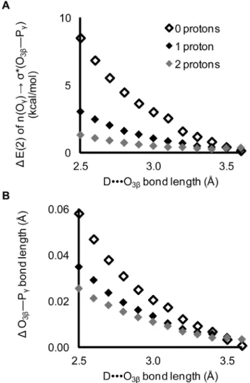
Density functional calculations on Structure 1 (Figure 2a) show that a single hydrogen bond, between N-methylacetamide and the O3b oxygen in methyl triphosphate, induces an increase in
hyperconjugation and a,0.05 A˚ elongation of the O3b—Pcbond
(Figure 6). This affirms the hypothesis that the O3b—Pc bond
length can, at least in principle, be weakened by O3bhydrogen
bonding acting through hyperconjugation.
Two factors determine the magnitude of hyperconjugation; the degree of donor and acceptor orbital overlap, and the orbital energy gap. The orbital overlap (Fij in Eq. 1) and the orbital energies of n(Oc) ands*(O3b—Pc) were calculated for Structure 1
(Figure 2a), as a function of hydrogen bond length (Figure 7). There is a strong dependence of the energy of thes*(O3b—Pc)
antibonding orbital upon strength of the hydrogen bond (Figure 7a). However, neither the energy of the non-bridgingc -oxygen lone pair orbitals (Figure 7b) nor the orbital overlap (Figure 7c) are affected. As the hydrogen bond is shortened (made stronger), the orbital energy gap decreases, increasing hypercon-jugation and elongating the O3b—Pcbond.
The relationship between O3b—Pc bond elongation and
hyperconjugation was confirmed by protonating the c-oxygens. Protonation of the c-oxygens decreases the hyperconjugative interaction: n(Oc)Rs*(O3b—Pc) [12,16]. We now find that
protonation also decreases the dependence of the O3b—Pcbond
length on the strength of the DNNNO3bhydrogen bond (Figure 8),
consistent with the impact of an O3bhydrogen bond upon O3b—
Hyperconjugation is also subject to other ‘‘secondary’’ pertur-bations that might be of greater biochemical relevance; hydrogen bonds added at the c-oxygens interfere with hyperconjugation [16]. The primary dependence of O3b—Pc elongation on the
Figure 4. Non-catalytic (blue) active sites have a preference positively charged hydrogen bond donors (a–b) whereas in catalytic (red) active sites neutral interactions are favored (c–d).Mean numbers of hydrogen bonds were measured for non-bridging (a & c) and bridging (b & d) oxygens. The catalytic group is composed of both O3b—Pcand Pa—O3astructure sets. Positive donors include Lys, Arg, and His side chains and neutral donors include Asn, Gln, Trp, Ser Thr,Tyr, Cys side chains, the nucleotide O29and O39oxygens and all backbone nitrogens (except Pro).
doi:10.1371/journal.pone.0108310.g004
Figure 5. Hyperconjugation in methyl triphosphate, a model for ATP.Electron density is transferred from a lone pair orbital on one of thec-oxygens (Oc), into the antibonding orbital (s*) of the O3b—Pc
bond. Red lettering indicates the atoms involved in the hyperconjuga-tive interaction and the red arrow represents electron density transfer. doi:10.1371/journal.pone.0108310.g005
Figure 6. The O3b—Pc bond length and hyperconjugation increase with decreasing DNNNO3b hydrogen bond length in Structure1. Calculated E(2) energy is of the n(Oc)Rs*(O3b—Pc)
hyperconjugative interaction. D is the N—H hydrogen bond donor in N-methylacetamide.
strength of the O3bhydrogen bond was then recalculated, after the
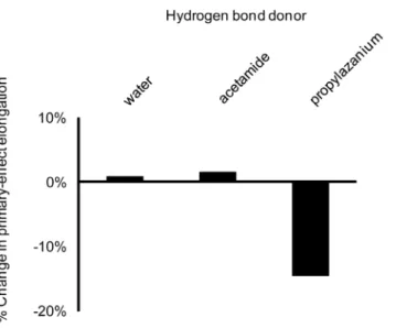
addition of (secondary) hydrogen bonds at c. Several c-oxygen hydrogen bond donors were considered: water, acetamide (mimicking an asparagine) and 1-propylaminium (mimicking a lysine; Figure 2). Hydrogen bonds to c-oxygens were fixed at 2.8 A˚ , while the hydrogen bond between N-methylacetamide and the O3b oxygen was varied from 2.5 A˚ to 3.1 A˚ in 0.2 A˚
increments.
Hydrogen bonds from neutral donors (water or acetamide) to a
c-oxygen have little effect (Figure 9). Only the salt bridge bond from a positively charged 1-propylaminium donor modulates the primary interactions. The effect is modest, reducing bond elongation by about 15%, but it is possible that multiple Ocsalt
bridges could have an additive effect.
Quantum mechanical analysis of interactions with non-bridging oxygens– Of interest is whether hyperconjugation can explain the higher frequencies of interactions with non-bridgingb-oxygens in O3b—Pccleaving active sites, relative to non-catalytic binding sites
(Figure 3). Similar to our study of O3b interactions, QM
calculations were performed for N-methylacetamide hydrogen-bonded to a non-bridging b-oxygen in Structure 7 (Figure 2g). Changes in the n(Oc)Rs*(O3b—Pc) hyperconjugative interaction
were calculated as the hydrogen bond length was varied from 2.5 A˚ to 3.5 A˚ in 0.2 A˚ increments.
The impact of a single hydrogen bond with a non-bridgingb -oxygen is smaller than that of an O3bhydrogen bond (Figure 10).
However, the database analysis revealed an average of four enzyme interactions summed across the two non-bridging b -oxygens (Figure 3a), nearly saturating the lone pairs. Multiple hydrogen bonds to a single oxygen in methyl triphosphate had an additive effect on hyperconjugation [16]. Thus the aggregate effect of interactions with non-bridgingb-oxygens could be commensu-rate with those of the O3b.
Figure 7. Effects of a hydrogen bond at O3b on orbital and interaction energies in Structure1.Shortening the hydrogen bond between N-methylacetamide and methyl triphosphate: A) decreases the orbital energy of thes*(O3b—Pc) anti-bonding orbital; while (B) leaving
unchanged both the n(Oc) donor orbital energies and (C) Fi,j, a measure
of the overlap between the n(Oc) lone pair orbitals ands*(O3b—Pc). D
denotes hydrogen bond donor.s* denotess*(O3b—Pc).
doi:10.1371/journal.pone.0108310.g007
Figure 8. Impact of protonation of thec-oxygens.Protonation at thec-oxygens decreases the effects of the O3bhydrogen bond on both A) the magnitude of hyperconjugation and B) the O3b—Pcbond length.
Changes are calculated relative to corresponding structures without a DNNNO3b hydrogen bond. The ability of interactions with the O3b to increase the scissile bond depends substantially on the presence of strong intrinsic hyperconjugation in the ligand.
In summary, hyperconjugation weakens the scissile O3b—Pc
bond. Hydrogen bonds to the phosphate oxygens can either amplify or reduce this effect, according to proximity with the scissile bond. The greatest weakening of the scissile bond comes from interactions with O3b, proximal to the scissile bond, with
lesser weakening from interactions at non-bridging Obthat are
two bonds removed. By contrast, hydrogen bonds toc-oxygens, vicinal to the scissile bond, interfere with the hyperconjugation, strengthening the scissile bond [16]. Changes in hydrogen bonding are inherent to the act of substrate binding, but of interest is whether particular types of interactions have been selected in enzyme evolution. Here we test the hypothesis that enzymes enhance hyperconjugation by maximizing binding to b- and minimizing binding toc-oxygens.
Noting that the desolvation of the non-bridging b- and c -oxygens on substrate binding would have opposite effects upon O3b_Pcbond elongation, the 2 A˚ structure database was queried
for the fractional saturation of lone pairs with enzyme or solvent interactions, comparing the total five lone pairs of non-bridgingb -oxygens to the 8 of non-bridgingc-oxygens. As predicted, the non-bridging b-oxygens retain higher numbers of interactions than either the non-bridginga- orc-oxygens (Figure 11, Table S7), and there are more numerous interactions with the bridging O3bthan
either the O3aor O59. The favoring of interactions with theb -phosphoryl group that we see in the database analysis is consistent not only with the QM-based prediction, but with the FTIR studies of Ras that showed stronger binding of theb-phosphoryl group [37].
Consistency of the observed disposition of active site interactions makes a circumstantial case for the importance to enzymes of hyperconjugation-mediated weakening of the O3b—Pcbond. The
ground state QM calculations show clearly that bond selectivity is impacted, so enzymes could be taking advantage of hyperconjuga-tion in specifying which of the phosphoanhydride bonds is to be cleaved. Understanding whether modulation of hyperconjugation has a role in rate enhancement is a more challenging question about which we cannot be as definitive. Bypassing the question of rate enhancement, even a role in bond selectivity alone could be of
sufficient selective advantage to explain the prevalence of hyperconjugation-mediating interactions in the structural data-base.
Ground state quantum mechanical modeling provides, through hyperconjugation, a unifying rationalization for interactions with the phosphate oxygens. The calculated changes in bond order and partial charge are qualitatively consistent with those inferred from kinetic isotope and FTIR studies [33,34], and are consistent with the dispositions of b- and c-oxygen interactions seen in the database analysis. Hydrogen bonding at the bridging O3b and
non-bridgingb-oxygens closes the energy orbital gap, increasing the transfer of electron density into thes* antibonding orbital and lengthening the O3b_Pc bond, while interactions at c-oxygens
have the opposite effect. Thus, differences in the interactions with
b- andc-oxygens can modulate the strength of the scissile bond in a mechanism mediated through hyperconjugation.
To affect rate, the activation barrier must be lowered by some combination of stabilizing the transition state (TS) or destabilizing the ground state. The (spontaneous) existence of hyperconjugation in isolated nucleotides means that it can be stabilizing the ground state. However, inter-molecular interactions that increase hyper-conjugation beyond its natural equilibrium level would destabilize the substrate. Yang and Cui examined whether the anomeric
Figure 9. Impact of (secondary) interactions withc-oxygens on O3b—Pc bond elongation induced by (primary) O3b interac-tions. Secondary interactions with c-oxygens have modest impact (much smaller than the direct effects characterized by Summerton et al. [16]) and are substantial only for charged donors.
doi:10.1371/journal.pone.0108310.g009
Figure 10. Impact of a hydrogen bond with a non-bridgingb -oxygen. An interaction between N-methylacetamide and a non-bridgingb-oxygen of methyl triphosphate decreases (A) the hypercon-jugation and (B) scissile bond length. Interactions with the non-bridging b-oxygen (filled diamonds, Structure 7) are compared to those for O3b (open diamonds, Structure 1). Changes are measured relative to methyl triphosphate absent a hydrogen bond.
effect in myosin destabilized the ground state [38]. A modest increase in ATP calculated potential energy, as the Pc—O3bbond
was lengthened, was deemed insufficient for substantial rate enhancement. However, the argument implicitly depends on a reciprocal relationship between bond length and hyperconjuga-tion, such that distortions of the bond induce a change in hyperconjugation to the corresponding level of the enzyme-bound substrate, a questionable assumption. A definitive assessment of the influence of hyperconjugation on transition state stabilization, has been stymied by inapplicability of NBO calculations to the TS, and by flat energy surfaces (calculated for myosin and RasGAP reactions) from which it is difficult to distinguish different possible mechanisms and estimate entropic terms [25,38]. These energy calculations did, however, imply that the phosphoryl transfer reaction was mixed associative/dissociative in character. Natural resonance theory calculations [39] for model associative and dissociative reactions showed that the hyperconjugation increased on approach to a dissociative transition state, and remained near constant for associative reactions [40]. Thus, for the Ras-Gap and other enzymes with a mixed mechanism, overall interactions that enhance hyperconjugation are predicted to stabilize the TS. Therefore the modulations characterized in this work could
plausibly be enhancing rate as well as bond selectivity. However, we lack a detailed understanding of how the interactions might change on progression to the TS, so caution is needed in speculating about rate enhancement. Furthermore, active site interactions commonly have multiple effects which are not comparatively evaluated in the current work. Thus, the current work adds hyperconjugation as one of the plausible rationaliza-tions of phosphoryl transfer rate enhancement, without ruling out the previously discussed electrostatic effects [5,24,25].
Experimental characterization of an arginine kinase mutant
Among the diverse enzymes shown by the survey to have O3b
interactions, the phosphagen kinases are of particular interest. Physiologically, these enzymes buffer cellular ATP levels through a reversible transfer of the c-phosphoryl to a guanidine substrate such as creatine or arginine [41]. The enzymes have no structural homology to those where O3b interactions were previously
implicated, and have not been reported to share mechanisms of catalysis. Indeed, in contrast to the dissociative mechanisms postulated for G-proteins and other enzymes with previously noted O3b interactions [2,5,20], a more associative mechanism was
suggested for phosphagen kinases [26,27]. A high (1.2 A˚ ) resolution crystal structure of an arginine kinase transition state analog complex added circumstantial evidence [42]. The nitrate, mimicking a transferring phosphoryl was situated between the guanidino N and nucleotide O3b which were separated by 6 A˚ ,
consistent with partial axial bonding of a pentavalent Pc, and the
angles of approach for guanidino N and O3bwere within a few
degrees of optimal for nucleophilic attack in an early stage of the reaction, before deprotonation of the guanidino N. This suggested an active site in which enzyme interactions were optimized for a significantly associative reaction. Such contrasts to the aforemen-tioned G proteins, together with the availability of high resolution structure, rendered arginine kinase particularly suitable as an experimental test of the predictive power of the database analysis. Below we report an experimental validation of the importance of O3binteractions highlighted by the database analysis, noting that
the steady state kinetics can be used to investigate the overall role of an interaction, but not detailed mechanism.
In the arginine kinase structure, the O3binteracts with Arg280 and Arg126. Arg126 interacts with both O3b and Oc, so Arg280,
which also interacts with an a-nonbridging oxygen of the nucleotide substrate (and an enzyme aspartate) was chosen for mutagenesis, because it did not have potentially confounding interactions with c-oxygens (Figure 12). Preliminary analysis showed that the activity of a R280K mutant was sufficient for steady-state kinetics analysis, but not that of a R280L mutant. Relative to the wild-type (WT), kcatof R280K was decreased from 104 s21 to 1 s21, whereas K
M values for the two substrates remained within 3-fold of WT (R280K: KM(Arg) = 0.8 mM;
KM(ATP) = 0.5 mM. WT KM(Arg) = 0.3 mM;
KM(ATP) = 0.3 mM) [43]. The much larger impact upon rate indicates that the effect of the mutation is predominantly upon the chemical reaction as opposed to substrate binding.
The arginine kinase R280K is analogous to the R291K mutant of the homologous enzyme, creatine kinase (CK) in rabbit muscle, which yielded similar kinetics (kcat= 2.2 s21 and KM(ATP) = 1.6 mM) [44]. It was suggested that this mutation abrogated catalysis by misaligning the substrates. However, near wild-type KM values argue against gross distortions of substrate-binding, although the effects of subtle modulations of alignment cannot be ruled out [45,46]. Our database survey now highlights the analogy of the O3binteraction with the corresponding ones in
Figure 11. Saturation of NTP oxygen lone pairs by enzyme or solvent interactions in O3b—Pc-cleaving active sites.Bars denote
standard errors.
G-proteins and other enzymes that have been discussed above. Thus, one should consider whether there could be a holistic rationalization of O3b interactions, common to phosphagen
kinases, G proteins and other enzymes.
The 100-fold rate reduction of R280K is commensurate with changes on conservative mutations of arginine kinase Glu225and Cys271 that are implicated respectively in base catalysis and electrostatic modulation of the guanidino substrate [45,47]. Thus, the R280K mutant adds to the picture of phosphagen kinases employing several means of enhancing rate, of which the O3b
interactions are an important, but not dominant component. Furthermore, it is noted that the wild-type turnover is not limited by chemistry, but by protein conformational change [17]. Thus any of these mutations could be having a greater effect upon the chemistry, because we are only able to measure the part of slowing beyond the 104 s21WT turnover.
Conclusions
Several messages can be taken from the current work. Firstly, top-down analysis of protein structure databases can reveal commonalities in the disposition of diverse active sites catalyzing analogous reactions. Secondly, O3binteractions figure
prominent-ly in a wide variety of phosphoryl transferases, not just those like the G proteins previously proposed to have a dissociative mechanism. Thirdly, modulation of hyperconjugation in selective bond destabilization can be used to rationalize the disposition of phosphate active site interactions, as a potential alternative to electrostatic rationales. Finally, we are reminded that interactions cause multiple effects within substrates, and that until more quantitative analysis allows prioritization of the various effects, stereolectronics should be considered alongside more classical explanations in understanding the workings of phosphoryl transfer enzymes.
Supporting Information
Figure S1 Exclusion of interactions with ion-coordi-nants.
(TIF)
Table S1 Protein Data Bank structures used in the study.
(XLSX)
Table S2 Structure selection criteria.
(XLSX)
Table S3 Pairwise t-tests between structure sets, no hydrogens.
(XLSX)
Table S4 Pairwise t-tests between structure sets, riding hydrogens added.
(XLSX)
Table S5 Structures with O3b nucleoside triphosphate interactions.
(XLSX)
Table S6 Salt bridges to nucleotidec-oxygens.
(XLSX)
Table S7 Pairwise t-tests for interactions between different oxygen types.
(XLSX)
Acknowledgments
Omar Davulcu is thanked for help in protein purification and enzyme kinetics and Nancy Meyer is thanked for help with generating figures. The authors would also like to thank Johannes Elferich and Kyle Ambert for help with R and Python scripting.
Author Contributions
Conceived and designed the experiments: JCS MSC JDE. Performed the experiments: JCS GMM. Analyzed the data: JCS. Contributed reagents/ materials/analysis tools: JCS MSC. Wrote the paper: JCS MSC JDE.
References
1. Matte A, Tari LW, Delbaere LT (1998) How do kinases transfer phosphoryl groups? Structure 6: 413–419.
2. Larsen TM, Benning MM, Rayment I, Reed GH (1998) Structure of the bis(Mg2+)-ATP-oxalate complex of the rabbit muscle pyruvate kinase at 2.1 A˚ resolution: ATP binding over a barrel. Biochemistry 37: 6247–6255. 3. Schweins T, Geyer M, Scheffzek K, Warshel A, Kalbitzer HR, et al. (1995)
Substrate-assisted catalysis as a mechanism for GTP hydrolysis of p21ras and other GTP-binding proteins. Nat Struct Biol 2: 36–44.
4. Bi X, Corpina RA, Goldberg J (2002) Structure of the Sec23/24-Sar1 pre-budding complex of the COPII vesicle coat. Nature 419: 271–277. 5. Maegley KA, Admiraal SJ, Herschlag D (1996) Ras-catalyzed hydrolysis of
GTP: a new perspective from model studies. Proc Natl Acad Sci U S A 93: 8160–8166.
6. Zhu G, Liu J, Terzyan S, Zhai P, Li G, et al. (2003) High resolution crystal structures of human Rab5a and five mutants with substitutions in the catalytically important phosphate-binding loop. J Biol Chem 278: 2452–2460. 7. Word JM, Lovell SC, Richardson JS, Richardson DC (1999) Asparagine and
glutamine: using hydrogen atom contacts in the choice of side-chain amide orientation. J Mol Biol 285: 1735–1747.
8. Fabiola F, Bertram R, Korostelev A, Chapman MS (2002) An improved hydrogen bond potential: impact on medium resolution protein structures. Protein Sci 11: 1415–1423.
9. Frisch MJ, Trucks GW, Schlegel HB, Scuseria GE, Robb MA, et al. (2009) Gaussian 09, Revision A.02. Wallingford CT: Gaussian, Inc.
10. Lee C, Yang W, Parr RG (1988) Development of the Colle-Salvetti correlation-energy formula into a functional of the electron density. Physical Review B: Condensed Matter 37: 785–789.
11. Miertus S, Scrocco E, Tomasi J (1981) Electrostatic Interaction of a Solute with a Continuum. A Direct Utilization of ab initio Molecular Potentials for the Prevision of Solvent Effects. Chem Phys 55: 117.
12. Ruben EA, Plumley JA, Chapman MS, Evanseck JD (2008) Anomeric effect in ‘‘high energy’’ phosphate bonds. Selective destabilization of the scissile bond and modulation of the exothermicity of hydrolysis. J Am Chem Soc 130: 3349–3358. 13. Weinhold F, Landis CR (2005) Valency and bonding : a natural bond and orbital donor-acceptor perspective. Cambridge: Cambridge University Press. ix, 749 p. p.
14. Glendening ED, Badenhoop JK, Reed AE, Carpenter JE, Bohmann JA, et al. (2001) NBO Version 5.9. 5.9 ed. Madison, Wisconsin: Theoretical Chemistry Institute, University of Wisconsin.
15. Dunning T (1989) Gaussian basis sets for use in correlated molecular calculations. I. The atoms boron through neon and hydrogen J Chem Phys 90: 1007.
16. Summerton JC, Evanseck JD, Chapman MS (2012) Hyperconjugation-Mediated Solvent Effects in Phosphoanhydride Bonds. The Journal of Physical Chemistry A 116: 10209–10217.
17. Davulcu O, Skalicky JJ, Chapman MS (2011) Rate-Limiting Domain and Loop Motions in Arginine Kinase. Biochemistry 50: 4011–4018.
18. Cepus V, Scheidig AJ, Goody RS, Gerwert K (1998) Time-resolved FTIR studies of the GTPase reaction of H-ras p21 reveal a key role for the beta-phosphate. Biochemistry 37: 10263–10271.
19. Leipe DD, Koonin EV, Aravind L (2003) Evolution and classification of P-loop kinases and related proteins. J Mol Biol 333: 781–815.
20. Hasemann CA, Istvan ES, Uyeda K, Deisenhofer J (1996) The crystal structure of the bifunctional enzyme 6-phosphofructo-2-kinase/fructose-2,6-bisphospha-tase reveals distinct domain homologies. Structure 4: 1017–1029.
21. Ban C, Junop M, Yang W (1999) Transformation of MutL by ATP binding and hydrolysis: a switch in DNA mismatch repair. Cell 97: 85–97.
22. Sengoku T, Nureki O, Nakamura A, Kobayashi S, Yokoyama S (2006) Structural basis for RNA unwinding by the DEAD-box protein Drosophila Vasa. Cell 125: 287–300.
23. Sullivan SM, Holyoak T (2008) Enzymes with lid-gated active sites must operate by an induced fit mechanism instead of conformational selection. Proc Natl Acad Sci U S A 105: 13829–13834.
24. Glennon TM, Villa J, Warshel A (2000) How does GAP catalyze the GTPase reaction of Ras? A computer simulation study. Biochemistry 39: 9641–9651. 25. Klahn M, Rosta E, Warshel A (2006) On the mechanism of hydrolysis of
phosphate monoesters dianions in solutions and proteins. J Am Chem Soc 128: 15310–15323.
26. Lowe G, Sproat BS (1980) Evidence for an associative mechanism in the phosphoryl transfer step catalyzed by rabbit muscle creatine kinase. J Biol Chem 255: 3944–3951.
27. Hansen DE, Knowles JR (1981) The Stereochemical Course of the Reaction Catalyzed by Creatine Kinase. Journal of Biological Chemistry 256: 5967–5969. 28. Ramon-Maiques S, Marina A, Gil-Ortiz F, Fita I, Rubio V (2002) Structure of acetylglutamate kinase, a key enzyme for arginine biosynthesis and a prototype for the amino acid kinase enzyme family, during catalysis. Structure 10: 329– 342.
29. Schumacher MA, Scott DM, Mathews II, Ealick SE, Roos DS, et al. (2000) Crystal structures of Toxoplasma gondii adenosine kinase reveal a novel catalytic mechanism and prodrug binding. J Mol Biol 298: 875–893.
30. Zhu Y, Huang W, Lee SSK, Xu W (2005) Crystal structure of a polyphosphate kinase and its implications for polyphosphate synthesis. EMBO Reports 6: 681– 687.
31. Andreassi JL, Vetting MW, Bilder PW, Roderick SL, Leyh TS (2009) Structure of the Ternary Complex of Phosphomevalonate Kinase: The Enzyme and Its Family. Biochemistry 48: 6461–6468.
32. Holzapfel G, Buhrman G, Mattos C (2012) Shift in the Equilibrium between On and Off States of the Allosteric Switch in Ras-GppNHp Affected by Small Molecules and Bulk Solvent Composition. Biochemistry 51: 6114–6126. 33. Allin C, Gerwert K (2001) Ras catalyzes GTP hydrolysis by shifting negative
charges from gamma- to beta-phosphate as revealed by time-resolved FTIR difference spectroscopy. Biochemistry 40: 3037–3046.
34. Du X, Sprang SR (2009) Transition state structures and the roles of catalytic residues in GAP-facilitated GTPase of Ras as elucidated by (18)O kinetic isotope effects. Biochemistry 48: 4538–4547.
35. Du X, Black GE, Lecchi P, Abramson FP, Sprang SR (2004) Kinetic isotope effects in Ras-catalyzed GTP hydrolysis: evidence for a loose transition state. Proc Natl Acad Sci U S A 101: 8858–8863.
36. Fleming I (2010) Molecular Orbitals and Organic Chemical Reactions. Chichester, West Sussex: John Wiley & Sons. 475 p.
37. Allin C, Ahmadian MR, Wittinghofer A, Gerwert K (2001) Monitoring the GAP catalyzed H-Ras GTPase reaction at atomic resolution in real time. Proc Natl Acad Sci U S A 98: 7754–7759.
38. Yang Y, Cui Q (2009) The hydrolysis activity of adenosine triphosphate in myosin: a theoretical analysis of anomeric effects and the nature of the transition state. J Phys Chem A 113: 12439–12446.
39. Glendening ED, Weinhold F (1998) Natural resonance theory: I. General formalism. Journal of Computational Chemistry 19: 593–609.
40. Ruben EA (2007) Stereoelectronic Effects in Phosphates [Ph.D. dissertation]. Tallahassee: Florida State University. 118 p.
41. Ellington WR (2001) Evolution and physiological roles of phosphagen systems. Annu Rev Physiol 63: 289–325.
42. Yousef MS, Fabiola F, Gattis J, Somasundaram T, Chapman MS (2002) Refinement of Arginine Kinase Transition State Analogue Complex at 1.2 A˚ resolution; mechanistic insights. Acta Crystallographica Section D: Biological Crystallography 58: 2009–2017.
43. Blethen SL (1972) Kinetic Properties of the Arginine Kinase Isoenzymes of Limulus polyphemus. Archives of Biochemistry and Biophysics 149: 244–251. 44. Jourden MJ, Geiss PR, Thomenius MJ, Horst LA, Barty MM, et al. (2005)
Transition state stabilization by six arginines clustered in the active site of creatine kinase. Biochim Biophys Acta 1751: 178–183.
45. Pruett PS, Azzi A, Clark SA, Yousef M, Gattis JL, et al. (2003) The putative catalytic bases have, at most, an accessory role in the mechanism of arginine kinase. J Biol Chem 29: 26952–26957.
46. Azzi A, Clark SA, Ellington WR, Chapman MS (2004) The role of phosphagen specificity loops in arginine kinase. Protein Sci 13: 575–585.
47. Gattis JL, Ruben E, Fenley MO, Ellington WR, Chapman MS (2004) The active site cysteine of arginine kinase: structural and functional analysis of partially active mutants. Biochemistry 43: 8680–8689.