Structure-Based Virtual Screening and
Discovery of New PPARδ/γ
Dual Agonist and
PPARδ
and
γ
Agonists
Vinicius G. Maltarollo1,2, Marie Togashi3, Alessandro S. Nascimento4, Kathia M. Honorio1,5*
1Centro de Ciências Naturais e Humanas, Universidade Federal do ABC, Santo André, São Paulo, Brasil 2Faculdade de Ciências Farmacêuticas, Universidade de São Paulo, São Paulo, São Paulo, Brasil,3 Faculdade de Ciências da Saúde, Universidade de Brasília, Brasília, Distrito Federal, Brasil,4Instituto de Física de São Carlos, Universidade de São Paulo, São Carlos, São Paulo, Brasil,5Escola de Artes Ciências e Humanidades, Universidade de São Paulo, São Paulo, São Paulo, Brasil
Abstract
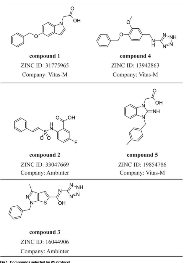
Peroxisome proliferator-activated receptors (PPARs) are involved in the control of carbohy-drate and lipid metabolism and are considered important targets to treat diabetes mellitus and metabolic syndrome. The available PPAR ligands have several side effects leading to health risks justifying the search for new bioactive ligands to activate the PPAR subtypes, in special PPARδ, the less studied PPAR isoform. Here, we used a structure-based virtual screening protocol in order to find out new PPAR ligands. From a lead-like subset of pur-chasable compounds, we identified 5 compounds with potential PPAR affinity and, from preliminaryin vitroassays, 4 of them showed promising biological activity. Therefore, from
ourin silicoandin vitroprotocols, new PPAR ligands are potential candidates to treat
metabolic diseases.
Introduction
Peroxisome proliferator-activated receptors (PPARs) constitute a subfamily of nuclear recep-tors involved in the transcription of genes related to the cellular proliferation and differentia-tion, immune responses and metabolism of carbohydrates and lipids [1–8]. From the
pathological point of view, these receptors are related to metabolic diseases (mainly, type 2 dia-betes mellitus, metabolic syndrome and dyslipidemia) [9–11], inflammatory process [12,13], neurodegeneration [14] and some kinds of cancer [15–17].
The treatment of type 2 diabetes mellitus (T2DM) and metabolic syndrome (MS) brings an important focus to the development of new PPAR agonists. There are at least two classes of pharmacological agents targeting the PPARs. The fibrates are known as PPARαligands used for the control of hypercholesterolemia, while the thiazolidinediones (TZDs) such as rosiglita-zone and pioglitarosiglita-zone are PPARγfull agonists used as insulin sensitizers in T2DM therapy. De-spite the clear efficacy to restore blood glucose levels, rosiglitazone was reported to cause important side effects, such as fluid retention, weight gain and increase in the chance of a
OPEN ACCESS
Citation:Maltarollo VG, Togashi M, Nascimento AS, Honorio KM (2015) Structure-Based Virtual Screening and Discovery of New PPARδ/γDual Agonist and PPARδandγAgonists. PLoS ONE 10(3): e0118790. doi:10.1371/journal.pone.0118790
Academic Editor:Paul Taylor, University of Edinburgh, UNITED KINGDOM
Received:June 30, 2014
Accepted:December 16, 2014
Published:March 13, 2015
Copyright:© 2015 Maltarollo et al. This is an open access article distributed under the terms of the
Creative Commons Attribution License, which permits unrestricted use, distribution, and reproduction in any medium, provided the original author and source are credited.
Data Availability Statement:All relevant data are within the paper and its Supporting Information files.
Funding:The authors would like to thank FAPESP (2011/18981-2, 2014/06565-2), CNPq and CAPES (Brazilian agencies) for funding. The funders had no role in study design, data collection and analysis, decision to publish, or preparation of the manuscript.
cardiovascular event [18]. These effects leaded some regulatory agencies around the world to restrict or suspend the use of rosiglitazone [19]. Interestingly, pioglitazone was shown to be a safer drug than rosiglitazone, raising two interesting considerations about the interactions be-tween PPARγand its agonists. As noted by Bruning and coworkers [18], small changes in the binding mode, as observed between rosiglitazone and pioglitazone, can lead to important changes in the pharmacological profile, including the side effects.
There is another class of PPAR agonists, fibrates, responsible for the activation of PPARα
that is well known to result in a decrease in the triglyceride levels and increase in HDL levels (considered important factors for the establishment of T2DM) [20]. On the other hand, PPARδ(the less studied receptor among the PPARs) was shown to be involved in anti-obesity effects [21] and anti-inflammatory processes as arthritis, eczema and psoriasis [22,23] reinforc-ing the potential beneficial effects of this receptor in the treatment of chronic and metabolic diseases. Most PPAR dual- and pan-agonists developed to date were discontinued during the clinical trials due to harmful effects observed in these assays, making unclear whether these ag-onists can actually exertin vivobeneficial effects [20]. Researches on these topics are very
im-portant have the main purpose of highlighting how relevant it is the development of new PPARδagonists that could be used as chemical probes for a better understanding on the
molec-ular mechanisms involved in the PPARδactivation.
Here, a virtual screening (VS) of a large chemical library, the ZINC database [24], based on the crystallographic structure of PPARδreceptor was used as a tool to identify new PPAR ligand candidates. PPAR agonists can be classified as full agonists (related to the stabilization ofα
-helix 12 [H12] by the interaction with polar residues and, this way, providing structural condi-tions to the recruitment of cofactors at the AF-2 region) and partial agonists (related to a subop-timal stabilization of H12 and also called H12 independent mechanism) [18]. Since the main commercial PPAR agonists (glitazones and fibrates) and the reference PPARδagonist
(GW501516) are full agonists [25,26], our VS protocol was carried out aiming to select the com-pounds by analyzing the interactions that characterize a full agonist. After several analyses of the main ligand-receptor interactions, five compounds were selected for preliminary biological assays and four novel ligands were identified as agonists acting on PPARδ/γand PPARγ. These
ligands are promising candidates to treat metabolic disorders, such as diabetes and metabolic syndrome.
Material and Methods
Molecular Docking
The‘clean-leads’subset of purchasable compounds library was chosen for virtual screening as available in ZINC (version of January-2010) [24]. This subset includes purchasable compounds with molecular weight between 250 and 350 Da, n-octanol/water partition coefficient values between 2.5–3.5 and number of rotatable bonds between 5 and 7 and this subset also excludes toxic compounds with aldehydes and thiol groups.
For the docking simulations, the crystal structure 3GZ9 containing PPARδLBD was chosen [27] and prepared in three steps. First, all non-amino acid atoms (waters, ions, ligands and oth-ers) were removed from the structure. Then, the missing hydrogen atoms were added and, fi-nally, the protonation state of the residues in the active site was automatically defined based on the local interactions. All water molecules were removed from the active site, since there are no structural water molecules mediating the main interactions responsible for the PPARδ
simulations were performed employing DOCK 3.5.54 [29–35] software. The ligand conformers were obtained from ZINC database [24].
50 molecules selected after several analyses of the docking results were used for redocking using docking [36] and GOLD 3.1 [37–39] with their default parameters. The Surflex-docking software employs the incremental search to generate Surflex-docking poses, whereas GOLD 3.1 software uses a genetic algorithm (GA) to generate the poses of ligands. Both Surflex-docking and GOLD programs generated 50 poses for each ligand and the top 5 ranked poses were analyzed. The ranking of the docked poses was performed using CScore and GOLD-Score, respectively. All the generated poses were analyzed with a rigorous visual inspection re-garding the main molecular interactions between some amino acid residues in the active site and the studied ligands. In this analysis, the polar interactions with Thr-289, His-323, His-449 and Tyr-473 and also stereochemical complementarities were inspected.
The main purpose of using three different docking programs to perform a consensus analy-sis is avoiding the pitfalls of each method. DOCK 3.5.54 and GOLD 3.1 programs use force-field based scoring function [34,39] while Surflex-docking uses a consensus scoring function (Chem-Score as empirical scoring function, D-score and G-score as force-field based scoring function and PMF-score as knowledge-based scoring function) [40]. These programs also have important differences in the generation of ligand conformers. The definition of active site (rect-angular box, sphere centered or amino acid residues) and the flexibility degree of ligand bonds/ angles can also affect the success rate in the molecular docking because it interferes in the search space of the docking algorithm. Finally, taking into account that the training set used to calibrate each docking engine is different from each other, it is expected that a docking pro-gram works better on a certain system than others [41]. Several studies indicate that it is too difficult to compare the efficiency of docking programs due to several factors such as the limita-tion of evalualimita-tion metrics (e. g. root mean squared devialimita-tion [RMSD] value could indicate an acceptable pose but the ligand orientation is wrong) [41–43]. Therefore, a consensus between the ranked poses and a careful visual inspection of the results obtained by the three different al-gorithms could improve the final results [44,45].
Transluciferase Assays
HeLa cells were cotransfected [46,47] by electroporation with expression vectors pcDNA3-PPARδor pcDNA3-PPARγ, report vector pGL3-PPARE and pRL. In addition, the cells were treated for 20h in the presence of the studied ligands. Bezafibrate was employed as positive con-trol to PPARδand rosiglitazone was employed as positive control to PPARγ. DMSO was em-ployed as vehicle to compounds 1–4 and the DMSO:EtOH (2:1 v/v) mixture was emem-ployed as vehicle for compound 5. The PPAR activation measurements were estimated by Luciferase Assay System (Promega) and were determined by the standard deviation of triplicate measure-ments. Unfortunately, the transactivation assays with PPARαwere not experimentally accessi-ble for us.
Molecular Dynamics (MD) Simulations
Before the MD simulations, we generated the initial conformation of the compound 1 in the PPARδandγbinding sites and compound 2 at PPARγbinding site using DOCK 3.4 software
with the same VS protocol. We used the 3GZ9 [27] structure as PPARδmodel and 1ZGY [48] as PPARγmodel.
water molecules were defined employing Simple Point Charge (SPC) model [51]. Protonation states of some amino acid residues were set according to pH 7.0 and counter ions were added to neutralize the system. The protonation states of histidine residues were set as default (epsi-lon form), because the initial protonation was tested only with docking protocol and for PPARδ. Gromos force field [52] was chosen to perform the MD simulations. The ligand
to-pologies were generated employing PRODRG2 Server [53], which also uses Gromos force field to parameterize charges and protonation states. The MD simulations were performed at constant temperature and pressure in a periodic truncated octahedral box, with a minimum distance between box edges and any protein atom equal to 2.0 nm.
Initially, an energy minimization using a steepest descent algorithm was performed. Then to equilibrate the system, 200 ps of MD were performed at 298 K with positional restraints ap-plied to the backbone atoms using LINCS algorithm. Finally, an unrestrained MD was per-formed at 298 K during 5 ns of simulation to assess the stability of the structures. During the simulations, temperature and pressure (1.0 bar) were maintained by the coupling to an external heat and an isotropic pressure bath. Finally, we generated all MD figures employing PyMOL 0.99c [54] software.
Results and Discussion
Structure-based Virtual Screening
From the 21 crystallographic structures of PPARδ[27,55–68] found in Protein Data Bank
(PDB), we selected the structure with PDB code 3GZ9 [27] due to its crystallographic quality (the lowest resolution value equals to 2Å, R-value and R-free values equal to 0.192 and 0.254, respectively) and the similarity of its crystallographic ligand to the compound dataset em-ployed in the enrichment-based model calibration stage. 51 PPARδligands synthesized and
tested by Wickenset al. [69] were employed as active compounds, while a subset of PPAR
de-coys (~ 3600 compounds) from Directory of Useful Dede-coys (DUD) [70] was employed as inac-tive compounds in the enrichment-based calibration. In this step, we performed the docking analysis of all actives ligands and decoys by varying several internal parameters related to li-gand orientation (distance tolerance; the number and histogram parameters of lili-gand-receptor spheres matching; the minimum and maximum number of ligand atoms to consider it as docked; distance and degrees of molecule initial translation and rotation, respectively) and the use of“chemical matching”function of DOCK 3.5.54. The chemical matching function con-sists in creating spheres related to a specific compatible chemical group (H-bond donor or ac-ceptor, charged groups and hydrophobic group) into the active site and trying to fit the correspondent groups of the ligand in the created spheres. In other words, chemical matching is a function with the idea of pharmacophore matching. The default settings of DOCK 3.5.54 without chemical matching function showed the best performance to distinguish the active compounds to decoys identifying all active compounds at 5% of all screened dataset.
Fig 1. Compounds selected by VS protocol.
Biological Assays
Initially, luciferase transactivation assays were carried out to verify the activity of the chosen compounds against PPARδandγin comparison to negative (vehicle) and positive (bezafibrate,
100μM for PPARδ; rosiglitazone, 100μM for PPARγ) controls (A Fig. inS1 File). For an initial screening, the compounds were tested in different concentrations, according to the ligand solu-bility in DMSO. The most promising results were observed for the compounds 1 (300μM) and 5 (10μM) which activated PPARδby 5.9 and 6.4 fold than vehicle and, surprisingly, by
com-pound 2 (3μM) which activated only PPARγby 6.9 fold than vehicle. The compound 1 also showed PPARγactivity (6.8 fold of activation) at the same concentration of the positive control
and compound 4 showed a small activity against PPARδandγ(2.5 and 2.8 fold in comparison to vehicle, respectively) at 10μM and 100μM, respectively.
After the first experimental assays, two compounds were selected for a deeper examination. Compound 1 was chosen due to its dual agonistic profile on PPARδandγ. On the other hand,
compound 2 was found as selective PPARγagonist. Dose-response curves were generated from the luciferase transcriptional activation for the compound 1 using PPARβ/δand PPARγ(B
Fig. inS1 File). EC50of 134.2μM and 18.1μM were found for PPARδand PPARγ, respectively,
confirming that the compound 1 is more potent as PPARγagonist than as PPARδagonist.
Compound 2 showed an EC50value of 190.8μM. Dose-response curves could not be generated
for the compound 5, since higher doses resulted in a marked decrease in the cellular activation. The compounds 1 and 2 showed EC50above than 100μM which is the limit to consider a
com-pound as a hit. On the other hand, all 5 selected comcom-pounds are negatively charged at
Fig 2. Final nanosecond snapshots of compound 1 molecular dynamic simulations in complex with PPAR subtypes.Main polar interactions between the compound 1 and the PPARδand PPARγduring the final nanosecond of MD simulation.
physiological pH due to its acid functions and, then, the low membrane permeability may in-fluence the compounds to reach the PPAR binding sites [72]. Then, the high obtained EC50
val-ues could be explained by this factor and the compounds 1 and 2, specifically, could be
explored by further SAR experiments in order to generate derived compounds with high PPAR binding affinity.
Molecular Dynamics Simulations
In order to study the binding mode of the most active compounds and their PPAR selectivity, we performed various molecular dynamics (MD) simulations. We selected the compounds 1, 2 and 5 to perform the simulations due to the significant PPAR activation in comparison to the vehicle and the positive control. The docking poses were selected as initial conformations in the MD simulations. We selected the compounds 1, 2 and 5 (the most active compounds) in complex with PPARδandγand we also made MD simulations for the PPAR unbound sub-types for comparison.
An equilibrium state was reached in all simulations after 4 ns, as observed by the RMSD val-ues, available in the Supplementary Material (C Fig. inS1 File). The RMSD values for the li-gands in the active sites and all H-bonds performed between the lili-gands and the PPAR receptors are shown in Supplementary Figs. (D and E Figs. inS1 File, respectively). From these
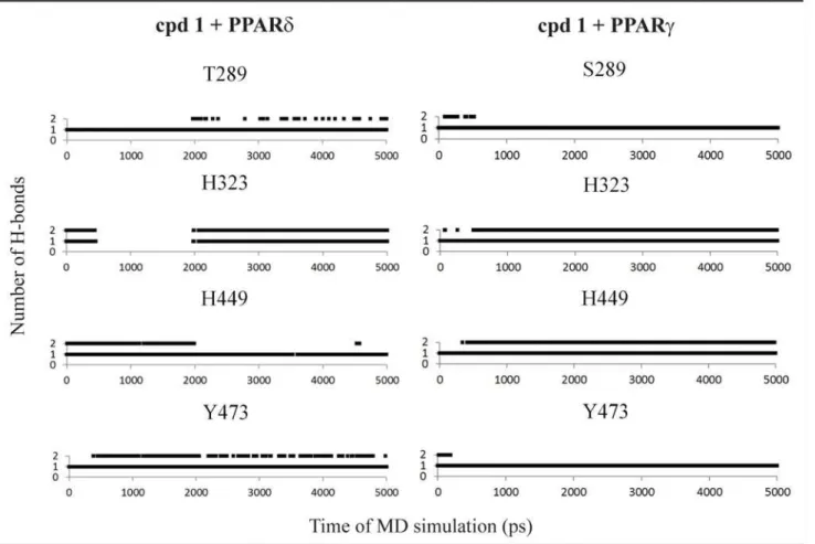
Fig 3. Hydrogen bonds diagram between compound 1 and polar residues of PPARδand PPARγ.Black squares indicate the presence of H-bonds and white ones correspond to the absence of H-bonds.
results, we analyzed the interactions between the selected ligands (1, 2 and 5) and the main res-idues in the active site of all PPAR subtypes after stabilization in order to understand their be-havior in the PPAR active sites, as well as their experimental selectivity. Finally, aiming to study the occupancy of the ligands into the H12 region, we analyzed the number of H-bonds between the compounds and some residues in each active complex.
Compound 1
For the [5-(benzyloxy)-1H-indol-1-yl]acetic acid (compound1,Fig. 2), H-bonds with all polar
residues in the PPAR active site for all subtypes (at least one His or Tyr residues) were observed in the MD simulations (Fig. 3). These polar interactions involve H323, H449, T289 and Y473 in PPARδ; and H323, H449, Y473 and S289 in PPARγ(Fig. 2). A similar binding mode was found to PPARδand PPARγ, a typical behavior for PPAR full agonists. An analysis of the
occu-pancy for the hydrogen bonds (Fig. 3) shows that the interaction of the compound 1 with the tyrosine residue located in H12 is more stable for PPARδand PPARγ[64].
Compound 2
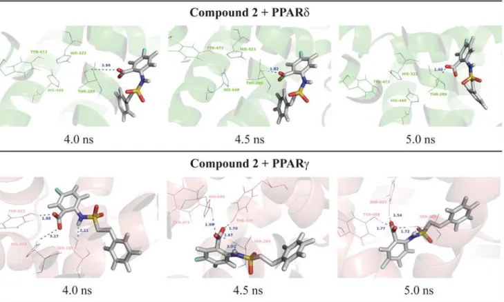
Surprisingly, unlike docking results obtained for PPARδ,
5-chloro-2-{[(2-phenylethyl)sulfo-nyl]amino}benzoic acid (compound2) showed only activation on PPARγ. Indeed, considering the interactions of this compound in the active site, this ligand is not able to maintain the polar interactions with PPARδ(Fig. 4), explaining its PPARγselectivity. At the PPARδbinding site,
the compound 2 forms H-bonds with some residues (Thr-289 of PPARδ) and is not able to
Fig 4. Final nanosecond snapshots of compound 2 molecular dynamic simulations in complex with PPAR subtypes.Main polar interactions between the compound 2 and the PPARδand PPARγduring the final nanosecond of MD simulation.
reach the polar cavity of both receptors (Fig. 5). This can also be explained due to the larger binding cavity of PPARγthanδ[6,64,73,74]. At the PPARγactive site, the compound 2 is able to perform and maintain polar interactions with Ser-289, His-323, His-449 and Tyr-473 along the 5ns of MD simulation according to Figs.4and5.
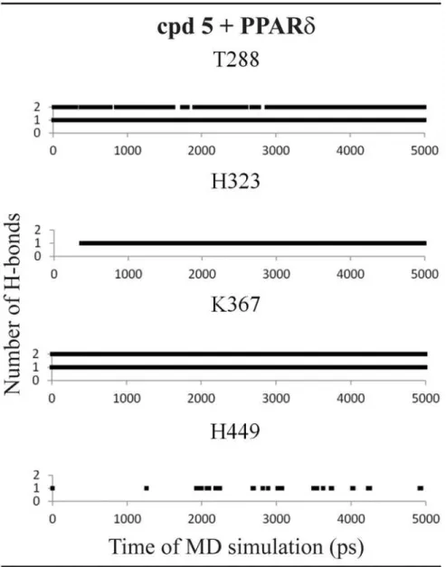
Compound 5
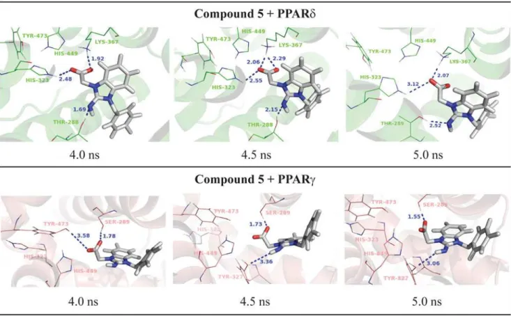
Finally, the compound 5 ([2-imino-3-(4-methylbenzyl)-2,3-dihydro-1H-benzimidazol-1-yl] acetic acid) showed PPARδactivation from our preliminary experimental assays. In line with the transactivation results, the MD simulations showed only loose interactions between this li-gand the PPARγbinding pocket. As shown inFig. 6, the compound 5 directly interacts with
Fig 5. Hydrogen bonds diagram between compound 2 and polar residues of PPARγ.Black squares indicate the presence of H-bonds and white ones correspond to the absence of H-bonds.
His-323 and Lys-367 and acts as an H-bond acceptor interacting with Thr-288 of PPARδ; on the other hand, in the PPARγactive site, the compound 5 interacts only with Ser-289. The lack
of strong interactions leads the ligand to move away from the active site even in short dynamics (5 ns). Therefore, one possible reason for a missing activity of the compound 5 on PPARγis
the lack of interactions with histidine or tyrosine residues as the compounds 1 and 2 perform, showing the importance of these residues in the PPARγactivation.
In the PPARδactive site, this ligand forms H-bonds with two histidine residues (His-323 and His-449) during all MD simulation and two additional interactions between Thr-288 and Lys-367 (Fig. 7).
In this study, we can conclude that the compounds selected by the virtual screening were able to perform the main polar contacts with PPARδandγ, which are a way to activate this nu-clear receptor providing the conditions to the gene transcription [73,75–81]. There are several experimental and theoretical studies indicating the different behavior of PPAR ligands [82– 85]. However, to the best of our knowledge, there are few studies involving PPARδligands,
proving the importance of our study. Here, the results obtained from the MD analyses are able to explain the behavior of the active compounds at the PPAR active sites. Based on the experi-mental and computational analyses, obtained in this study, we can conclude that our structure-based studies were successfully carried out and a new scaffold of PPAR ligands was found out.
Fig 6. Final nanosecond snapshots of compound 5 molecular dynamic simulations in complex with PPAR subtypes.Main polar interactions between the compound 5 and the PPARδand PPARγduring the final nanosecond of MD simulation.
Conclusion
In this study, we performed a structure-based virtual screening using a PPARδstructure aiming
to find out new molecular entities with PPAR affinity. Then, the clean-leads ZINC subset was employed as ligand database and the docking analyses were performed with DOCK program. The binding energies and visual inspections were used to rank the compound library. Finally, a consensus analysis using GOLD and Surflex-Dock programs was carried out and 5 substances with potential PPAR affinity were selected. From the 5 purchased compounds, 4 of them pre-sented potential biological activity: compounds 1 and 4 showed PPARδ/γactivity; compound 2
displayed a significant PPARγactivation and; finally, compound 5 presented as a PPARδ
agonist. In addition to the new found scaffold, it is important to mention that the tetrazole group is present in the compound 4 (with low levels of activation) as well in 13 of the first 50
Fig 7. Hydrogen bonds diagram between compound 5 and polar residues of PPARδ.Black squares indicate the presence of H-bonds and white ones correspond to the absence of H-bonds.
compounds ranked by DOCK (A Table inS1 File). Indeed, the tetrazole moiety is a well-known bioisosteric replacement for acids [86]. So, the presence of tetrazole in our findings is not surprisingly, but the experimental evidences for the activity of the compound 4 support the use of this ligand in further investigations. Therefore, the tetrazole group can act as the polar head present in the typical PPAR ligands and can be explored in future SAR studies.
Supporting Information
S1 File. Structure of the selected compounds, results from the biological assays and MD simulations. A Fig. Luciferase assays. Activation of PPARδ(A) and PPARγ(B) at the single concentration of the 5 selected ligands.B Fig. EC50values of the compounds 1 and 2.C Fig.
RMSD values for the protein backbone during the MD simulation.D Fig. RMSD values for the ligand atoms during the MD simulation.E Fig. Number of H-bonds between the selected
li-gands and the protein atoms during the MD simulation.A Table. 50 compounds selected by DOCK. These compounds were employed in the redocking analyses using GOLD and Surflex programs.
(ZIP)
Author Contributions
Conceived and designed the experiments: VGM MT ASN KMH. Performed the experiments: VGM MT ASN KMH. Analyzed the data: VHM MT ASN KMH. Contributed reagents/materi-als/analysis tools: VGM MT ASN KMH. Wrote the paper: VGM MT ASN KMH.
References
1. McEwan I (2009) Nuclear Receptors: One Big Family. In: McEwan I, editor. The Nuclear Receptor Su-perfamily: Humana Press. pp. 3–18.
2. Michalik L, Auwerx J, Berger JP, Chatterjee VK, Glass CK, Gonzales FJ et al. (2006) International Union of Pharmacology. LXI. Peroxisome proliferator-activated receptors. Pharmacol Rev. 58: 726– 741. PMID:17132851
3. Dreyer C, Krey G, Keller H, Givel F, Helftenbein G, Wahli W. (1992) Control of the Peroxisomal Beta-Oxidation Pathway by a Novel Family of Nuclear Hormone Receptors. Cell. 68: 879–887. PMID:
1312391
4. Issemann I, Green S (1990) Activation of a Member of The Steroid-Hormone Receptor Superfamily by Peroxisome Proliferators. Nature. 347: 645–650. PMID:2129546
5. Schmidt A, Endo N, Rutledge SJ, Vogel R, Shinar D, Rodan GA. (1992) Identification of a New Member of The Steroid-Hormone Receptor Superfamily that is Activated by a Peroxisome Proliferator and Fatty-Acids. Mol Endocrinol. 6: 1634–1641. PMID:1333051
6. Zoete V, Grosdidier A, Michielin O (2007) Peroxisome proliferator-activated receptor structures: Ligand specificity, molecular switch and interactions with regulators. BBA-Mol Cell Biol L. 1771: 915–925. PMID:17317294
7. Berger J, Moller DE (2002) The mechanisms of action of PPARs. Annu Rev Med. 53: 409–435. PMID:
11818483
8. Robinson E, Grieve DJ (2009) Significance of peroxisome proliferator-activated receptors in the cardio-vascular system in health and disease. Pharmacol Therapeut. 122: 246–263.
9. Jialal I, Smith G (2012) Managing the Dyslipidemia of Metabolic Syndrome: Beyond Statin Therapy. Metab Syndr Rela D. 10: 159–160. doi:10.1089/met.2012.1500PMID:22568574
10. Wang Y, Jacome-Sosa MM, Ruth MR, Lu Y, Shen J, Reaney MJ, et al. (2012) The intestinal bioavail-ability of vaccenic acid and activation of peroxisome proliferator-activated receptor-αand-γin a rodent model of dyslipidemia and the metabolic syndrome. Mol Nutr Food Res. 56: 1234–1246. doi:10.1002/ mnfr.201100517PMID:22714958
12. Buss ZdS, Medeiros YS, Froede TS (2012) PPAR-Gamma Agonist Rosiglitazone Attenuates the In-flammation Caused by Carrageenan in the Mouse Model of Pleurisy. InIn-flammation. 35: 280–288. doi:
10.1007/s10753-011-9316-6PMID:21465278
13. Yu Y, Zhang Z- H, Wei S- G, Weiss RM, Felder RB (2012) Peroxisome Proliferator-Activated
Receptor-γRegulates Inflammation and Renin-Angiotensin System Activity in the Hypothalamic Paraventricular Nucleus and Ameliorates Peripheral Manifestations of Heart Failure. Hypertension. 59: 477–484. doi:
10.1161/HYPERTENSIONAHA.111.182345PMID:22083161
14. Chen YC, Wu JS, Tsai HD, Huang CY, Chen JJ, Sun GY, et al. (2012) Peroxisome Proliferator-Activat-ed Receptor Gamma (PPAR-γ) and Neurodegenerative Disorders. Mol Neurobiol. 46: 114–124. doi:
10.1007/s12035-012-8259-8PMID:22434581
15. Bolden A, Bernard L, Jones D, Akinyeke T, Stewart LV (2012) The PPAR Gamma Agonist Troglitazone Regulates Erk 1/2 Phosphorylation via a PPARγ-Independent, MEK-Dependent Pathway in Human Prostate Cancer Cells. PPAR Res. 2012. doi:10.1155/2012/929052PMID:22448169
16. Robbins GT, Nie D (2012) PPAR gamma, bioactive lipids, and cancer progression. Front Biosci. 17: 1816–1834. PMID:22201838
17. Rogenhofer S, Ellinger J, Kahl P, Stoehr C, Hartmann A, Engehausen D, et al. (2012) Enhanced Ex-pression of Peroxisome Proliferate-activated Receptor Gamma (PPAR-γ) in Advanced Prostate Can-cer. Anticancer Res. 32: 3479–3483. PMID:22843934
18. Bruning JB, Chalmers MJ, Prasad S, Busby SA, Kamenecka TM, He Yet al. (2007) Partial agonists acti-vate PPARgamma using a helix 12 independent mechanism. Structure. 15: 1258–1271. PMID:
17937915
19. Pouwels KB, van Grootheest K (2012) The rosiglitazone decision process at FDA and EMA. What should we learn? Int J Risk Saf Med. 24: 73–80. doi:10.3233/JRS-2012-0559PMID:22751189
20. Balakumar P, Rose M, Ganti SS, Krishan P, Singh M (2007) PPAR dual agonists: are they opening Pandora’s Box? Pharmacol Res. 56: 91. PMID:17428674
21. Evans JL, Lin JJ, Goldfine ID (2005) Novel approach to treat insulin resistance, type 2 diabetes, and the metabolic syndrome: simultaneous activation of PPARalpha, PPARgamma, and PPARdelta. Curr Diabetes Rev. 1: 299–307. PMID:18220606
22. Kilgore KS, Billin AN (2008) PPARbeta/delta ligands as modulators of the inflammatory response. Curr Opin Investig Drugs. 9: 463–469. PMID:18465655
23. Fan Y, Wang Y, Tang Z, Zhang H, Qin X, Zhu Y, et al. (2008) Suppression of pro-inflammatory adhe-sion molecules by PPAR-delta in human vascular endothelial cells. Arterioscler Thromb Vasc Biol. 28: 315–321. PMID:18048767
24. Irwin JJ, Sterling T, Mysinger MM, Bolstad ES, Coleman RG (2012) ZINC: A Free Tool to Discover Chemistry for Biology. J Chem Inf Model. 52: 1757–1768. doi:10.1021/ci3001277PMID:22587354
25. Krishnamurthy Praveen T, Joghee Nanjan Chandrasekar M, Joghee Nanjan M (2013) Novel Glita-zones with Diverse Peroxisome Proliferator Activated Receptor Modulatory Potential. Curr Bioact Compd. 9: 221–234.
26. Pereira R, Gaudon C, Iglesias B, Germain P, Gronemeyer H, Lera AR (2006) Synthesis of the PPARβ/
δ-selective agonist GW501516 and C4-thiazole-substituted analogs. Bioorg Med Chem Lett. 16: 49– 54. PMID:16242326
27. Connors RV, Wang Z, Harrison M, Zhang A, Wanska M, Hiscock S. et al. (2009) Identification of a PPARδagonist with partial agonistic activity on PPARγ. Bioorg Med Chem Lett. 19: 3550–3554. doi:
10.1016/j.bmcl.2009.04.151PMID:19464171
28. Pettersen EF, Goddard TD, Huang CC, Couch GS, Greenblatt DM, Meng EC, et al. (2004) UCSF Chi-mera—A visualization system for exploratory research and analysis. J Comput Chem. 25: 1605–1612. PMID:15264254
29. Ewing TJA, Kuntz ID (1997) Critical evaluation of search algorithms for automated molecular docking and database screening. J Comput Chem. 18: 1175–1189.
30. Gschwend D, Kuntz ID (1996) Orientational sampling and rigid-body minimization in molecular docking revisited: On-the-fly optimization and degeneracy removal. J Comput-Aided Mol Des. 10: 123–132. PMID:8741016
31. Kuntz ID, Blaney JM, Oatley SJ, Langridge R, Ferrin TE (1982) A Geometric Approach to Macromole-cule-Ligand Interactions. J Mol Biol. 161: 269–288. PMID:7154081
32. Makino S, Kuntz ID (1997) Automated flexible ligand docking method and its application for database search. J Comput Chem. 18: 1812–1825.
34. Shoichet BK, Kuntz ID (1991) Protein Docking and Complementarity. J Mol Biol. 221: 327–346. PMID:
1920412
35. Shoichet BK, Bodian DL, Kuntz ID (1992) Molecular Docking Using Shape Descriptors. J Comput Chem. 13: 380–397.
36. Jain AN (2003) Surflex: Fully automatic flexible molecular docking using a molecular similarity-based search engine. J Med Chem. 46: 499–511. PMID:12570372
37. Jones G, Willett P, Glen RC, Leach AR, Taylor R (1997) Development and validation of a genetic algo-rithm for flexible docking. J Mol Biol. 267: 727–748. PMID:9126849
38. Nissink JWM, Murray C, Hartshorn M, Verdonk ML, Cole JC, Taylor R. (2002) A new test set for validat-ing predictions of protein-ligand interaction. Proteins. 49: 457–471. PMID:12402356
39. Verdonk ML, Cole JC, Hartshorn MJ, Murray CW, Taylor RD (2003) Improved protein-ligand docking using GOLD. Proteins. 52: 609–623. PMID:12910460
40. Wang R, Lai L, Wang S (2002) Further development and validation of empirical scoring functions for structure-based binding affinity prediction. J Comput-Aided Mol Des. 16:11–26. PMID:12197663
41. Cole JC, Murray CW, Nissink JWM, Taylor RD, Taylor R (2005) Comparing protein–ligand docking pro-grams is difficult. Proteins. 60: 325–332. PMID:15937897
42. Kroemer RT, Vulpetti A, McDonald JJ, Rohrer DC, Trosset JY, Giordanetto F, et al. (2004) Assessment of Docking Poses: Interactions-Based Accuracy Classification (IBAC) versus Crystal Structure Devia-tions. J Chem Inf Comp Sci. 44: 871–881. PMID:15154752
43. Sousa SF, Fernandes PA, Ramos MJ (2006) Protein–ligand docking: Current status and future chal-lenges. Proteins. 65: 15–26. PMID:16862531
44. Houston DR, Walkinshaw MD (2013) Consensus Docking: Improving the Reliability of Docking in a Vir-tual Screening Context. J Chem Inf Model. 53: 384–390. doi:10.1021/ci300399wPMID:23351099
45. Teramoto R, Fukunishi H (2007) Supervised Consensus Scoring for Docking and Virtual Screening. J Chem Inf Model. 47: 526–534. PMID:17295466
46. Masters JR (2002) HeLa cells 50 years on: the good, the bad and the ugly. Nat Rev Cancer. 2: 315– 319. PMID:12001993
47. Gey G, Coffman WD, Kubicek MT (1952) Tissue culture studies of the proliferative capacity of cervical carcinoma and normal epithelium. Cancer Res. 12: 264–265.
48. Li Y, Choi M, Suino K, Kovach A, Daugherty J, Kliewer SA, et al. (2005) Structural and biochemical basis for selective repression of the orphan nuclear receptor liver receptor homolog 1 by small heterodi-mer partner. P Natl Acad Sci USA. 102: 9505–9510. PMID:15976031
49. Berendsen HJC, Vanderspoel D, Vandrunen R (1995) GROMACS—A Message-Passing Parallel Mo-lecular Dynamics Implementation. Comput Phys Commun. 91: 43–56.
50. Lindahl E, Hess B, van der Spoel D (2001) GROMACS 3.0: a package for molecular simulation and tra-jectory analysis. J Mol Model. 7: 306–317.
51. Berendsen HJC, Postma JPM, van Gunsteren WF, Hermans J. Interaction models for water in relation to protein hydration. In: Pullman B, editor. Intermolecular Forces. Springer Netherlands; 1981. pp. 331–342.
52. van Gunsteren WF, Billeter SR, Eising AA, Hünenberger PH, Krüger P, Mark AE, et al. (1996) Biomo-lecular Simulation: The GROMOS96 manual and userguide. Hochschuleverlag AG an der ETH Zürich, Switzerland; 1996.
53. Schuttelkopf AW, van Aalten DMF (2004) PRODRG: a tool for high-throughput crystallography of pro-tein-ligand complexes. Acta Crystallogr D. 60: 1355–1363. PMID:15272157
54. Scientific D (2006) PyMOL v0.99c. South San Francisco, California, USA.
55. Fyffe SA, Alphey MS, Buetow L, Smith TK, Ferguson MAJ, Sørensen MD, et al. (2006) Recombinant Human PPAR-β/δLigand-binding Domain is Locked in an Activated Conformation by Endogenous Fatty Acids. J Mol Biol. 356: 1005–1013. PMID:16405912
56. Iwashita A, Muramatsu Y, Yamazaki T, Muramoto M, Kita Y, Yamazaki S, et al. (2007) Neuroprotective Efficacy of the Peroxisome Proliferator-Activated Receptorδ-Selective Agonists in Vitro and in Vivo. J Pharmacol Exp Ther. 320: 1087–1096. PMID:17167170
57. Artis DR, Lin JJ, Zhang C, Wang W, Mehra U, Perreault M, et al. (2009) Scaffold-based discovery of indeglitazar, a PPAR pan-active anti-diabetic agent. P Natl Acad Scie USA. 106: 262–267.
59. Xu HE, Lambert MH, Montana VG, Parks DJ, Blanchard SG, Brown PJ, et al. (1999) Molecular recogni-tion of fatty acids by peroxisome proliferator-activated receptors. Mol Cell. 3: 397–403. PMID:
10198642
60. Jin L, Lin S, Rong H, Zheng S, Jin S, Wang R, et al. (2011) Structural Basis for Iloprost as a Dual Peroxi-some Proliferator-activated Receptorα/δAgonist. J Biol Chem. 286: 31473–31479. doi:10.1074/jbc. M111.266023PMID:21775429
61. Keil S, Matter H, Schönafinger K, Glien M, Mathieu M, Marquette JP, et al. (2011) Sulfonylthiadiazoles with an Unusual Binding Mode as Partial Dual Peroxisome Proliferator-Activated Receptor (PPAR)γ/δ
Agonists with High Potency and in vivo Efficacy. Chem Med Chem. 6: 633–653. doi:10.1002/cmdc. 201100047PMID:21400663
62. Evans KA, Shearer BG, Wisnoski DD, Shi D, Sparks SM, Sternbach DD, et al. (2011) Phenoxyacetic acids as PPAR delta partial agonists: Synthesis, optimization, and in vivo efficacy. Bioorg Med Chem Lett. 21: 2345–2350. doi:10.1016/j.bmcl.2011.02.077PMID:21414782
63. Takada I, Yu RT, Xu HE, Lambert MH, Montana VG, Kliewer SA, et al. (2000) Alteration of a single amino acid in peroxisome proliferator-activated receptor-alpha (PPAR alpha) generates a PPAR delta phenotype. Mol Endocrinol. 14: 733–740. PMID:10809235
64. Oyama T, Toyota K, Waku T, Hirakawa Y, Nagasawa N, Kasuga JI, et al. (2009) Adaptability and selec-tivity of human peroxisome proliferator-activated receptor (PPAR) pan agonists revealed from crystal structures. Acta Crystallogr D. 65: 786–795. doi:10.1107/S0907444909015935PMID:19622862
65. Pettersson I, Ebdrup S, Havranek M, Pihera P, Korinek M, Mogensen JP, et al. (2007) Design of a par-tial PPAR delta agonist. Bioorg Med Chem Lett. 17: 4625–4629. PMID:17560785
66. Epple R, Azimioara M, Russo R, Xie Y, Wang X, Cow C, et al. (2006) 3,4,5-Trisubstituted isoxazoles as novel PPARδagonists. Part 2. Bioorg Med Chem Lett. 16: 5488–5492. PMID:16931011
67. Shearer BG, Patel HS, Billin AN, Way JM, Winegar DA, Lambert MH, et al. (2008) Discovery of a novel class of PPAR delta partial agonists. Bioorg Med Chem Lett. 18: 5018–5022. doi:10.1016/j.bmcl.2008. 08.011PMID:18722772
68. Luckhurst CA, Stein LA, Furber M, Webb N, Ratcliffe MJ, Allenby G, et al. (2011) Discovery of isoindo-line and tetrahydroisoquinoisoindo-line derivatives as potent, selective PPAR delta agonists. Bioorg Med Chem Lett. 21: 492–496. doi:10.1016/j.bmcl.2010.10.117PMID:21094606
69. Wickens P, Zhang C, Ma X, Zhao Q, Amatruda J, Bullock W, et al. (2007) Indanylacetic acids as
PPAR-δactivator insulin sensitizers. Bioorg Med Chem Lett. 17: 4369–4373. PMID:17601734
70. Huang N, Shoichet BK, Irwin JJ (2006) Benchmarking Sets for Molecular Docking. J Med Chem. 49: 6789–6801. PMID:17154509
71. Tripos Inc. (2008) Sybyl 8.1. St. Louis, MO, USA.
72. Lipinski CA (2000) Drug-like properties and the causes of poor solubility and poor permeability. J Phar-macol Toxicol. 44: 235–249. PMID:11274893
73. Nascimento AS (2010) Structural requirement for PPAR gamma binding revealed by a meta analysis of holo-crystal structures. Biochimie. 92: 499–506. doi:10.1016/j.biochi.2010.01.018PMID:20138109
74. Xu HE, Lambert MH, Montana VG, Plunket KD, Moore LB, Collin JL, et al. (2001) Structural determi-nants of ligand binding selectivity between the peroxisome proliferator-activated receptors. P Natl Acad Sci USA. 98: 13919–13924. PMID:11698662
75. Batista FAH, Trivella DBB, Bernardes A, Gratieri J, Oliveira PSL, Figueira ACM, et al. (2012) Structural Insights into Human Peroxisome Proliferator Activated Receptor Delta (PPAR-Delta) Selective Ligand Binding. PloSOne. 7: 1–7. doi:10.1093/jscr/2012.7.1PMID:24960726
76. Garcia TS, Honorio KM (2011) Two-Dimensional Quantitative Structure-Activity Relationship Studies on Bioactive Ligands of Peroxisome Proliferator-Activated Receptor delta. J Braz Chem Soc. 22: 65– 72.
77. Maltarollo VG, Homem-de-Mello P, Honorio KM (2011) Role of physicochemical properties in the acti-vation of peroxisome proliferator-activated receptor delta. J Mol Model. 17: 2549–2558. doi:10.1007/ s00894-010-0935-xPMID:21207086
78. Maltarollo VG, Silva DC, Honorio KM (2012) Advanced QSAR studies on PPAR´ ligands related to met-abolic diseases. J Braz Chem Soc. 23: 78–84.
79. Maltarollo VG, Honorio KM (2012) Ligand- and Structure-Based Drug Design Strategies and PPAR delta/alpha Selectivity. Chem Biol Drug Des. 80: 533–544. doi:10.1111/j.1747-0285.2012.01424.x
PMID:22672760
81. Giaginis C, Theocharis S, Tsantili-Kakoulidou A (2009) A QSAR Study on Indole-Based PPAR-gamma Agonists in Respect to Receptor Binding and Gene Transactivation Data. QSAR Comb Sci. 28: 802– 805.
82. Puhl AC, Bernardes A, Silveira RL, Yuan J, Campos JLO, Saidemberg DM, et al. (2012) Mode of Perox-isome Proliferator-Activated Receptor gamma Activation by Luteolin. Mol Pharmacol. 81: 788–799. doi:10.1124/mol.111.076216PMID:22391103
83. Nevin DK, Lloyd DG, Fayne D (2011) Rational Targeting of Peroxisome Proliferating Activated Recep-tor Subtypes. Curr Med Chem. 18: 5598–5623. PMID:22172067
84. Yue L, Ye F, Xu X, Shen J, Chen K, Shen X, et al. (2005) The conserved residue Phe273(282) of PPAR alpha(gamma), beyond the ligand-binding site, functions in binding affinity through solvation effect. Bio-chimie. 87: 539–550. PMID:15935279
85. Liu XY, Wang RL, Xu WR, Tang LD, Wang SQ, Chou KC (2011) Docking and Molecular Dynamics Sim-ulations of Peroxisome Proliferator Activated Receptors Interacting with Pan Agonist Sodelglitazar. Protein Peptide Lett. 18: 1021–1027. PMID:21592078