Characterization of the Apoptotic Response
Induced by the Cyanine Dye D112: A
Potentially Selective Anti-Cancer Compound
Ning Yang1, Paul Gilman2, Razmik Mirzayans3, Xuejun Sun3, Nicolas Touret1, Michael Weinfeld3, Ing Swie Goping1,3*
1Department of Biochemistry, University of Alberta, Edmonton, Alberta, Canada,2Independent Consultant, Retired from Research Laboratories, Eastman Kodak Company. Rochester, New York, United States of America,3Department of Oncology, University of Alberta, Edmonton, Alberta, Canada
*igoping@ualberta.ca
Abstract
Chemotherapeutic drugs that are used in anti-cancer treatments often cause the death of both cancerous and noncancerous cells. This non-selective toxicity is the root cause of un-toward side effects that limits the effectiveness of therapy. In order to improve chemothera-peutic options for cancer patients, there is a need to identify novel compounds with higher discrimination for cancer cells. In the past, methine dyes that increase the sensitivity of pho-tographic emulsions have been investigated for anti-cancer properties. In the 1970's, Kodak Laboratories initiated a screen of approximately 7000 dye structural variants for selective toxicity. Among these, D112 was identified as a promising compound with elevated toxicity against a colon cancer cell line in comparison to a non-transformed cell line. Despite these results changing industry priorities led to a halt in further studies on D112. We decided to re-vive investigations on D112 and have further characterized D112-induced cellular toxicity. We identified that in response to D112 treatment, the T-cell leukemia cell line Jurkat showed caspase activation, mitochondrial depolarization, and phosphatidylserine externalization, all of which are hallmarks of apoptosis. Chemical inhibition of caspase enzymatic activity and blockade of the mitochondrial pathway through Bcl-2 expression inhibited D112-in-duced apoptosis. At lower concentrations, D112 inD112-in-duced growth arrest. To gain insight into the molecular mechanism of D112 induced mitochondrial dysfunction, we analyzed the in-tracellular localization of D112, and found that D112 associated with mitochondria. Interest-ingly, in the cell lines that we tested, D112 showed increased toxicity toward transformed versus non-transformed cells. Results from this work identify D112 as a potentially interest-ing molecule warrantinterest-ing further investigation.
Introduction
Programmed cell death, or apoptosis, is vital for proper development and homeostasis in virtu-ally all tissues [1]. Two well-studied signaling processes termed the intrinsic and extrinsic
a11111
OPEN ACCESS
Citation:Yang N, Gilman P, Mirzayans R, Sun X, Touret N, Weinfeld M, et al. (2015) Characterization of the Apoptotic Response Induced by the Cyanine Dye D112: A Potentially Selective Anti-Cancer Compound. PLoS ONE 10(4): e0125381. doi:10.1371/journal.pone.0125381
Academic Editor:Suzie Chen, Rutgers University, UNITED STATES
Received:January 12, 2015
Accepted:March 23, 2015
Published:April 30, 2015
Copyright:© 2015 Yang et al. This is an open access article distributed under the terms of the
Creative Commons Attribution License, which permits unrestricted use, distribution, and reproduction in any medium, provided the original author and source are credited.
Data Availability Statement:All relevant data are within the paper and its Supporting Information files.
pathways represent the major mechanisms of apoptosis [2]. Extrinsic apoptosis involves extra-cellular ligand-mediated activation of plasma membrane-localized death receptors, while in-trinsic apoptosis is initiated by intracellular stresses, such as hypoxia, DNA damage, oxidative stress, and anti-cancer therapies [3]. These apoptotic stimuli lead to mitochondrial membrane permeabilization [4] and the release of apoptotic factors, such as cytochrome c [5], from the mitochondrial inter-membrane space. The intrinsic pathway is tightly regulated by the Bcl-2 family of proteins [6,7] that comprise anti-apoptotic (such as Bcl-2, Bcl-XL, Bcl-w) and pro-apoptotic proteins (such as Bax, Bak, Bid, Bad, Bim) [8]. Pro-apoptotic proteins stimulate for-mation of the Bax/Bak pore [9] or regulate the existing mitochondrial permeability transition pore [10] to release mitochondrial apoptosis-inducing proteins. On the other hand, anti-apoptotic proteins, such as Bcl-2 and Bcl-XL [11,12], inhibit mitochondrial dysfunction, thus blocking apoptosis. When pro-apoptotic Bcl-2 proteins prevail, the released cytochrome c stimulates formation of an oligomeric structure named the‘apoptosome’including apoptotic protease activating factor-1 and initiator caspase, caspase-9. This structure triggers the caspase cascade by activating downstream effector caspases such as caspase 3.
Apoptosis is widely blocked in cancer. Cancer is the second most common cause of death, accounting for nearly 1 in every 4 deaths in North America [13,14]. Apoptosis deregulation often manifests as clinical anticancer treatment resistance. Additionally, off-target damage to non-cancerous tissue is a major toxicity to many drug regimens. Hence identifying drugs that more selectively target cancer cells and spare healthy tissue is the focus of many drug
discovery initiatives.
Our studies focused on elucidating the molecular mechanism of toxicity of a cyanine mole-cule, D112, and evaluating its cancer cell selectivity. D112 was identified by the Eastman Kodak Company through a cancer drug-screening program initiated at the Dana Farber Insti-tute in the1970’s [15]. Initial observations deduced that the electrochemical reduction potential of a dye influenced the ability of the dye to inhibit the growth of healthy sea urchin eggs [16]. Using this rationale, the Kodak Laboratory employed electrochemical reduction potential to se-lect drugs for screening of anti-cancer properties. Approximately 7000 dye structural variants were tested and D112 (Fig 1A), emerged as a lead compound with higher cytotoxic activity against a cancer cell line versus a non-transformed cell line [15]. Specifically, the IC50 of D112 to the normal monkey kidney epithelial cell line CV-1 was 9μg/ml, compared to 0.01μg/ml to
the human colon cancer cell line CX-1, thus achieving a killing ratio (IC50 CV-1/ IC50 CX-1) of 900 [15]. Furthermore, D112 showed enhanced selectivity compared to the widely used clical chemotherapeutic agent, adriamycin. Despite these promising results, due to changes in in-dustry priorities, investigations into D112 were not further pursued.
Currently, there is still a pressing need to identify new drugs that can discriminate cancer cells from normal cells, so we decided to revive studies on D112. Our study investigated the cy-totoxicity of D112 in cancer cells, characterized D112-induced apoptosis, and further explored its possible selective activity against cancer in comparison to normal cells.
Results
D112 induces apoptosis in Jurkat cells
To investigate whether D112 can induce apoptosis, we treated Jurkat cells (a human T-cell leu-kemia sub-line, named Jneo [17]) with D112 at increasing concentrations for 24 h and mea-sured the apoptotic hallmarks of phosphatidylserine externalization, mitochondrial
depolarization, and caspase activation. We first assessed cell death with the apoptotic marker of phosphatidylserine (PS) exposure. PS is normally restricted to the inner leaflet of the plasma membrane bilayer, and is externalized in response to caspase activation [18]. To measure
analysis, decision to publish, or preparation of the manuscript.
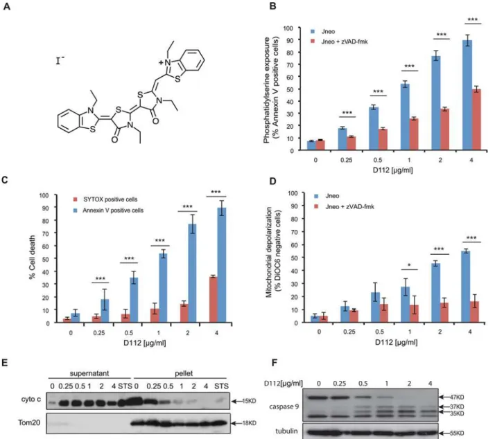
whether D112 induced PS exposure, we harvested cells after D112 treatment and measured fluorescent-annexin V binding by flow cytometry. The addition of D112 increased Annexin V positive cells in a dose-dependent manner (Fig 1B). To determine whether PS exposure was caspase-dependent, we co-incubated cells with the pan-caspase inhibitor zVAD-fmk and estab-lished that D112-mediated PS externalization was caspase-dependent. Of note, at higher Fig 1. D112 induces apoptosis in Jurkat cells.(A) D112 structure. (B) Jneo cells were treated with the indicated concentrations of D112 for 24 h in the presence or absence of the pan-caspase inhibitor zVAD-fmk (20μM). Phosphatidylserine exposure indicated by Alexa Fluor 647-annexin V positive cells is
shown as a percent of total cells as determined by flow cytometry. Mean±SD of three independent experiments performed in triplicate are shown,*P<0.05, **P<0.01,***P<0.001. (C) Necrosis detection. Cells were treated as above and were incubated with SYTOX Green stain or Alexa Fluor 647-annexin V.
Mean±SD of three independent experiments performed in triplicate are shown,*P<0.05,**P<0.01,***P<0.001. (D) Mitochondria dysfunction. Cells were
treated as above and incubated with DiOC6. Loss of electrochemical potential was determined by loss of fluorescence in the FL-1 channel as measured by
flow cytometry. Mean±SD of three independent experiments performed in triplicate are shown,*P<0.05,**P<0.01,***P<0.001. (E) Cytochrome c release. Cells were incubated with D112 at indicated concentrations for 24 h and then harvested and fractionated. Supernatant and pellet were loaded separately and subjected to western blot analysis. The experiment was performed independently three times and a representative blot is shown. (F) Caspase 9 cleavage. Cells were incubated for 24 h with increasing concentrations of D112 as indicated. Whole cell lysates were subjected to western blot analysis with the indicated antibodies. The experiment was performed independently three times and a representative blot is shown.
concentrations of D112, a significant amount of PS exposure was observed in the presence of zVAD-fmk, relative to non-D112-treated cells. These results suggested that D112 either in-duced an alternative caspase-independent necrotic pathway, or that secondary necrosis (in vitro membrane damage) accounted for Annexin V positivity at higher concentrations of D112. To distinguish between these two possibilities, we measured plasma membrane disrup-tion through uptake of the vital DNA-binding dye, SYTOX green (Fig 1C). Membrane damage was only evident at D112 concentrations that were higher than the amount required to detect PS exposure indicating that apoptosis and not necrosis was the initiating event. To directly measure secondary necrosis, we simultaneously labeled D112-treated cells with Alexa Fluor 647 Annexin V and SYTOX green in a time course experiment (S1andS2Figs). SYTOX green-only positive cells were undetectable. Instead, cells became Annexin V positive first and only at later time points became double positive for Annexin V and SYTOX green. Moreover, addition of the pan caspase inhibitor zVAD-fmk, was not associated with production of SYTOX green positive cells indicating that zVAD-fmk and D112 did not induce primary ne-crosis as has been demonstrated in other systems [19].
Since mitochondria are central regulators of the apoptotic pathway, we also examined mito-chondrial electrochemical depolarization in response to D112 treatment. Mitomito-chondrial depo-larization was measured by the loss of fluorescence of the potentiometric dye DiOC6(3) (3,3'-Dihexyloxacarbocyanine Iodide) (Fig 1D). Our study showed that the proportion of DiOC6 negative cells increased in response to D112 treatment. Furthermore, incubation with zVAD-fmk inhibited D112-induced mitochondrial depolarization, indicating that mitochondrial elec-trochemical potential loss was downstream of caspase activation. We next tested whether D112 induced the release of cytochrome c from the mitochondria. We incubated cells with increasing concentrations of D112 and fractionated cell components into heavy membrane and cytosolic fractions (Fig 1E). D112 induced the release of cytochrome c from the mitochondrial pellet fraction into the cytosolic supernatant fraction. Mitochondrial involvement and release of cyto-chrome c suggested that D112 would induce activation of the initiator caspase 9. In support of this, we observed activation-associated cleavage of caspase 9 in response to D112 incubation (Fig 1F). These results are consistent with a model whereby D112 induces cell death via the mi-tochondrial intrinsic apoptosis pathway.
Caspase 9 activation classically leads to executioner caspase 3 activation thus we tested the requirement for, caspase 3. Incubation with the caspase 3-selective inhibitor zDEVD-fmk sig-nificantly reduced D112 induced apoptosis (Fig 2A). We then assessed the activation profile for caspase 3. Caspases are present as inactive pro-enzymes and are activated by proteolytic cleavage [20]. In response to increasing concentrations of D112, we observed the cleavage of pro-caspase 3 from 35 KD to the subunits p17/19 (Fig 2B). Addition of zVAD-fmk inhibited the D112-dependent production of p17/19 and instead produced an intermediate p20 frag-ment. To assess whether the p20 subunit was associated with enzymatic activity, we incubated cell lysates with the caspase 3 fluorogenic substrate AC-DEVD-AFC and measured fluorescence of free AFC. D112-treated cells harbored significantly increased caspase 3 enzymatic activity, while cells that had also been co-incubated with zVAD-fmk had background levels of enzymat-ic activity (Fig 2C). These results indicated that D112 induced the cleavage of caspase 3 to p20. However, the p20 product was not catalytically active, with subsequent caspase activity gener-ated by the production of active p19/17 subunits of caspase 3.
All together, our results indicated that D112 induced apoptosis as evidenced by caspase acti-vation. Caspase activity induced both PS externalization and mitochondrial dysfunction. Therefore, we next decided to assess the functional requirement of mitochondrial dysfunction for D112-induced cytotoxicity.
Anti-apoptotic Bcl-2 blocks D112-induced cell death
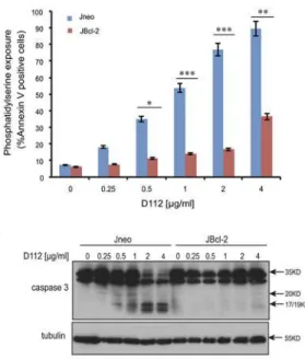
To explore the role of mitochondrial dysfunction in D112-induced apoptosis, we examined D112 sensitivity in Jurkat cells ectopically expressing Bcl-2 (JBcl-2). We observed that both D112-induced mitochondrial depolarization (Fig 3A) and PS exposure (Fig 3B) were signifi-cantly inhibited in JBcl-2 cells. Since caspase activation was critically important for PS exposure and mitochondrial depolarization, we directly measured enzymatic activity. As expected, we found that D112-treated JBcl-2 cells had background levels of caspase 3 activity (Fig 3C). Fur-ther, analysis of caspase cleavage patterns demonstrated that JBcl-2 cells showed no production Fig 2. D112 induces caspase activation in Jurkat cells.(A) Phosphatidylserine exposure. Jneo cells were treated with indicated concentrations of D112 for 24 h in the presence or absence of the caspase 3 inhibitor z-DEVD-fmk (20μM), stained as indicated and analyzed by flow cytometry. Mean±SD of three
independent experiments performed in triplicate are shown,*P<0.05,**P<0.01,***P<0.001. (B) Caspase cleavage. Cells were incubated for 24 h with
increasing concentrations of D112 as indicated. Whole cell lysates were subjected to western blot analysis with the indicated antibodies. The experiment was performed independently three times and a representative blot is shown. (C) Caspase 3 enzymatic activation. Jneo cells were treated with D112 in the presence or absence of z-VAD-fmk (20 uM) for 24 h prior to lysis. Cell lysates were incubated with the caspase 3 specific fluorometric substrate, Ac-DEVD-AFC, for 1 h and caspase 3 activity was measured at 405 nm. Mean±SD of three independent experiments performed in triplicate are shown,
*P<0.05,**P<0.01,***P<0.001.
Fig 3. D112-induced apoptosis is blocked by Bcl-2.Jneo and JBcl-2 cells were treated with the indicated amounts of D112 for 24 h. Apoptotic cell death was measured by (A) mitochondrial dysfunction, (B) phosphatidylserine exposure, (C) caspase 3 enzymatic activation, and (D) caspase cleavage. All of these apoptotic hallmarks were quantitated as described previously. (E) Bax/Bak expression is required for D112-induced cell death. Jneo and JR cells were treated with indicated concentrations of D112 for 24 h and phosphatidylserine positivity was recorded as done previously. (F) Inhibition of the apoptosis pathway protects cell from D112-induced cell death. Cells were treated with 62.5 ng/ml D112 for 24 h. Cells then were incubated in fresh medium and counted daily. Cell survival was detected by trypan blue staining. (G) Cell proliferation is inhibited by D112. Cells were treated with 62.5 ng/ml D112 for 24 h. Cells then were incubated in fresh medium and total live cell number was counted. The expression of Bcl-2, Bax and Bak were verified in western blot insets in A and E. In all experiments, the mean±SD of three independent experiments performed in triplicate are shown,*P<0.05,**P<0.01,***P<0.001.
of active subunits of caspase 3 (Fig 3D). These results confirmed that the mitochondrial path-way was critical to induce caspase activation in response to D112.
As a complementary approach, the involvement of mitochondrial dysfunction in
D112-induced apoptosis was also confirmed by examining PS exposure in a sub-line of Jurkat cells (named JR) with diminished levels of Bax and Bak proteins [23,24]. In response to apo-ptotic stimuli, pro-apoapo-ptotic proteins Bax and Bak form pores in the mitochondrial outer membrane, causing the release of mitochondrial proteins inducing apoptosis [9,25]. Given that Bcl-2 blocked D112-induced cytotoxicity, we reasoned that loss-of-function of the pro-apoptotic proteins Bax and Bak would have a similar effect. Indeed in JR cells, PS exposure was not induced by D112, indicating that Bax/Bak was required for D112-induced cell death (Fig 3E). D112 did not appear to require the Permeability Transition Pore, as cyclosporin A did not inhibit D112-induced cell death (S4 Fig). These results confirmed the crucial role of mitochon-drial dysfunction in D112-induced apoptosis and more specifically, that D112 induced a Bax/ Bak-dependent mitochondrial apoptotic pathway triggering caspase activation.
To determine whether the extrinsic pathway contributed to D112-induced apoptosis, we ex-amined caspase 8 activation by western blotting using a caspase 8 cleavage-specific antibody (S5 Fig). D112 induced caspase 8 cleavage, however, this was blocked by Bcl-2, indicating that caspase 8 activation was downstream of mitochondrial dysfunction and not dependent on ex-trinsic apoptotic mediators. To assess whether caspase 8 activation contributed to D112-in-duced apoptosis, we assayed Jurkat cells that stably expressed the rabbitpox virus-encoded caspase inhibitor SPI-2 that are resistant to extrinsic-mediated apoptosis [26]. These cells were equally sensitive to D112 treatment as control cells indicating that caspase 8 is dispensable for D112-mediated apoptosis.
We next assessed the effects of D112 treatment on cell viability. A four-fold lower dose than was used in the previous apoptosis assays showed a gradual decrease in viability of Jneo cells over 5 days, as determined by trypan blue staining. In contrast, both the presence of Bcl-2 or the depletion of Bax/Bak inhibited D112-induced cell death (Fig 3F). To assess whether D112 also affected proliferation, we counted the total number of live cells daily. D112 inhibited the proliferation of the JBcl-2 and JR cells, with significant difference to Jneo cells by day 5 (Fig 3G). Therefore, low doses of D112 induced apoptosis dependent on mitochondrial dys-function and arrested cell proliferation.
D112 co-localizes with mitochondria
The transfected cells were incubated with D112 for 4 h, and live images were captured with a confocal microscope. We observed that D112 co-localized with neither Rab5 nor Rab7, sug-gesting that endocytosis was not responsible for D112 cellular uptake. Because endocytosis is an early event, we also recorded D112 localization at earlier time points (S6andS7Figs). Even at the 5-minute time point, D112 was not significantly localized with either endosomal marker. We next analyzed D112 localization with respect to the mitochondrial outer membrane protein mEmerald-Tom 20 (Fig 4C). We observed that the D112 signal strongly overlapped with the mitochondrial marker. Linescan analysis of each fluorescent channel indicated an association of D112 with mitochondria and this co-localization was confirmed as statistically significant by Mander’s correlation analysis (Fig 4D). Thus D112 associates with mitochondria and this prox-imity may contribute to the D112-induced activation of the mitochondrial apoptotic pathway. Fig 4. D112 co-localizes with mitochondria.SK-BR-3 cells were transiently transfected with the indicated plasmids expressing either mEmerald-Tom20, Rab5-GFP or Rab7-GF. Cells were treated with 0.25μg/ml
D112 for 4 h and then live imaging was performed with confocal microscopy. (A) D112 did not co-localize with Rab5. (B) D112 did not co-localize with Rab7. (C) D112 co-localized with Tom 20. The experiment was performed independently twice and representative images are shown. (D) Summary of the Mander’s correlation coefficients of Rab5, Rab7 or Tom20 and D112 in ten SK-BR-3 cells separately.
D112 shows increased toxicity to breast cancer cells and metastatic
melanoma cells
We first became interested in studying D112 based on the initial reports that D112 had selec-tive toxicity to a human colon cancer cell line in comparison to a normal monkey kidney cell line. However, common anti-cancer treatments are less effective against solid tumors versus haematological malignancies (as exemplified by Jurkat cells) and show a non-apoptotic mecha-nism of action [28–30]. Thus we examined whether D112 induced apoptosis in cell lines de-rived from solid tumors and whether D112 showed any selectivity for transformed or metastatic cancer cells. Accordingly, we first analyzed the paired cell lines of Hs 578T and Hs 578Bst. Hs 578T is a human breast carcinoma cell line and Hs 578Bst is a normal cell line de-rived from non-transformed adjacent tissue from the same patient. The carcinoma cell line was significantly more sensitive to D112-induced apoptosis in comparison to the normal cell line (Fig 5A). We then expanded this observation to other breast cell lines (Fig 5B). We observed that the non-transformed human mammary epithelial cell line MCF-10A showed no signifi-cant dose-dependent response to D112 treatment. On the other hand, the mammary carcino-ma lines, SK-BR-3 and MDA-MB-468, were significantly more sensitive to D112-induced apoptosis. We next took advantage of a progressive metastasis model that had been established in the mouse B16 cell lines [31]. The B16-F0 cell line is a mouse melanoma cell line, while B16-F10 is an aggressive metastatic cell line generated from the B16-F0 line through a ten-time
in vivoselective procedure. The B16-F0 cells were more resistant to D112-induced apoptosis
than the highly metastatic B16-F10 line (Fig 5C). Thus, in the cell lines that we tested, D112 triggered an increased apoptotic response in transformed versus normal cells and increased cy-totoxicity to a metastatic versus a weakly metastatic line.
We next evaluated the effect of D112 on these adherent cell lines by the conventional clono-genic survival assay, which provides a read-out of the sum of cytotoxic (e.g. apoptosis) and cy-tostatic (growth arrest) responses [28,29]. Cell lines were treated with D112 for 24 h and the number of clonal colonies (>50 cells) that formed over a span of 2 weeks was recorded (Fig 5D). MCF10A cells were three times more resistant to D112 treatment than SK-BR-3 or MDA-MB-468 cells, based on the IC50 of 15 versus 5 ng/ml. Additionally, weakly-metastatic B16-F0 showed a 2-fold greater resistance than the metastatic B16-F10 cell line (Fig 5E). Since this low dose of D112 was below the threshold to detect apoptosis, it appeared that, similar to Jneo cells, low doses of D112 induced proliferative arrest. Nevertheless, these assays suggest that both high and low doses of D112 have selective activity against the transformed/metastatic cell lines that we tested.
Discussion
D112 was identified by Kodak as a cyanine dye that had higher toxicity against a cancer cell line than a non-transformed cell line. Whether this selectivity could be applied to other cancer cell lines was not known. Furthermore, the mechanism of D112-induced killing remained un-clear. In this study, we investigated D112-mediated cell killing in a number of cell lines and identified that relatively high doses of D112 triggered the mitochondrial apoptotic pathway and lower doses induced cell cycle arrest.
Fig 5. D112 shows selective cytotoxicity against transformed cells.(A) Paired human normal breast cell line Hs 578Bst and human breast carcinoma cell line Hs 578T. (B) Non-transformed human mammary epithelial cell lines MCF-10A in comparison to the mammary carcinoma cell lines SK-BR-3 and MDA-MB-468. (C) Melanoma parental cell line B16-F0 and melanoma metastatic cell line B16-F10. All cell lines were treated with the indicated amounts of D112 for 24 h and apoptosis was indicated by PS exposure, which was measured as described previously. (D) (E) Long-term colony formation of non-transformed cell lines and cancer cell lines. Cell lines were treated with the indicated amounts of D112 for 24 h, and all cells were harvested, counted and seeded in 24-well plates. Colonies were stained with crystal blue and counted after 7–14 days incubation. Mean±SD of three independent experiments performed in triplicate are shown,*P<0.05,**P<0.01,***P<0.001.
Non-caspase proteases [34] and a wide range of stressors such as ER stress, IL-3-deprivation [35] or staurosporine [36] can also produce a p20 fragment that becomes apparent in the pres-ence of zVAD-fmk. In many cases, the enzyme responsible for the generation of p20 is unclear, although in some cases chymotrypsin-like serine proteases can generate a p20 fragment [35]. Caspase 3 autocatalysis then produces the active subunits p19/17 [33]. As we observed that D112 produced similar caspase 3 proteolytic products, D112 likely stimulates an apoptotic pathway that is shared with other stress-inducers. Furthermore, we determined that elevated Bcl-2 levels inhibited not only caspase activity, but also the production of p20, indicating that Bcl-2 functioned upstream of both caspases and the initial caspase 3-protease. As Bcl-2 blocks mitochondrial dysfunction by inhibiting Bax/Bak pore formation, it is possible that the uniden-tified caspase 3-protease is localized to the mitochondria and is released to the cytosol through a Bax/Bak pore.
The second hallmark of apoptosis that we tested was mitochondrial depolarization. We ob-served that D112 induced mitochondrial depolarization was downstream of caspase activation. Mitochondrial membrane depolarization has been originally postulated to be an early event in the apoptotic pathway. However, more recent evidence suggests that it can also be a subsequent event in the apoptotic pathway, downstream of caspase activation. Mitochondrial potential was disrupted after caspase-3-mediated cleavage of protein kinase C delta (PKCδ), which allowed it to translocate to the mitochondria [37]. In another study, Fas-induced loss of mitochondrial potential occurred after the activation of caspase-1-like caspases, and the addition of caspase inhibitors inhibited mitochondrial membrane depolarization [38,39]. Similarly, our study showed that the caspase pan-inhibitor z-VAD-fmk inhibited mitochondrial membrane depo-larization, suggesting that mitochondrial depolarization was a downstream event in our case.
It is noteworthy that mitochondrial membrane depolarization was rescued by caspase inhib-itors, whereas PS externalization was only partly inhibited in D112 induced cell death. For ex-ample, in the case of cells treated with 1μg/ml D112, 20% more Annexin V-positive cells were
detected under this condition even in the presence of zVAD-fmk. Conceivably, other non-caspase-dependent factors, for example Apoptosis-Inducing-Factor (AIF), and/or ATP-depletion may contribute to PS externalization [8]. In support of this, both caspase- and AIF-mediated PS exposures are dependent on mitochondrial dysfunction and are blocked by Bcl-2 [18]. Also, PS exposure is proposed to be a consequence of ATP depletion [40]. In healthy cells, ATP-dependent translocases maintain PS on the inside of the cell. During apoptosis, ATP lev-els decrease and translocase activity diminishes, leading to PS externalization [41]. In our study, D112 co-localized with mitochondria, and it is possible that D112 inhibits ATP produc-tion by interfering with mitochondrial respiraproduc-tion, hence contributing to PS exposure in a cas-pase-independent manner.
we are not able to predict whether this difference in sensitivity could translate into a selective anticancer agent.
Our current work demonstrated that D112 localizes to mitochondria and induces Bax/Bak-dependent mitochondrial outer membrane permeabilization, cytochrome c release and activa-tion of caspases 9 and 3. Mitochondrial depolarizaactiva-tion was dependent on caspase activaactiva-tion, suggesting a positive amplification loop for mitochondrial dysfunction. We speculate that this loop may involve activity of caspase-cleaved Bid and is unlikely to require Permeability Transi-tion Pore, since preliminary studies with cyclosporin A that blocks PTP activaTransi-tion did not de-crease apoptosis. With respect to upstream signaling pathways, it is not clear how D112 triggers mitochondrial effects. Perhaps, due to its redox potential, D112 may inhibit the elec-tron transport chain and elevate reactive oxygen species that trigger mitochondrial damage and apoptosis. Further studies will address the mechanism of increased toxicity of the trans-formed versus non-transtrans-formed cell lines that we have shown here. These future studies will determine whether D112 may serve as a prototype for optimization of novel molecules with even more enhanced selective activity.
Materials and Methods
Cell culture and chemicals
Stable transfected human T-cell leukemia Jurkat cell lines Jneo and JBcl2 were kindly provided by Dr. Chris Bleackley (University of Alberta), JR cells were kindly provided by Dr. Hannah Rabinowich (University of Pittsburgh). The human normal breast epithelial cell lines MCF-10A was obtained from ATCC and maintained as previously described [42]. B16-F0 (ATCC, CRL-6322), B16-F10 (ATCC, CRL-6475), Hs578BST (ATCC, HTB-125) and Hs578T (ATCC, HTB-126) cells were obtained from ATCC (Manassas, VA, USA). B16-F0, B16-F10 and Hs578T were maintained in Dulbecco's Modified Eagle's Medium, high glucose, with 10% FCS; Hs578BST was maintained in Dulbecco's Modified Eagle's Medium, high glucose, with 10% FCS and 30ng/ml EGF. Z-VAD-fmk and z-DEVD-fmk were purchased from BD Pharmingen (Mississauga, ON, Canada), SYTOX Green death stain and Alexa Fluor 647 Annexin V conju-gate were all obtained from Invitrogen (Carlsbad, CA, USA). All chemicals were purchased from Sigma-Aldrich (St Louis, MO, USA) unless indicated otherwise.
Western blotting
Jurkat cells were treated with 0, 0.25, 0.5, 1, 2 or 4μg/ml D112 for 24 h. All cells were harvested
and lysed in lysis buffer (50 mM Tris pH 8, 150 mM NaCl, 1% NP-40, 0.5% sodium deoxycho-late, 1mMβ-mercaptoethanol). The whole cell lysates were then mixed with 6X SDS-PAGE gel loading buffer and boiled for 10 min. Protein concentration was measured using Pierce BCA protein assay kit from Thermo Fisher Scientific Inc (Waltham, MA). 15μg proteins from cell
lysates were loaded onto 14% SDS polyacrylamide gels and subjected to SDS-PAGE analysis. Proteins were resolved at 100 V and subsequently transferred to PVDF membranes by use of Bio-Rad Mini-Gel Box Electrotransfer for 1 h at 300 mA. Membranes were blocked in phos-phate-buffered saline (PBS) containing 0.1% Tween 20 and 5% skimmed milk for 1 h. Proteins were visualized with specific primary antibodies, followed by goat anti—mouse or rabbit HRP-conjugated secondary antibodies.
(556433) was from BD Pharmingen (San Diego, CA) and antibody against Tubulin (T5168) was from Sigma-Aldrich.
Determination of apoptosis
Apoptosis was determined by analyzing phosphatidylserine exposure using Alexa Fluor 647 Annexin V conjugate staining and mitochondria dysfunction indicated by loss of DiOC6 fluo-rescence. Briefly, cells were washed 3 times with PBS after D112 treatment. For Annexin V 647 staining, cells were resuspended in 100μL annexin-binding buffer (10 mM HEPES, 140 mM
NaCl, and 2.5 mM CaCl2, pH 7.4) with 2.5μL Alexa Fluro 647 Annexin V conjugate, and
incu-bated for 15 min at room temperature. After washing with PBS, the percentage of cells binding Alexa Fluor 647 Annexin V was determined using an Accuri C6 flow cytometer (BD Accuri, Ann Arbor, MI, USA) in the FL-4 channel. For DiOC6staining, cells were stained with 40 ng/ ml DiOC6in PBS for 10 min at room temperature, and loss of electrochemical potential was determined by loss of fluorescence in the FL-1 channel as measured by flow cytometry. D112 was measured in the FL-2 channel.
Caspase 3 enzymatic activation assay
2x10⁷cells were treated with D112 at indicated concentrations for 24 h. All cells were harvested and washed twice with PBS, then resuspended in 100μl lysis buffer (50 mM HEPES, 100 mM
NaCl, 0.1% CHAPS, 1 mM DTT, 100 mM EDTA, pH 7.4) on ice for 20 min. Protein concen-tration was measured using a Pierce BCA protein assay kit. 100μg protein from each assay was
diluted in 50μl cell lysis buffer and mixed with 50μl of 2X Reaction Buffer (50 mM HEPES,
100 mM NaCl, 0.1% CHAPS, 10 mM DTT, 100 mM EDTA, 10% glycerol, pH 7.4). 5μl of the 4
mM DEVD-AFC substrate (200μM final concentration) was added to the mixture and
incu-bated at 37°C for 1 h. Caspase 3 activity was determined at 405 nm in a 96-well plate on a Mul-tiskan Ascent plate reader (Thermo Fisher Scientific Inc).
Detection of necrosis
Cell death was detected by SYTOX Green death stain. Briefly, cells were harvested after treat-ment, and resuspended in binding buffer containing 1μl/ml of SYTOX Green stain solution.
Next, cells were incubated for 20 min at room temperature before analysis. Signals were collect-ed in in the FL-1 channel as measurcollect-ed by flow cytometry.
Cell viability assay
Cell viability was determined by trypan blue exclusion method. Briefly, Jneo, JBcl-2 and JR cells were treated with D112, and then cells were harvested and stained with 0.4% trypan blue solution. The number of blue staining cells and total cells were counted, and cell survival rate was calculated.
Mitochondria fractionation
Mitochondrial fractionation was conducted as described previously [34]. Jneo cells were treated with D112 or staurosporine (as a positive control) at indicated concentrations for 24 h. 2 × 10⁶ cells per sample were harvested and cell pellets was then re-suspended in 100μl of digitonin
Triton X-100, 25 mM Tris pH 8.0). Both supernatant and pellet were mixed with SDS loading buffer and subjected to western blotting.
Transient transfection
SK-BR-3 cells were seeded in an 8-well Nunc Lab-Tek chambered coverglass (Sigma) and reached 70% confluence for transfection. To form the transfection complex, 10μl pre-warmed
Opti-MEMI Reduced-Serum Medium (Life Technologies, Burlington, ON, Canada) with 0.2μg plasmid DNA was mixed with an equal volume of Opti-MEMI Reduced-Serum Medium
with 0.4μl TransIT-LT1 reagent (Mirus, Madison, WI, USA). After 20 min incubation at room
temperature, the complex was added dropwise to different areas of the wells and the cells were incubated for 6 h for delivery. Complex was then replaced by fresh complete growth medium for gene expression and membrane recovery before the next treatment. Plasmid mEmerald-Tom20 was kindly supplied by Dr. Robert E. Campbell (Department of Chemistry, University of Alberta, Canada).
Confocal imaging of living cells
For confocal live cell microscopy, SK-BR-3 cells transfected with plasmids were treated with 0.25μg /ml D112 for 4 h. Images were then directly taken on a Zeiss LSM 710 inverted confocal
microscope fitted with a 40x 1.4 NA Oil DIC Plan-Apochromat objective (Carl Zeiss Canada, Toronto, ON, Canada). After acquisition, images were visualized using ZEN 2009 Light Edition (Carl Zeiss) and MetaXpress (Version 5.2) (Molecular Devices) software.
Image analysis
Image analysis and processing was performed in MetaXpress and Fiji (online open-source,
http://fiji.sc/Fiji). Linescan analysis was performed on MetaXpress. Calculation of Mander’s cor-relation coefficient for colocalization analysis was performed on Fiji. In brief, a region of interest (ROI) was applied to transfected cells. A manual threshold was applied to each channel using ‘dark background and the Mander’s correlation calculation was run by Coloc 2 method of Fiji software (http://fiji.sc/Colocalization_Analysis#Colocalization_analysis_using_Coloc_2).
Cell proliferation
Jneo, JBcl-2, JR cells were treated with 62.5ng/ml D112 for 24 h. All cells were washed and re-suspended in fresh medium. 104cells (per well) were seeded in 24-well plates. Cell number was counted every 24 h.
Colony formation
Multiple cell lines were treated with indicated doses of D112 for 24 h. All cells were harvested and seeded in 12-well plates for colony formation. Considering non-transformed cell lines showed lower colony forming ability than cancer cell lines under normal conditions, for MCF-10A cell line, 500 cells were seeded, while for SK-BR-3, MDA-MB-468, B16-F0 and B16-F10 cell lines, 100 cells were seeded. Cells were incubated for 7–14 days. Colonies were then fixed and stained with crystal violet solution with 0.05% (w/v) crystal violet, 4% formaldehyde in 1 X PBS.
Statistical analysis
with equal variance. For statistical analysis of multiple groups (Fig5Band5D), the one-way Analysis of Variance (ANOVA) test was performed and p-values were obtained by Tukey’s
Post Hoc test.
Supporting Information
S1 Fig. D112 induced apoptosis and secondary necrosis.Jneo cells were treated with 2μg/ml
of D112 for the indicated time points and then double labeled with SYTOX green and Alexa Fluro 647 Annexin V. Cells were analyzed by flow cytometry. Shown is a representative of one of three experiments performed in triplicate.
(TIFF)
S2 Fig. D112 induced apoptosis and secondary necrosis.Jneo cells were treated with 2μg/ml
of D112 in the presence of zVAD-fmk (20μM) for the indicated time points and then double
labeled with SYTOX green and Alexa Fluro 647 Annexin V. All cells were analyzed by flow cy-tometry. Shown is a representative of one of three experiments performed in triplicate. (TIFF)
S3 Fig. BH3-only proteins were examined by western blotting.Jneo cells were incubated for 24 h with increasing concentrations of D112 as indicated. Whole cell lysates were subjected to western blotting analysis with the indicated antibodies. The experiment was performed inde-pendently three times and a representative blot is shown. Bid (2002), Puma (12450) and Bim (2819) antibodies were from Cell Signaling; Bik (sc1710) antibody was from Santa Cruz; Noxa (ab13654) antibody was from Abcam.
(EPS)
S4 Fig. Permeability Transition Pore was not involved in D112-induced cell death.Jneo cells were treated with the indicated concentrations of D112 in the presence or absence of 5μM
cyclosporine A (CsA) for 24 h. Jneo cells were treated with H2O2(200μM and 400μM) for 4
hours, as a positive control [43]. Phosphatidylserine exposure indicated by Alexa Fluor 647-annexin V positive cells is shown as a percent of total cells as determined by flow cytome-try. In all experiments, the mean ± SD of three independent experiments performed in tripli-cate are shown,P<0.05,P<0.01,P<0.001.
(EPS)
S5 Fig. The extrinsic pathway did not contribute to D112-induced apoptosis.(A) caspase 8 cleavage was examined by western blotting. Cleaved caspase 8 antibody (9496) was from cell signaling. (B) Jneo and Spi-2 expressing Jurkat cells were treated with the indicated concentra-tions of D112 for 24 h. Phosphatidylserine exposure indicated by Alexa Fluor 647-annexin V positive cells is shown as a percent of total cells as determined by flow cytometry. In all experi-ments, the mean ± SD of three independent experiments performed in triplicate are shown,
P<0.05,P<0.01,P<0.001. (EPS)
S6 Fig. Endocytosis was not involved in D112 intracellular uptake.SK-BR-3 cells were tran-siently transfected with Rab5-GFP. Cells were treated with 0.25μg/ml D112 for 5 min, 15 min,
S7 Fig. Endocytosis was not involved in D112 intracellular uptake.SK-BR-3 cells were tran-siently transfected with Rab7-GFP. Cells were treated with 0.25μg/ml D112 for 5 min, 15 min,
30 min or 60 min, and then live imaging was performed with confocal microscopy. Summary of the Mander’s correlation coefficients of Rab7 and D112 in ten SK-BR-3 cells was showed at the bottom. Mean ± SD of three independent experiments performed in triplicate are shown. (TIF)
Acknowledgments
We thank Dr. Jack Williams (now deceased) and Dr. Gregory Taylor (University of Alberta) for bringing D112 to our attention. We thank Drs. Hannah Rabinowich (University of Pitts-burgh) and Chris Bleackley for the kind gifts of JR, and Jneo/Bcl-2 cell lines, respectively. Plas-mid mEmerald-Tom20 (54282, Addgene) was kindly supplied by Dr. Robert E. Campbell (University of Alberta). We thank Rachel Montpetit for editorial input, Geraldine Barron for microscopy advice and Carolina Ortiz for help with image analysis. Finally, we are grateful to Dr. Thomas Simmen (University of Alberta) and all members of the Goping lab for
helpful discussions.
Author Contributions
Conceived and designed the experiments: NY PG RM MW ISG. Performed the experiments: NY. Analyzed the data: NY RM MW ISG. Contributed reagents/materials/analysis tools: NY XJS NT ISG. Wrote the paper: NY RM MW ISG.
References
1. Ning Y, Goping IS. Apoptosis. Nabi IR, editor: Morgan & Claypool Life Sciences; 2013. 101 p. 2. Portt L, Norman G, Clapp C, Greenwood M, Greenwood MT. Anti-apoptosis and cell survival: a review.
Biochimica et biophysica acta. 2011; 1813(1):238–59. Epub 2010/10/26. doi:10.1016/j.bbamcr.2010. 10.010PMID:20969895.
3. Wong RS. Apoptosis in cancer: from pathogenesis to treatment. Journal of experimental & clinical can-cer research: CR. 2011; 30:87. Epub 2011/09/29. doi:10.1186/1756-9966-30-87PMID:21943236; PubMed Central PMCID: PMCPmc3197541.
4. Estaquier J, Vallette F, Vayssiere JL, Mignotte B. The mitochondrial pathways of apoptosis. Advances in experimental medicine and biology. 2012; 942:157–83. Epub 2012/03/09. doi: 10.1007/978-94-007-2869-1_7PMID:22399422.
5. Liu X, Kim CN, Yang J, Jemmerson R, Wang X. Induction of apoptotic program in cell-free extracts: re-quirement for dATP and cytochrome c. Cell. 1996; 86(1):147–57. Epub 1996/07/12. PMID:8689682. 6. Martinou JC, Youle RJ. Mitochondria in apoptosis: Bcl-2 family members and mitochondrial dynamics.
Dev Cell. 2011; 21(1):92–101. doi:10.1016/j.devcel.2011.06.017PMID:21763611; PubMed Central PMCID: PMC3156409.
7. Gross A, McDonnell JM, Korsmeyer SJ. BCL-2 family members and the mitochondria in apoptosis. Genes & development. 1999; 13(15):1899–911. Epub 1999/08/13. PMID:10444588.
8. Susin SA, Lorenzo HK, Zamzami N, Marzo I, Snow BE, Brothers GM, et al. Molecular characterization of mitochondrial apoptosis-inducing factor. Nature. 1999; 397(6718):441–6. Epub 1999/02/16. doi:10. 1038/17135PMID:9989411.
9. Wei MC, Zong WX, Cheng EH, Lindsten T, Panoutsakopoulou V, Ross AJ, et al. Proapoptotic BAX and BAK: a requisite gateway to mitochondrial dysfunction and death. Science. 2001; 292(5517):727–30. Epub 2001/04/28. doi:10.1126/science.1059108PMID:11326099; PubMed Central PMCID: PMCPmc3049805.
10. De Giorgi F, Lartigue L, Bauer MK, Schubert A, Grimm S, Hanson GT, et al. The permeability transition pore signals apoptosis by directing Bax translocation and multimerization. FASEB journal: official publi-cation of the Federation of American Societies for Experimental Biology. 2002; 16(6):607–9. Epub 2002/03/29. PMID:11919169.
12. Yasuhara N, Sahara S, Kamada S, Eguchi Y, Tsujimoto Y. Evidence against a functional site for Bcl-2 downstream of caspase cascade in preventing apoptosis. Oncogene. 1997; 15(16):1921–8. doi:10. 1038/Sj.Onc.1201370PMID:WOS:A1997YA87200006.
13. Society AC. Cancer Facts & Figures 2014 2014. Available:http://www.cancer.org/research/ cancerfactsstatistics/cancerfactsfigures2014/index.
14. Society CC. National statistics at a glance from Canadian Cancer Statistics 2013. Available:http:// www.cancer.ca/en/cancer-information/cancer-101/canadian-cancer-statistics-publication/?region = bc. 15. Gilman P, Parton R, Lenhard J, inventors; Eastman Kodak Company, assignee. Correlation of
anti-can-cer activity of dyes with redox potentials. USA patent US20060099712 A1. 2006 May 11,2006. 16. Zigman S, Gilman PB. Biological Influence of Dyes and Anti-cancer Effects. Science. 1980; 208:188.
PMID:7361115
17. Goping IS, Barry M, Liston P, Sawchuk T, Constantinescu G, Michalak KM, et al. Granzyme B-induced apoptosis requires both direct caspase activation and relief of caspase inhibition. Immunity. 2003; 18 (3):355–65. Epub 2003/03/22. PMID:12648453.
18. Borisenko GG, Matsura T, Liu SX, Tyurin VA, Jianfei J, Serinkan FB, et al. Macrophage recognition of externalized phosphatidylserine and phagocytosis of apoptotic Jurkat cells—existence of a threshold. Archives of biochemistry and biophysics. 2003; 413(1):41–52. Epub 2003/04/23. PMID:12706340. 19. Vercammen D, Brouckaert G, Denecker G, Van de Craen M, Declercq W, Fiers W, et al. Dual signaling
of the Fas receptor: initiation of both apoptotic and necrotic cell death pathways. The Journal of experi-mental medicine. 1998; 188(5):919–30. Epub 1998/09/09. PMID:9730893; PubMed Central PMCID: PMCPmc2213397.
20. Shi Y. Mechanisms of caspase activation and inhibition during apoptosis. Molecular cell. 2002; 9 (3):459–70. doi:10.1016/S1097-2765(02)00482-3PMID:11931755.
21. Lomonosova E, Chinnadurai G. BH3-only proteins in apoptosis and beyond: an overview. Oncogene. 2008; 27 Suppl 1:S2–19. Epub 2009/07/31. doi:10.1038/onc.2009.39PMID:19641503; PubMed Cen-tral PMCID: PMCPmc2928556.
22. Hartman ML, Czyz M. Pro-apoptotic activity of BH3-only proteins and BH3 mimetics: from theory to po-tential cancer therapy. Anti-cancer agents in medicinal chemistry. 2012; 12(8):966–81. Epub 2012/01/ 24. PMID:22263800.
23. Wang GQ, Wieckowski E, Goldstein LA, Gastman BR, Rabinovitz A, Gambotto A, et al. Resistance to granzyme B-mediated cytochrome c release in Bak-deficient cells. The Journal of experimental medi-cine. 2001; 194(9):1325–37. Epub 2001/11/07. PMID:11696597; PubMed Central PMCID:
PMCPmc2195982.
24. Han J, Goldstein LA, Gastman BR, Rabinovitz A, Wang GQ, Fang B, et al. Differential involvement of Bax and Bak in TRAIL-mediated apoptosis of leukemic T cells. Leukemia. 2004; 18(10):1671–80. Epub 2004/09/10. doi:10.1038/sj.leu.2403496PMID:15356645.
25. Karch J, Kwong JQ, Burr AR, Sargent MA, Elrod JW, Peixoto PM, et al. Bax and Bak function as the outer membrane component of the mitochondrial permeability pore in regulating necrotic cell death in mice. eLife. 2013; 2:e00772. Epub 2013/08/31. doi:10.7554/eLife.00772PMID:23991283; PubMed Central PMCID: PMCPmc3755340.
26. Barry M, Heibein JA, Pinkoski MJ, Lee SF, Moyer RW, Green DR, et al. Granzyme B short-circuits the need for caspase 8 activity during granule-mediated cytotoxic T-lymphocyte killing by directly cleaving Bid. Molecular and cellular biology. 2000; 20(11):3781–94. Epub 2000/05/11. PMID:10805722; PubMed Central PMCID: PMCPmc85698.
27. Bareford LA, Swaan PW. Endocytic mechanisms for targeted drug delivery. Advanced Drug Delivery Reviews. 2007; 59(8):748–58. doi:10.1016/J.Addr.2007.06.008. WOS:000249935000006. PMID:
17659804
28. Mirzayans R, Andrais B, Scott A, Wang YW, Murray D. Ionizing radiation-induced responses in human cells with differing TP53 status. International journal of molecular sciences. 2013; 14(11):22409–35. Epub 2013/11/16. doi:10.3390/ijms141122409PMID:24232458; PubMed Central PMCID: PMCPmc3856071.
29. Brown JM, Wouters BG. Apoptosis, p53, and tumor cell sensitivity to anticancer agents. Cancer re-search. 1999; 59(7):1391–9. Epub 1999/04/10. PMID:10197600.
30. Brown JM, Attardi LD. The role of apoptosis in cancer development and treatment response. Nat Rev Cancer. 2005; 5(3):231–7. Epub 2005/03/02. doi:10.1038/nrc1560PMID:15738985.
32. Fernandes-Alnemri T, Litwack G, Alnemri ES. CPP32, a novel human apoptotic protein with homology to Caenorhabditis elegans cell death protein Ced-3 and mammalian interleukin-1 beta-converting en-zyme. The Journal of biological chemistry. 1994; 269(49):30761–4. Epub 1994/12/09. PMID:7983002. 33. Han Z, Hendrickson EA, Bremner TA, Wyche JH. A sequential two-step mechanism for the production
of the mature p17:p12 form of caspase-3 in vitro. The Journal of biological chemistry. 1997; 272(20): 13432–6. Epub 1997/05/16. PMID:9148968.
34. Heibein JA, Goping IS, Barry M, Pinkoski MJ, Shore GC, Green DR, et al. Granzyme B-mediated cyto-chrome c release is regulated by the Bcl-2 family members bid and Bax. The Journal of experimental medicine. 2000; 192(10):1391–402. Epub 2000/11/21. PMID:11085742; PubMed Central PMCID: PMCPmc2193181.
35. Egger L, Schneider J, Rheme C, Tapernoux M, Hacki J, Borner C. Serine proteases mediate apopto-sis-like cell death and phagocytosis under caspase-inhibiting conditions. Cell Death Differ. 2003; 10(10):1188–203. Epub 2003/09/23. doi:10.1038/sj.cdd.4401288PMID:14502242.
36. Johnson VL, Ko SC, Holmstrom TH, Eriksson JE, Chow SC. Effector caspases are dispensable for the early nuclear morphological changes during chemical-induced apoptosis. Journal of cell science. 2000; 113 (Pt 17):2941–53. Epub 2000/08/10. PMID:10934034.
37. Denning MF, Wang Y, Tibudan S, Alkan S, Nickoloff BJ, Qin JZ. Caspase activation and disruption of mitochondrial membrane potential during UV radiation-induced apoptosis of human keratinocytes re-quires activation of protein kinase C. Cell Death Differ. 2002; 9(1):40–52. Epub 2002/01/23. doi:10. 1038/sj.cdd.4400929PMID:11803373.
38. Wang L, Kinnear C, Hammel JM, Zhu W, Hua Z, Mi W, et al. Preservation of mitochondrial structure and function after cardioplegic arrest in the neonate using a selective mitochondrial KATP channel opener. The Annals of thoracic surgery. 2006; 81(5):1817–23. Epub 2006/04/25. doi:10.1016/j. athoracsur.2005.11.029PMID:16631678.
39. Susin SA, Zamzami N, Castedo M, Daugas E, Wang HG, Geley S, et al. The central executioner of apo-ptosis: multiple connections between protease activation and mitochondria in Fas/APO-1/CD95- and ceramide-induced apoptosis. The Journal of experimental medicine. 1997; 186(1):25–37. Epub 1997/ 07/07. PMID:9206994; PubMed Central PMCID: PMCPmc2198951.
40. Castedo M, Hirsch T, Susin SA, Zamzami N, Marchetti P, Macho A, et al. Sequential acquisition of mito-chondrial and plasma membrane alterations during early lymphocyte apoptosis. Journal of immunology (Baltimore, Md: 1950). 1996; 157(2):512–21. Epub 1996/07/15. PMID:8752896.
41. Bevers EM, Williamson PL. Phospholipid scramblase: an update. FEBS letters. 2010; 584(13):2724–30. Epub 2010/03/23. doi:10.1016/j.febslet.2010.03.020PMID:20302864.
42. Debnath J, Muthuswamy SK, Brugge JS. Morphogenesis and oncogenesis of MCF-10A mammary epi-thelial acini grown in three-dimensional basement membrane cultures. Methods (San Diego, Calif). 2003; 30(3):256–68. Epub 2003/06/12. PMID:12798140.