Sentinel Lymph Node Detection Using
Carbon Nanoparticles in Patients with Early
Breast Cancer
Xiufeng Wu1☯*, Qingzhong Lin1☯, Gang Chen3☯, Jianping Lu3, Yi Zeng1, Xia Chen1, Jun Yan2*
1Department of Surgery, Fujian Provincial Tumor Hospital, Teaching Hospital of Fujian Medical University, Fuzhou 350014, Fujian, People’s Republic of China,2Department of General Surgery, Nanfang Hospital, Southern Medical University, Guangzhou 510515, Guangdong, People’s Republic of China,3Department of Pathology, Fujian Provincial Tumor Hospital, Teaching Hospital of Fujian Medical University, Fuzhou 350014, Fujian, People’s Republic of China
☯These authors contributed equally to this work.
*[email protected](XFW); [email protected](JY)
Abstract
Purpose
Carbon nanoparticles have a strong affinity for the lymphatic system. The purpose of this study was to evaluate the feasibility of sentinel lymph node biopsy using carbon nanoparti-cles in early breast cancer and to optimize the application procedure.
Methods
Firstly, we performed a pilot study to demonstrate the optimized condition using carbon nanoparticles for sentinel lymph nodes (SLNs) detection by investigating 36 clinically node negative breast cancer patients. In subsequent prospective study, 83 patients with clinically node negative breast cancer were included to evaluate SLNs using carbon nanoparticles. Another 83 SLNs were detected by using blue dye. SLNs detection parameters were com-pared between the methods. All patients irrespective of the SLNs status underwent axillary lymph node dissection for verification of axillary node status after the SLN biopsy.
Results
In pilot study, a 1 ml carbon nanoparticles suspension used 10–15min before surgery was associated with the best detection rate. In subsequent prospective study, with carbon nano-particles, the identification rate, accuracy, false negative rate was 100%, 96.4%, 11.1%, respectively. The identification rate and accuracy were 88% and 95.5% with 15.8% of false negative rate using blue dye technique. The use of carbon nanoparticles suspension showed significantly superior results in identification rate (p = 0.001) and reduced false-neg-ative results compared with blue dye technique.
OPEN ACCESS
Citation:Wu X, Lin Q, Chen G, Lu J, Zeng Y, Chen X, et al. (2015) Sentinel Lymph Node Detection Using Carbon Nanoparticles in Patients with Early Breast Cancer. PLoS ONE 10(8): e0135714. doi:10.1371/ journal.pone.0135714
Editor:Xiaoan Liu, The First Affiliated Hospital with Nanjing Medical University, CHINA
Received:December 8, 2014
Accepted:July 24, 2015
Published:August 21, 2015
Copyright:© 2015 Wu et al. This is an open access article distributed under the terms of theCreative Commons Attribution License, which permits unrestricted use, distribution, and reproduction in any medium, provided the original author and source are credited.
Data Availability Statement:All relevant data are within the paper.
Conclusion
Our study demonstrated feasibility and accuracy of using carbon nanoparticles for SLNs mapping in breast cancer patients. Carbon nanoparticles are useful in SLNs detection in institutions without access to radioisotope.
Introduction
Sentinel lymph node biopsy (SLNB) is regarded as standard of care for axillary nodal staging in clinically axillary node-negative breast cancer patients [1]. SLNB will ensure the need for axillary lymph node dissection (ALND) in patients with positive sentinel lymph nodes (SLNs) and is equivalent to ALND in terms of correct staging but is associated with less-extensive morbidity than ALND [2–4]. Currently, SLNB is dependent on injection of blue dye, radioactive colloid, the combination of both or indocyanine green (ICG). The identification rates vary with blue dye (68–
86%), radioisotope (86–99%), combined technique (89–97%), ICG (73.8–99%) [5–11]. Despite high rates of sentinel lymph node (SLN) detection with these techniques, there is no general con-sensus about the optimal technique [12]. Radioactive colloid method results in some concerns about limited availability and cost of radio colloids, and radiation exposure [13]. In addition, lym-phoscintigraphy with a radioactive colloid cannot provide real-time visual during the surgical pro-cedure. Both blue dye and ICG, due to its relatively small diameter, permit flow through the SLNs to higher tier nodes, which results in incorrectly identifying sentinel nodes [14,15]. Moreover, the ICG technique requires special equipment in the operating room to enable this procedure [16].
Carbon nanoparticles are a synthetic tracer via the specific modification of small activated carbon particles with an average diameter of 150 nm, which is widely used in the field of cancer diagnosis and therapy [17]. They have received considerable interest in recent years, especially with respect to their potential utilization of lymphatic mapping. Carbon nanoparticles selec-tively enter the lymphatic vessels rather than blood capillaries due to the molecular size and permeability. Upon injection into the tissues around the tumor, carbon nanoparticles are rap-idly engulfed by macrophages and then pass through the lymphatic vessels to the SLNs, thus staining them black. This technique facilitates the vital staining of tumor-draining lymph nodes, and has been applied in the detection of sentinel lymph nodes (SLNs) in colorectal and thyroid cancers [18,19]. Carbon nanoparticles have no toxic side effects on the human body due to less access to the blood circulation. Because of safety and strong affinity for the lym-phatic system, carbon nanoparticles were approved for SLN mapping in gastric cancer by Chi-nese Food and Drug Administration. Therefore the feasibility of carbon nanoparticles for the identification of SLNs in early breast cancer must be investigated. In our study, the assessment of SLN detection using carbon nanoparticles is performed. We compare the identification rate, accuracy, false negative rate using carbon nanoparticles with those using blue dye to determine whether this method can be used to guild SLN biopsy and to assess its potential for SLN detec-tion in early breast cancer. This is, to our best knowledge, the first demonstradetec-tion of the use of carbon nanoparticles to detect SLNs in breast cancer.
Methods
Study Design
There were two steps in this research. Firstly, a pilot study was performed to determine the optimized condition using carbon nanoparticles for SLNs detection. Then, a prospective study Competing Interests:The authors have declared
was conducted to compare the sensitivity, specificity, and accuracy of using carbon nanoparti-cles for SLN mapping in breast cancer patients with those using blue dye.
Ethics Statement
Patients with clinically node negative breast cancer were recruited to participate in this study, which was approved by Institutional Review Board of Fujian Provincial Tumor Hospital. Writ-ten informed consent was obtained prior to study participation.
Patients
This study was designed to evaluate prospectively the feasibility of SLNs detection with carbon nanoparticles after peri-areolar intradermal injection of carbon nanoparticles. Inclusion crite-ria were: pathological diagnosis of breast cancer, maximum tumor diameter 3 cm, with indica-tions for mastectomy or breast-conserving surgery, and no clinically positive axillary lymph nodes. Axillary lymph node status was assessed before surgery by ultrasonography. Exclusion criteria comprised palpable axillary lymph nodes, tumor diameter>3cm, multicentric tumor. Demographic and clinicopathological data such as age, tumor size, grade, tumor histology, hormone receptor, HER2 status were recorded prospectively. The identification rate, sensitiv-ity, specificsensitiv-ity, accuracy, false negative rate, negative predictive value, positive predictive value was obtained after planned ALND.
Carbon Nanoparticles Suspension
Carbon nanoparticles were purchased from Chongqing LUMMY Pharmaceutical Co (Chong-qing, China) in the form of a standard carbon nanoparticles suspension (1ml: 50mg). This sta-ble suspension of carbon pellets of 150 nm in diameter does not enter the blood circulation, causing no toxic side effects on the human body. A small amount of tiny carbon particles may be captured by macrophages, and are excreted through the lungs and intestines after a few months. Carbon nanoparticles do not cause acute systemic toxicity, either.
Sentinel Lymph Node Biopsy
was considered as sentinel lymph node and excised along with perinodal fat. The specimen excised was sent for detailed pathological examination by paraffin fixation processing. All pro-cedures of SLNB were finished within 30–45 minutes. Due to inherent cultural barriers and cancer fatalism in Chinese women, all patients chose complete axillary node dissection upon diagnosed with breast cancer. Therefore, a complete axillary lymph node clearance was done and the specimen was sent for histopathological examination. The information of full axillary clearance was used for validating the results of sentinel lymph node biopsy. The sentinel lymph nodes were formalin fixed and paraffin embedded. All SLNs were evaluated by Haematoxylin and Eosin (H&E) staining and those with negative histology for metastasis, immunohisto-chemical staining with monoclonal antibody against cytokeratin. Macrometastases were defined as a tumor size>2 mm and micrometastases as a tumor size between 0.2 and 2 mm. Tumors<0.2 mm were regarded as isolated tumor cell (ITC). A sentinel node was defined as positive if a macrometastases, micrometastases or ITC was identified.
SLNs Identification Using Blue Dye. Another 83 SLNB procedures were performed by using blue dye. Similarly, a 1% solution of blue dye (1 ml) was intradermally injected into the periareolar region in 4 (clockwise) quadrants of the breast. The whole breast was massaged for about 5 minutes. The blue stained lymph nodes were considered SLNs, which were harvested and then were sent for detailed pathological examination by paraffin fixation processing. A complete axillary lymph node clearance was done following SLNB and the specimen was sent for histopathological examination.
Statistical Analysis
In pilot study, the detection rates of SLNs using different dose and the detection rates of non-SLNs in three injection timing were compared using the chi-squared test. In prospective study, the false negative rate (FNR), sensitivity, negative predictive value (NPV), positive predictive value (PPV), specificity, and accuracy of SLN biopsy with carbon nanoparticles and blue dye were calculated by using the following formulas:
False negative rate
¼number of false negative SLNs=ðtrue positive þ false negative nodesÞx100
Sensitivity¼number of true positive SLNs=ðtrue positive þ false negative nodesÞx100
Negative predictive value
¼number of true negative SLNs=ðtrue negative þ false negative nodesÞx100
Positive predictive value
¼number of true positive SLNs=ðtrue positive þ false positive nodesÞx100
Specificity¼number of true negative SLNs=ðfalse positive þ true negative nodesÞx100
Accuracy¼ ðtrue positive þ true negative nodesÞ=total nodes x100
Results
Injection Dosage and Timing of Application in Pilot Study
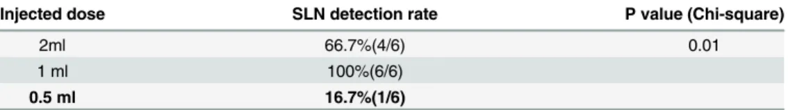
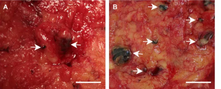
In pilot study, 36 clinically node negative breast cancer patients were investigated. We used 2 ml carbon nanoparticles suspenstion at the beginning of study. We found non-SLNs had been dyed black with this dose, which affected correct SLN staging. Then we decreased the dose to 1 ml and 0.5 ml. The SLNs detection rate with 2 ml of carbon nanoparticles was 66.7% inferior to 100% using 1 ml of carbon nanoparticles. However, when the amount of carbon nanoparticles was decreased to 0.5 ml, the SLNs detection rate was reduced to 16.7% because carbon nanoparticles could not clearly show SLNs with this dose. There was statistically signifi-cant difference in the SLNs detection rate in these three doses (P = 0.01) (Table 1). When com-pared one by one, the SLNs identification rate was significantly higher in 1ml than in 0.5ml (p = 0.02). There was no difference in the identification rate between 1ml and 2ml (p = 0.46), nor was that between 2ml and 0.5ml (p = 0.24). Timing of application also affected the detec-tion efficiency. With 1 ml of carbon nanoparticles, no black-stained non-SLNs were visible in patients who were injected 10–15 minutes before surgery (0/6) during complete axillary node dissection. In contrast, the black-stained non-SLNs detection rate was 83.3% (5/6) and 100% (6/6) in patients who were injected 1 day (Fig 1A) or 2 days (Fig 1B) before surgery, respec-tively. There was statistically significant difference in the non-SLNs detection rate in timing of application (P = 0.001) (Table 2). In terms of comparison of one by one, there was significant difference in non-SLNs detection rate between 10–15 minutes and 1day before surgery (p = 0.02), so was that between 10–15 minutes and 2day before surgery (p = 0.002). No statisti-cally significant differences in non-SLNs detection rates were seen between 1day and 2day before surgery (p = 1.0).
Patients and Tumor Characteristics in Prospective Study
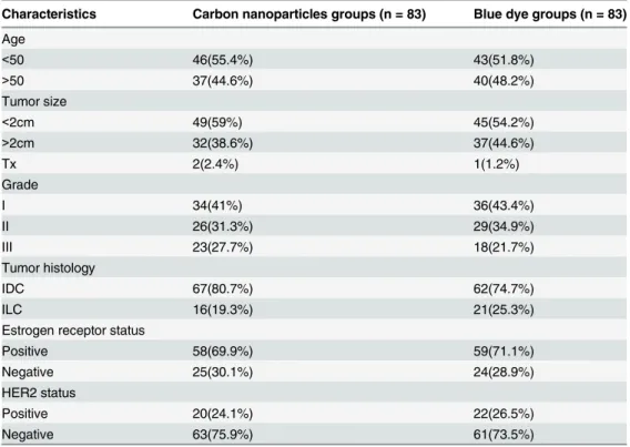
Eighty three women with operable primary breast cancer took part in carbon nanoparticles groups. Their median age was 51 years (range 28–75 years). Of these 83 patients, 46 were pre-menopausal and 37 were postpre-menopausal. Among these 83 patients, 49 had tumor less than 2 cm, 32 had tumor larger than 2 cm and 2 were Tx tumors (previous surgical excision at outside our institution). There were another 83 cases of operable primary breast cancers using blue dye technique. Their median age was 49.2 years (range 24–72 years). Of these 83 patients, 43 were premenopausal and 40 were postmenopausal. Patient data in prospective study are shown in
Table 3.
Sentinel Lymph Node Biopsy in Prospective Study
The carbon nanoparticles technique was found to be as easy to use as blue dye method by the surgeons. This method did not require special equipment in the operating room to enable this procedure. In prospective study, SLNs were successfully identified in all patients (100%) using carbon nanoparticles method. All SLNs had been stained black by carbon nanoparticles
Table 1. Comparison of SLNs detection rates using carbon nanoparticles between different doses in pilot study.
Injected dose SLN detection rate P value (Chi-square)
2ml 66.7%(4/6) 0.01
1 ml 100%(6/6)
0.5 ml 16.7%(1/6)
(Fig 2). The mean number of sentinel nodes per patient was 2.9 (range, 1–10). None of the 83 patients experienced adverse effects in response to carbon nanoparticles. Of the 83 SLNB pro-cedures using blue dye, 73 had SLNs successfully identified, an identification rate of 88% (73/ 83). The average number of SLNs detected using blue dye was 2.0 (range, 1–6). We observed no allergic reactions to blue dye during the study.
Histopathology in Prospective Study
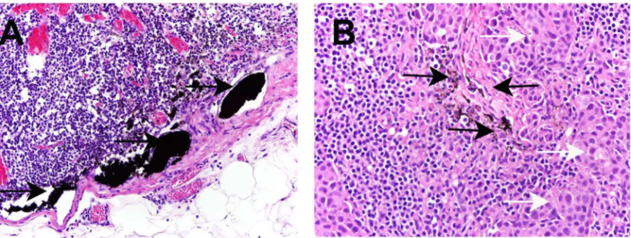
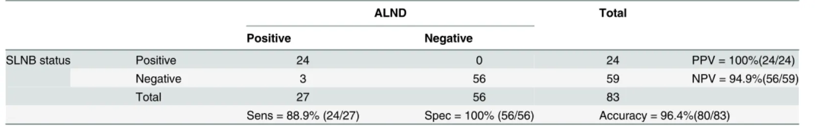
Carbon nanoparticles were seen in lymphatic vessels, and lymphoid sinus in negative SLNs. Similarly, carbon nanoparticles were seen in positive lymph nodes (Fig 3). Of the 83 patients with sentinel nodes using carbon nanoparticles, 24 (28.9%) had a tumor-positive SNLB speci-men. Among these 24 patients, 19 (79.2%) had at least 1 macrometastasis, and 3 (12.5%) had at least 1 micrometastasis as the largest metastatic deposit. Another 2 (8.3%) had individual tumor cells. Immunohistochemistry was performed in patients with negative histology for metastasis. Of the 59 patients who had negative sentinel nodes, all axillary nodes were negative in 56 cases. The remaining three patients with disease-free sentinel nodes were tumor-positive in other axillary nodes. Thus, with carbon nanoparticles technique, the negative predictive value was 94.9% (56/59), the sensitivity = 88.9% (24/27), false negative rate = 11.1% (3/27), specificity = 100% (56/56), the positive predictive value = 100% (24/24), and accuracy being 96.4% (80/83) (Table 4). Of the 83 SLNB procedures using blue dye, 73 had SLNs successfully identified, an identification rate of 88% (73/83). Among these 73 patients, 16 (21.9%) had a tumor-positive SNLB specimen including 12 with at least 1 macrometastasis, 3 with at least 1 Fig 1. A Black-dyed lymph nodes (as indicated by arrows) were visible 1 day after injection of carbon nanoparticles.B Increased non SLNs (as indicated by arrows) were stained black by carbon nanoparticles when injected 2 days before surgery. Scale bar is 1cm.
doi:10.1371/journal.pone.0135714.g001
Table 2. Comparison of non-SLNs detection rates using carbon nanoparticles between different injec-tion timing in pilot study.
Timing of injection Non-SLN detection rate P value (Chi-square)
10–15min before surgery 0%(0/6) 0.001
1day before surgery 83.3%(5/6)
2day before surgery 100%(6/6)
micrometastasis. Another one had individual tumor cells. 3 patients with positive non-SLNs had no SLNs involvement according to the final histology; Thus, the false negative rate was15.8% (3/19), the sensitivity = 84.2% (16/19), specificity = 100% (54/54), the positive pre-dictive value = 100% (16/16), and accuracy being 95.9% (70/73) (Table 5). The SLNs identifica-tion rate using carbon nanoparticles was significantly higher than that using blue dye
(p = 0.001). In terms of accuracy and false negative rate, there were no statistically significant differences in between the two methods (Table 6). However, there was a decrease in the false negative rate, from 15.8% with blue dye to 11.1% using carbon nanoparticles.
Discussion
Since the first introduction in the 1990s, sentinel lymph node biopsy has been the standard of care for breast cancer patients with clinically negative axilla, which is less invasive, less morbid but with an efficacy equal to ALND [2–4]. Current existing techniques, including blue dye, radioisotope, combined technique, and ICG, are useful in identification of SLNs. However, the optimal modality remains challenging because of certain disadvantages with these techniques.
The present study confirmed that carbon nanoparticles can be used successfully for SLNs identification in patients with early breast cancer. Carbon nanoparticles have a strong affinity for the lymphatic system. Once injected into the subareolar tissue, they were taken up specifically by lymphatic vessels and delivered to the sentinel lymph nodes. The lymph nodes then turned black (Fig 2), which facilitated their identification during surgery. Carbon nanoparticles are a popular nanomaterial and readily available. It rarely produces side effects and we observed no allergic reactions during the study. The identification rate with carbon nanoparticles technique was better Table 3. Patients and tumor characteristics in prospective study.
Characteristics Carbon nanoparticles groups (n = 83) Blue dye groups (n = 83)
Age
<50 46(55.4%) 43(51.8%)
>50 37(44.6%) 40(48.2%)
Tumor size
<2cm 49(59%) 45(54.2%)
>2cm 32(38.6%) 37(44.6%)
Tx 2(2.4%) 1(1.2%)
Grade
I 34(41%) 36(43.4%)
II 26(31.3%) 29(34.9%)
III 23(27.7%) 18(21.7%)
Tumor histology
IDC 67(80.7%) 62(74.7%)
ILC 16(19.3%) 21(25.3%)
Estrogen receptor status
Positive 58(69.9%) 59(71.1%)
Negative 25(30.1%) 24(28.9%)
HER2 status
Positive 20(24.1%) 22(26.5%)
Negative 63(75.9%) 61(73.5%)
IDC: Invasive ductal carcinoma; ILC: Invasive lobular carcinoma; HER2: Human epithelial growth factor receptor2.
than using blue dye in our study, although the accuracy of detection between both techniques was comparable (p = 1.0). In addition, carbon nanoparticles were superior to a combination of gamma probe and blue dye in SLNs identification rate. The latter was currently reported to obtain the best SLNs detection rate [20]. The false negative rate is another common index of sen-tinel node mapping success. Although the false negative rates were not statistically different between the two methods in this study (Table 5), the reduced false negative rate with the use of carbon nanoparticles may provide favorable clinical benefit. With increasing experience of SLNs detection using carbon nanoparticles, this method may hold great promise for SLNs detection due to improved identification rates and lower false-negative rates.
An optimal lymphatic tracer should have size (in the range of 50–200 nm) small enough to enter the lymphatic capillaries and transport rapidly to the SLNs, yet large enough to retain in the sentinel nodes long enough for imaging and SLNs identification without prematurely migrating to higher tier nodes [21–23]. Nanosized carbon particles with an average diameter of Fig 2. A black-stained SLN (as indicated by black arrow) with afferent lymph vessel (as indicated by white arrow) and efferent lymph vessel (as indicated by red arrow).Scale bar is 1cm.
doi:10.1371/journal.pone.0135714.g002
150 nm, which ensures that these particles pass through the lymphatic capillaries and accumu-late in the lymph nodes long enough for the SLNs to be identified during surgery. In contrast, the blue dye molecules are rather small (<2 nm), and thus they can quickly transport through the sentinel lymph nodes, causing color fading of blue dye and a high possibility of false nega-tive rate [24], as is the case with ICG [15]. Therefore, it should be more easy applying carbon nanoparticles than using blue dye or ICG in SLN biopsy due to its longer presentation time in SLNs. This has important clinical implications. Because the dyes quickly diffuse through SLNs, a‘blue’node may not be the true sentinel node, but instead a level II or even level III, non-sen-tinel node. Therefore, non-sennon-sen-tinel lymph nodes could incorrectly be identified as SLNs, caus-ing more nodes than necessary to be excised and a false-negative stagcaus-ing. Currently,
approximately 1% of the nodes are undetectable when using a radiotracer and a blue dye dur-ing sentinel lymph node biopsy [25], probably because radiotracer or blue dye flows through the SLNs to higher tier nodes. A better retention in the SLNs using carbon nanoparticles is most likely to reduce this false negative detection. In this case, carbon nanoparticles detection was more reliable and stable than blue dye or ICG because the dye distribution in SLNs subse-quent to injection of carbon nanoparticles was more likely to last longer.
The timing of the application differs with different tracer. With the patent blue dye, the maximum coloring is obtained the tenth minute after injection. After that, the coloring gradu-ally fades, indicating SLNB procedure using blue dye should be finished within ten minutes. In our pilot study, we had injected the carbon nanoparticles at 10–15 minutes, 1 day, 2 days before surgery. We found that 10–15 minutes before surgery is the best time for maximum coloring of SLNs, and black SLNs could be identified very clearly during the surgery (Table 2). Following SLNB which was finished within 20–30 minutes, no black-stained non-SLNs were visible dur-ing complete axillary node dissection. However, the time lapse of carbon nanoparticles reten-tion in SLNs needs to be further investigated. Notably, increased non sentinel lymph nodes were stained black 1 day or 2 days after injection of carbon nanoparticles (Fig 1). In contrast, Table 4. Accuracy of SLNB using carbon nanoparticles in early breast cancer.
ALND Total
Positive Negative
SLNB status Positive 24 0 24 PPV = 100%(24/24)
Negative 3 56 59 NPV = 94.9%(56/59)
Total 27 56 83
Sens = 88.9% (24/27) Spec = 100% (56/56) Accuracy = 96.4%(80/83) Sens: Sensitivity; Spec: Specificity; PPV: Positive predictive value; NPV: Negative predictive value.
doi:10.1371/journal.pone.0135714.t004
Table 5. Accuracy of SLNB using blue dye in early breast cancer.
ALND Total
Positive Negative
SLNB status Positive 16 0 16 PPV = 100%(16/16)
Negative 3 54 57 NPV = 94.7%(54/57)
Total 19 54 73
Sens = 84.2%(16/19) Spec = 100% (54/54) Accuracy = 95.9%(70/73) Sens: Sensitivity; Spec: Specificity; PPV: Positive predictive value; NPV: Negative predictive value.
data from Yuan et al.'s study [26] demonstrated all the patent blue-dyed nodes lost the color rapidly when the timing of injection was more than 6 h before surgery. Another critical issue in SLNs identification is injection dose. In our pilot study, we used 2 ml carbon nanoparticles sus-penstion at the beginning of study. We found non-SLNs had been dyed black with this dose and then we decreased the dose gradually. On the contrary, 0.5 milliliter carbon nanoparticles sometimes could not clearly show SLNs, which affected detection efficiency. Therefore, a 1 ml carbon nanoparticles suspension was sufficient and recommended for identification of SLNs (Table 1).
Of the 59 patients with a negative SLN using carbon nanoparticles suspension, metastasis was found in a non-sentinel node in three patients. We reviewed the characteristics of these patients. One had a 30-mm grade 3 invasive ductal carcinoma which has more likely to skip the SLNs and metastasize to high-level station lymph nodes. One tumor was located medially and preferentially drained to the internal mammary nodes (IMNs) instead of the axillary nodes. In this study, lymph nodes existing outside the axilla were not examined. The other patient had undergone previous surgery which could have disrupted lymphatic drainage to the axillary nodes. Therefore, tumor size, localization, previous surgery could have negative effects on the accuracy of the SLN biopsy [27–29].
This study used gold standard- pathological analysis, to distinguish the positive and negative SNLs with an aim at investigation of the feasibility of using carbon nanoparticles to detect SLNs in early breast cancer. Our results showed that carbon nanoparticles could be an excellent candidate to stably and reliably detect SLNs in patients with early breast cancer.
The detection rate using carbon nanoparticles was 100%, with lower false negative rate than blue dye. This demonstrated that SLNs detection with our system was practical and applicable, especially in country where the incidence of newly diagnosed breast cancer is rising and the use of the sentinel lymph node biopsy procedure is limited due to less access to radioisotopes.
The use of carbon nanoparticles does have some limitations. For instance, carbon nanopar-ticles cannot be seen through skin and fatty tissue, and permit only limited visualization of afferent lymphatic vessels and the SLNs. However, fluorescent carbon nanoparticles obtained by conjugating carbon nanoparticles and ICG, holds great promise for SLNs mapping, which allows accurate localization of SLNs. Another disadvantage of carbon nanoparticles is tattooing of the breast which was observed in 10 cases, with complete resolution within 2 months in each case.
In summary, this prospective study demonstrated feasibility and accuracy of using carbon nanoparticles for SLN mapping in breast cancer patients. The carbon nanoparticles method would be particularly useful in institutions without access to radioisotope.
Acknowledgments
This work was supported by the National Natural Science Foundation of China (81272574), the Natural Science Foundation of Fujian Province (2014J01300, 2014J05086), the Medical Innovation Program of Fujian Province (2012-CXB-7), the Program from Education Bureau of Table 6. Comparison of SLN detection rates between using carbon nanoparticles and blue dye in prospective study.
carbon nanoparticles Blue dye P value
SLN identification rate 100% 88% 0.001
Accuracy 96.4% 95.9% 1.0
False negative rate 11.1% 15.8% 0.68
Fujian Province (JB13127), and the Scientific Research Foundation for High Level Talents in Nanfang Hospital of Southern Medical University.
Author Contributions
Conceived and designed the experiments: JY XFW. Performed the experiments: QZL GC JPL ZY XC. Analyzed the data: XFW JY XC. Contributed reagents/materials/analysis tools: XFW JY. Wrote the paper: XFW JY.
References
1. Veronesi U, Paganelli G, Galimberti V, Viale G, Zurrida S, Bedoni M, et al. (1997) Sentinel-node biopsy to avoid axillary dissection in breast cancer with clinically negative lymph-nodes. Lancet 349:1864–7. PMID:9217757
2. Krag DN, Anderson SJ, Julian TB, Brown AM, Harlow SP, Costantino JP, et al. (2010) Sentinel-lymph-node resection compared with conventional axillary-lymph-Sentinel-lymph-node dissection in clinically Sentinel-lymph-node-negative patients with breast cancer: overall survival findings from the NSABP B-32 randomised phase 3 trial. Lancet Oncol 11:927–933. doi:10.1016/S1470-2045(10)70207-2PMID:20863759
3. Ashikaga T, Krag DN, Land SR, Julian TB, Anderson SJ, Brown AM, et al. (2010) Morbidity results from the NSABP B-32 trial comparing sentinel lymph node dissection versus axillary dissection. J Surg Oncol 102:111–118. doi:10.1002/jso.21535PMID:20648579
4. Mansel RE, Fallowfield L, Kissin M, Goyal A, Newcombe RG, Brown AM, et al. (2006) Randomized multicenter trial of sentinel node biopsy versus standard axillary treatment in operable breast cancer: the ALMANAC Trial. J Natl Cancer Inst 98: 599–609. PMID:16670385
5. Wang L, Yu JM, Wang YS, Zuo WS, Gao Y, Fan J, et al. (2007) Preoperative lymphoscintigraphy pre-dicts the successful identification but is not necessary in sentinel lymph nodes biopsy in breast cancer. Ann Surg Oncol 14:2215–2220. PMID:17522946
6. Kitai T, Inomoto T, Miwa M, Shikayama T. (2005) Fluorescence navigation with indocyanine green for detecting sentinel lymph nodes in breast cancer. Breast Cancer 12: 211–215. PMID:16110291
7. Tagaya N, Yamazaki R, Nakagawa A, Abe A, Hamada K, Kubota K, et al. (2008) Intraoperative identifi-cation of sentinel lymph nodes by near-infrared fluorescence imaging in patients with breast cancer. Am J Surg 195: 850–853. doi:10.1016/j.amjsurg.2007.02.032PMID:18353274
8. Goyal A, Newcombe RG, Chhabra A, Mansel RE; ALMANAC Trialists Group. (2006) Factors affecting failed localisation and false-negative rates of sentinel node biopsy in breast cancer–results of the ALMANAC validation phase. Breast Cancer Res Treat 99:203–208. PMID:16541308
9. Veronesi U, Paganelli G, Viale G, Galimberti V, Luini A, Zurrida S, et al. (1999) Sentinel lymph node biopsy and axillary dissection in breast cancer: results in a large series. J Natl Cancer Inst 91:368–373. PMID:10050871
10. Martin RC, Edwards MJ, Wong SL, Tuttle TM, Carlson DJ, Brown CM, et al. (2000) Practical guidelines for optimal gamma probe detection of sentinel lymph nodes in breast cancer: results of a multi-institu-tional study. For the University of Louisville Breast Cancer Study Group. Surgery 128:139–144. PMID:
10922983
11. Krag DN, Anderson SJ, Julian TB, Brown AM, Harlow SP, Ashikaga T, et al. (2007) Technical outcomes of sentinel-lymph-node resection and conventional axillary-lymph-node dissection in patients with clini-cally node-negative breast cancer: results from the NSABP B-32 randomised phase III trial. Lancet Oncol 8:881–888. PMID:17851130
12. Tuttle TM, Zogakis TG, Dunst CM, Zera RT, Singletary SE. (2002) A review of technical aspects of sen-tinel lymph node identification for breast cancer. J Am Coll Surg 195: 261–268. PMID:12168974
13. Stratmann SL, McCarty TM, Kuhn JA. (1999) Radiation safety with breast sentinel node biopsy. Am J Surg 178: 454–457. PMID:10670851
14. Rob L, Strnad P, Robova H, Charvat M, Pluta M, Schlegerova D, et al. (2005) Study of lymphatic map-ping and sentinel node identification in early stage cervical cancer. Gynecol Oncol 98:281–8 PMID:
15961145
16. Verbeek FP, Troyan SL, Mieog JS, Liefers GJ, Moffitt LA, Rosenberg M, et al. (2014) Near-infrared fluo-rescence sentinel lymph node mapping in breast cancer: a multicenter experience. Breast Cancer Res Treat 143:333–342. doi:10.1007/s10549-013-2802-9PMID:24337507
17. Modugno G, Ménard-Moyon C, Prato M, Bianco A. (2014) Carbon nanomaterials combined with metal nanoparticles for theranostic applications. Br J Pharmacol Oct 17.
18. Yan J, Xue F, Chen H, Wu X, Zhang H, Chen G, et al. (2014) A multi-center study of using carbon nano-particles to track lymph node metastasis in T1-2 colorectal cancer. Surg Endosc 28:3315–21. doi:10. 1007/s00464-014-3608-5PMID:24935202
19. Hao RT, Chen J, Zhao LH, Liu C, Wang OC, Huang GL, et al. (2012) Sentinel lymph node biopsy using carbon nanoparticles for Chinese patients with papillary thyroid microcarcinoma. Eur J Surg Oncol 38:718–24. doi:10.1016/j.ejso.2012.02.001PMID:22521260
20. Kim T, Giuliano AE, Lyman GH. (2006) Lymphatic mapping and sentinel lymph node biopsy in early stage breast Carcinoma-A meta-analysis. Cancer 106:4–16. PMID:16329134
21. Cong L, Takeda M, Hamanaka Y, Gonda K, Watanabe M, Kumasaka M, et al. (2010) Uniform silica coated fluorescent nanoparticles: synthetic method, improved light stability and application to visualize lymph network tracer. PLoS One 5:e13167. doi:10.1371/journal.pone.0013167PMID:20976187
22. Johnson L, Charles-Edwards G, Douek M. (2010) Nanoparticles in sentinel lymph node assessment in breast cancer. Cancers 2:1884–94. doi:10.3390/cancers2041884PMID:24281206
23. Cousins A, Thompson SK, Wedding AB, Thierry B. (2014) Clinical relevance of novel imaging technolo-gies for sentinel lymph node identification and staging. Biotechnol Adv 32:269–79. doi:10.1016/j. biotechadv.2013.10.011PMID:24189095
24. Sohrabnezhad S, Pourahmad A, Sadjadi MA. (2007)New methylene blue incorporated in mordenite zeolite as humidity sensor material. Mater. Lett 61:2311–2314.
25. Kang T, Yi M, Hunt KK, Mittendorf EA, Babiera GV, Kuerer H, et al. (2010) Does Blue dye contribute to success of sentinel node mapping for breast cancer? Ann Surg Oncol 17: 280–285. doi:10.1245/ s10434-010-1235-5PMID:20853047
26. Yuan SH, Xiong Y, Wei M, Yan XJ, Zhang HZ, Zeng YX, et al. (2007) Sentinel lymph node detection using methylene blue in patients with early stage cervical cancer. Gynecol Oncol 106:147–52. PMID:
17499345
27. Feldman SM, Krag DN, McNally RK, Moor BB, Weaver DL, Klein P. (1999) Limitation in gamma probe localization of the sentinel node in breast cancer patients with large excisional biopsy. J Am Coll Surg 188: 248–54. PMID:10065813
28. Johnson N, Soot L, Nelson J, Franzini MD, Vea H, Gruner S, et al. (2000) Sentinel node biopsy and internal mammary lymphatic mapping in breast cancer. Am J Surg 179:386–8. PMID:10930486