UNIVERSIDADE DE LISBOA
FACULDADE DE MEDICINA
Strategies for early detection of renal injury in HIV-infected
patients: the new troponin
Sara Sofia dos Santos Brilha
UNIVERSIDADE DE LISBOA
FACULDADE DE MEDICINA
Strategies for early detection of renal injury in HIV-infected
patients: the new troponin
Sara Sofia dos Santos Brilha
Supervisor: Prof. Doutora Emília Valadas, Faculdade de Medicina de Lisboa
All information mentioned in this manuscript is the author’s exclusive responsibility and therefore, the Faculdade de Medicina da Universidade de Lisboa is independent from any accountability regarding its contents.
Mestrado em Doenças Infecciosas Emergentes 2011
“(...) All our science, measured against reality, is primitive and childlike and yet it is the
most precious thing we have.”
Abstract
Background: HIV-infected patients have a known increased risk of kidney disease.
For that reason, a biomarker that enables reliable detection of early and mild kidney dysfunction would be advantageous. Cystatin C is considered to be a better marker of kidney function than creatinine.
Objectives: This study aimed to evaluate if serum cystatin C was a better marker
than creatinine in a HIV-infected population.
Material and Methods: This was an observational study of HIV-infected patients that
attend the Infectious Diseases Department, at the Hospital Santa Maria, Lisbon. Patients with known kidney disease or HIV-2 infection were excluded. Clinical and demographic data was recorded and serum creatinine and cystatin C levels were determined.
Results: A total of 242 patients were included. Cystatin C is an independent factor of
age, gender, ethnicity and body mass index. Of the 17 patients with elevated levels of cystatin C but with an estimated creatinine clearance within normal range, 11 were on antiretroviral therapy with tenofovir and/or atazanavir. Fourteen patients were smokers and 10 patients had hepatitis C virus co-infection, which are known causes of inflammation.
In a matched control group of 27 patients, with cystatin C within normal range, the majority of patients also were on an antiretroviral regimen with tenofovir and/or atazanavir. However, none of them had hepatitis C virus co-infection. Moreover, a
statistical association was found between high levels of cystatin C and alanine transaminase.
Differences between biomarkers’ levels and patients on combinations with tenofovir, ritonavir boosted atazanavir or both were also studied. Although it was not found any statistically significant difference in creatinine and estimated creatinine clearance, with cystatin C, patients that were on atazanavir or tenofovir and had a higher level of cystatin C, compared with patients that never were on these drugs.
Conclusion: Cystatin C levels may be increased in inflammatory conditions.
Therefore, in HIV-infected patients, where chronic systemic inflammation is present, often associated to other inflammation sources, such as chronic hepatitis C virus co-infection, the use of cystatin C to monitor kidney function may overestimate kidney impairment.
In what concerns to nephrotoxicity, it was found that patients on atazanavir had higher levels of cystatin C. It is important to develop prospective studies, in order to assess the real long-time impact of the more recent antiretroviral drugs on kidney function.
Keywords:
Resumo
Introdução: Os doentes infectados por VIH possuem um risco aumentado para o
desenvolvimento de doença renal. Por esta razão, seria importante utilizar um marcador que permitisse uma detecção precoce de alterações da função renal. A cistatina C tem sido referida na literatura como um melhor marcador da função renal, comparativamente com a creatinina.
Objectivos: O objectivo deste estudo é analisar se a cistatina C sérica constitui um
melhor marcador da função renal que a creatinina, para a população de doentes infectados por VIH.
Material e métodos: Este é um estudo observacional de doentes infectados por
VIH, seguidos no Serviço de Doenças Infecciosas do Hospital de Santa Maria, Lisboa. Os doentes com doença renal documentada e infecção por VIH-2 foram excluídos deste estudo.
Foram recolhidos dados clínicos e demográficos e foram determinados os níveis séricos de creatinina e cistatina C.
Resultados: Um total de 242 doentes foi incluído neste estudo. Os níveis de
cistatina C mostraram-se independentes da idade, sexo, etnia e índice de massa corporal. Dos 17 doentes com níveis elevados de cistatina C mas com clearance da creatinina dentro dos valores de referência, 11 doentes estavam sob terapêutica antirretroviral com tenofovir e/ou atazanavir potenciado com ritonavir. Catorze doentes eram fumadores e 10 estavam co-infectados por vírus da hepatite C, que
com cistatina C dentro dos valores normais, a maioria dos doentes também se encontrava sob terapêutica antirretroviral com tenofovir e/ou atazanavir. No entanto, nenhum possuía co-infecção por vírus da hepatite C.
Foram também analisadas diferenças entre os níveis dos marcadores e doentes em combinações com tenofovir, atazanavir potenciado com ritonavir ou com ambos. Embora não tenha sido encontrada nenhuma diferença estatisticamente significativa nos níveis de creatinina e clearence da creatinina estimado, com a cistatina C, doentes sob atazanavir ou tenofovir e atazanavir possuíam níveis mais elevados deste marcador, comparativamente com os doentes que nunca estiveram sob estes antirretrovirais.
Conclusão: Os níveis de cistatina C podem aumentar com factores inflamatórios.
Por esta razão, no caso dos doentes infectados por VIH, onde existe uma inflamação sistémica crónica, muitas vezes associada a outras fontes de inflamação, como a co-infecção por vírus da hepatite C, a utilização exclusiva da cistatina C para a monitorização da função renal pode conduzir a uma sobrestimação de uma lesão renal.
Em termos de nefrotoxicidade, verificou-se que doentes sob atazanavir possuíam níveis mais elevados de cistatina C. É de extrema importância, o desenvolvimento de novos estudos prospectivos, de forma a permitir a análise do impacto a longo prazo destes antirretrovirais mais recentes na função renal.
Palavras-chave:
Acknowledgments
I dedicate the present dissertation to my grandparents, who I will always carry in my heart. Thank you for everything you taught me.
A warm thanks to my beloved family and dearest friends. Thank you for the everlasting support and understanding.
Here I express my deepest gratitude to my mother, who is my reference of strength and conduct. Thank you for always being present and, more than anybody, for always believing in me.
I wish to express my sincere gratitude to my supervisor, Professor Emília Valadas, for all the support and guidance, all the given opportunities and also for the important life lessons during these almost two years at the Laboratório de Diagnóstico Molecular de Doenças Infecciosas, Faculdade de Medicina de Lisboa.
A kind thanks to Professor Thomas Hänscheid, from whom I have also learned so much, and helped and guided me during my last year of the undergraduate degree. If I am now so passionate about research in infectious diseases, it is also due to his mentoring.
I would also want to give a special thank to Professor Francisco Antunes, Director of the Infectious Diseases Department, Hospital Santa Maria, for consenting with the realization of this study.
A sincere thanks to Dr. Robert Badura, Nurse Bárbara Silva, Ana Ferrão and Sandra Sousa, from the Clínica Universitária de Doenças Infecciosas e Parasitárias. Without their help and support this work would not be possible.
I also want to thank Dr. Maria João and Dr. António Santos from the Instituto de Formação Avançada, for always being available to help me during this time as a student of the Master in Emergent Infectious Diseases.
Table of contents
FIGURES INDEX ... 13
TABLES INDEX ... 15
ABBREVIATIONS LIST ... 17
INTRODUCTION ... 21
1. EPIDEMIOLOGY OF HIV INFECTION ... 21
2. MECHANISM OF KIDNEY FUNCTION ... 24
3. ACUTE KIDNEY INJURY ... 26
4. CHRONIC KIDNEY DISEASE ... 32
5. MECHANISM OF HIV INFECTION OF KIDNEY CELLS ... 37
5. COMBINATION ANTIRETROVIRAL THERAPY ... 38
5.1. cART associated kidney disease ... 40
6. KIDNEY FUNCTION ASSESSMENT: TRADITIONAL METHODS VERSUS NEW BIOMARKERS...44
6.2 Interleukin 18 ... 47
6.3 Kidney injury molecule 1 ... 48
6.4 Liver fatty acid binding protein ... 49
6.5 Lipocalin-2 or Neutrophil gelatinase-associated lipocalin ... 51
STUDY OBJECTIVES ... 57
MATERIAL AND METHODS ... 59
Study settings ... 59
Study population ... 60
Kidney function markers measurements ... 61
Statistical analysis ... 62 RESULTS ... 64 DISCUSSION ... 81 CONCLUDING REMARKS ... 91 BIBLIOGRAPHY ... 95 ANNEXES ... 123
Figures Index
FIGURE 1- GENERAL PERSPECTIVE OF HIV INFECTION IN THE WORLD
(1990-2008) ... 21
FIGURE 2- CONDITIONS REPORTED IN US DEATH CERTIFICATES THAT
MENTIONED HIV INFECTION, IN 2006 ... 23
FIGURE 3- PHYSIOLOGY OF A NEPHRON ... 25
FIGURE 4 – RIFLE CLASSIFICATION SCHEME ... 27
FIGURE 5 - PROXIMAL TUBULAR CELL TRANSPORT PATHWAY FOR
TENOFOVIR ... 42
FIGURE 6- RIBBON DIAGRAM OF HUMAN CYSTATIN C STRUCTURE ... 46
FIGURE 7 – RIBBON DIAGRAM OF HUMAN INTERLEUKIN-18 STRUCTURE ... 47
FIGURE 8 – RIBBON DIAGRAM OF KIDNEY INJURY MOLECULE 1 STRUCTURE.48
FIGURE 10 - NGAL CRYSTAL STRUCTURE WITH THE BACTERIAL
ENTEROCHELIN LIGAND ... 52
FIGURE 11- SAMPLES DISTRIBUTION PER CREATININE LEVEL ... 54
FIGURE 12 – CORRELATION BETWEEN CR, CYST C AND ESTIMATED CRCL ... 71
FIGURE 13- BOX-PLOT OF CYSTATIN C LEVELS IN PATIENTS WITH HIV/HCV
CO-INFECTION, ACCORDING WITH THE LEVELS OF ALT ... 75
FIGURE 14- BOX-PLOT OF CYST C AND ECRCL LEVELS PER CART GROUP .... 77
Tables Index
TABLE 1. HIV-RELATED ACUTE KIDNEY DISEASES. ... 30
TABLE 2. STAGES OF CHRONIC KIDNEY DISEASE. ... 33
TABLE 3. CAUSES AND CHARACTERISTICS OF CKD IN HIV-INFECTED
PATIENTS. ... 35
TABLE 4. ANTIRETROVIRAL DRUG ASSOCIATED KIDNEY INJURY ... 41
TABLE 5. DEMOGRAPHIC AND LABORATORY DATA OF THE STUDY
POPULATION (N=242) ... 65
TABLE 6. CURRENT CART REGIMENS. ... 66
TABLE 7. KIDNEY FUNCTION ACCORDING TO ANTIRETROVIRAL THERAPY .. 68
TABLE 8. KIDNEY FUNCTION ACCORDING TO THE DURATION OF INFECTION.68
TABLE 9. KIDNEY FUNCTION ACCORDING WITH THE NUMBER OF YEARS ON
TABLE 11. MULTIPLE LINEAR REGRESSION MODEL FOR AGE, GENDER,
ETHNICITY AND BMI. ... 72
TABLE 12. RESULTS OF ECRCL AND CYSTATIN C IN 242 HIV-INFECTED
PATIENTS. ... 73
TABLE 13. COMPARISON BETWEEN TWO SUBGROUPS OF PATIENTS: WITHIN
Abbreviations List
ACE- Angiotensin-converting enzyme ADQI - Acute Dialysis and Quality Initiative AIDS- Acquired immunodeficiency syndrome ALT- Alanin transaminase
AKI- Acute kidney injury
AKIN- Acute Kidney Injury Network AKF- Acute kidney failure
ART- Antiretroviral therapy ATN- Acute tubular necrosis
ATIN- Acute tubule interstitial nephritis ATV/r – Atazanavir boosted with ritonavir cART- Combination antiretroviral therapy CHLN- Centro Hospitalar Lisboa Norte Cr – Creatinine
CrCl- Creatinine clearance Cyst C– Cystatin C
CKD- Chronic kidney disease CSF- Cefalorraquidian fluid
eCrCl – Estimated creatinine clearance eGFR- Estimated glomerular filtration rate ELISA- Enzyme-linked immunosorbent assay
FABPs - Fatty acid binding proteins GFR- Glomerular filtration rate GN- Glomerulonephritis
HBV- Hepatitis B virus HCV- Hepatitis C virus
H-FABP- Heart fatty acid binding protein HIV- Human immunodeficiency virus
HIVICK- Human immunodeficiency virus immune complex-mediated kidney disease HSM- Hospital Santa Maria
HUS- Haemolytic uremic syndrome ICU – Intensive Care Unit
IDV- Indinavir
IgA- Immunoglobulin A IL-8– Interleukin 8 IQR- Inter-quartile range
KIM-1- Kidney injury molecule 1
L-FABP- Liver fatty acid binding protein MDRD- Modified Diet in Renal Disease
NGAL- Neutrophil gelatinase-associated lipocalin NNRTI- Non nucleoside reverse transcriptase inhibitor NRTI- Nucleoside reverse transcriptase inhibitors NSAID- Nonsteroidal anti-inflammatory drug PIs- Protease inhibitors
RIFLE– Risk, injury, fail, lost, end-stage kidney disease classification RTV- Ritonavir
SD- Standard deviation
sNGAL- Serum neutrophil gelatinase-associated lipocalin TDF- Tenofovir disoproxil fumarate
uNGAL- Urinary neutrophil gelatinase-associated lipocalin UO- Urinary output
Introduction
Objectives
Material and Methods
Results
Discussion
Concluding remarks
Bibliography
Annexes
Introduction
1. Epidemiology of HIV infection
Human immunodeficiency virus (HIV) infection continues to be an important global epidemic, with an estimate of 2.7 million people newly infected and two million deceased people due to AIDS related conditions, in the year of 20081.
Figure 1- General perspective of HIV infection in the world (1990-2008).
Number of people living with HIV, number of people newly infected with HIV and number of AIDS deaths in the world (in Millions), between the years of 1990 and 20081.
Nowadays, the number of HIV infected individuals is higher than in any previous year, as the advent of the combination antiretroviral therapy (cART) had a dramatic impact on morbidity and mortality. In a similar way, the number of AIDS-related deaths has declined by over 10% during the last five years1.
Since the introduction of cART in 1996, developed countries are witnessing changes in morbidity and mortality in HIV-infected patients. Opportunistic infections are being replaced by kidney, liver and cardiac diseases which are, in developed countries, the leading causes of death in this population2 (figure 2). According to the American death certificates, that mentioned HIV infection, in the year 2006, kidney disease were, in fact, the forth cause of death, only surpassed by pneumonia, liver disease and sepis2.
It is known that a considerable proportion of all HIV-infected patients develop at least transient changes in the kidney function, during the course of HIV infection. Studies have shown that 20-30% of infected individuals have kidney dysfunctions 3,4, whereas others reported that 15% of these patients develop chronic kidney disease (CKD) or end-stage kidney disease (ESKD)5. These patients not only have a risk of HIV-associated kidney disease, but also an increased risk of other diseases, such as thrombotic microangiopathy, associated to the immune system6,7, associated with hepatitis virus B or C (HBV/HCV) co-infection8, drug induced nephrotoxicity and electrolytic changes. Furthermore, as HIV infection is becoming more frequent in older patient groups, age-associated conditions such as diabetes, hypertension or metabolic syndrome are now more common. These facts led, in 2005, to the publication of guidelines, for Chronic Kidney Disease treatment in HIV-infected patients, by the Infectious Diseases Society of America4.
Figure 2- Conditions reported in U.S. death certificates that mentioned HIV
infection, in 2006.
Adapted from: Adih et al. JIAPAC 2010 [ahead of print]
NHL- Non-Hodgkin’s lymphoma; KS- Kaposi sarcoma
2. Mechanism of Kidney Function
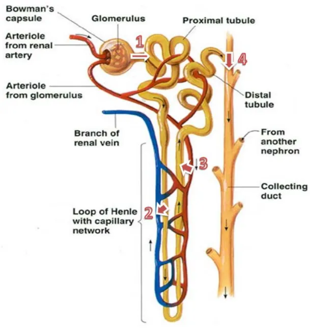
The volume of urine excreted per day (around 1.5L) is a result of two opposing processes namely, ultrafiltration and reabsorption. In the first process, 180 L or more fluid per day is filtered by glomerular capillaries9. The glomeruli filter the blood, leaving blood cells and large proteins (such as albumin), while other material, such as water, small proteins and waste products are passed through Bowman’s capsule, and then into the tubule (figure 3)10.
On the reabsorption process, more than 99% of this ultrafiltrate is sent back to the bloodstream9. Different parts of the tubules perform different functions in this process. The proximal convulated tubule processes and reabsorbs the substances that are still necessary (water, electrolytes, glucose, amino acids and minerals) passing them back to the bloodstream. At the same time that this occurs, other substances such as hydrogen ions, ammonia, creatinine and metabolic products of drugs are secreted from the bloodstream directly to the tubule10. More water is reabsorbed in the distal tubule, which passes the excess water and waste products to the collecting tubule, which travels through the ureter and into the bladder10.
The kidney also secretes hormones namely, erythropoietin, renin, and calcitriol. Erythropoietin stimulates the bone marrow to produce new red blood cells (RBCs). Renin helps regulate blood pressure, and calcitriol is a form of vitamin D and it is important in maintaining bones and the level of calcium in the body10.
Figure 3- Physiology of a nephron.
Legend: (1)Filtration; (2)Reabsorption; (3) Secretion; (4) Excretion Adapted from: www.cic-caracas.org/departments/science/Topic12.php
Although the kidney is a resilient organ, kidney disorders can occur and be either glomerular, causing problems in the filtration process, or tubular, affecting the
by a progressive distortion of kidney morphology and architecture. At an advanced stage of the disease, usually called kidney failure, the kidney loses the capacity to remove and regulate water and other products and, waste products (causing uremia) and excess water (causing swelling) start to build up9. The various kidney diseases are categorized according to whether they are acute and potentially reversible or chronic and generally irreversible, as well as the site of the damage (either glomerular or tubulointerstitial).
3. Acute Kidney Injury
Acute kidney failure (AKF) is a generic term for an abrupt and sustained decrease of kidney function leading to retention of nitrogenous, such as urea and creatinine, and non-nitrogenous waste products11.
Despite the advances in treatment strategies and comprehension of the pathogenesis, until recently there was no clear definition of AKF. This definition still remains subjected to controversy and there is a lack of consensus. In fact, there are more than 30 definitions in the literature for AKF. The definitions varied from a 25% increase over baseline serum creatinine to the need for dialysis12.
The term acute kidney injury (AKI) has been recently proposed to replace AKF, since an acute decline in kidney function is generally secondary to an injury which leads to functional and structural changes in the kidney13. The term AKI is also intended to reflect the entire spectrum of AKF.
Figure 4 – RIFLE classification scheme.
In the risk, injury, fail, loss and end-stage kidney disease (RIFLE) classification the criteria (GFR or UO) that lead to the worst possible classification should be used. The Failure class is present even if the increase in serum creatinine is below threefold, as long as the serum creatinine is 4.0 mg per 100 ml or above in the setting of an acute increase of at least 0.5 mg per 100 ml. The shape of the figure indicates that more patients (high sensitivity) will be included in the upper category, including some without actually having kidney failure (less specificity). In contrast, at the bottom, the criteria are strict (high specificity), but some patients will be missed (less sensitivity).
GFR-glomerular filtration rate; UO-urinary output AKF-acute kidney failure; ESKD- End-stage kidney disease.
In order to meet the need of an uniform definition, the Acute Dialysis and Quality Initiative (ADQI) group published, in 2004, a new classification which includes several parameters: the risk, injury, failure, loss and end-stage kidney disease (RIFLE) classification, in which are defined three classes of severity- risk (R), injury (I) and failure (F) classes - and two outcomes – loss (L) and end-stage kidney disease (ESKD)14.
The proposed classification of AKI by the RIFLE criteria is shown in figure 4.
Some studies have applied the RIFLE classification to evaluate the occurrence rate and/or outcome of AKI. More recently, this classification has been applied to evaluate clinical characteristics and its predictive ability in intensive care unit (ICU) patients, one of them specifically directed for critically ill HIV-infected patients15-18. Those studies reported a good discriminative ability and in a Portuguese study, Lopes et al. reported that the RIFLE criteria represent a helpful tool in the prognosis of ICU HIV-infected patients18.
In order to further improve the AKI definition, the Acute Kidney Injury Network (AKIN) was created, proposing a modified version of RIFLE classification, known as AKIN criteria19. The most important modifications of AKIN criteria include:
a) the removal of the outcome criteria and the severity criteria are designated as stage 1, 2 and 3;
b) the broadening of the risk category to include an increase in serum creatinine of at least 0.3 mg/dl, even if it this does not reach the 50% threshold;
d) the categorization of any patient treated with kidney replacement therapy as stage 3 regardless of serum creatinine and urine output.
The AKIN criteria were found to improve detection of AKI sensitivity when compared with the RIFLE classification by Lopes et al .20. On the other hand, a retrospective study of the Australian and New Zealand Intensive Care Society database did not reveal any significant difference21.
AKI has been reported in up to 20% of all hospitalized HIV-infected patients22 and etiologies of AKI in HIV-infected patients are generally similar to what is seen in non-infected hospitalized patients23.
Acute deterioration into kidney failure can be prerenal (hypoperfusion), renal (intrinsic to kidney tissue) or postrenal (obstructive) (table 1).
Prerenal azotemia is characterized by increased levels of nitrogen-containing compounds and in the blood caused by a decrease in blood flow to the kidneys, and therefore by an insufficient filtering of the blood. This condition is the most common cause of AKI in HIV-infected patients24,25. This type of hemodynamic kidney failure includes hypovolumetric states, often as a result of gastrointestinal fluid loss, associated with profuse vomiting and/or diarrhea. Disordered kidney regulation of salt and water balance can also contribute to intravascular volume depletion in these patients. Similarly, central or nephrogenic diabetes insipidus in these patients often causes unregulated water loss, which leads to dehydration26. Prerenal azotemia can also result from sepsis which develops from a combination of endotoxin-associated systemic vasodilatation, arterial hypotension, capillary leakage and kidney arteriolar vasoconstriction associated with vasopressor drug therapy26.
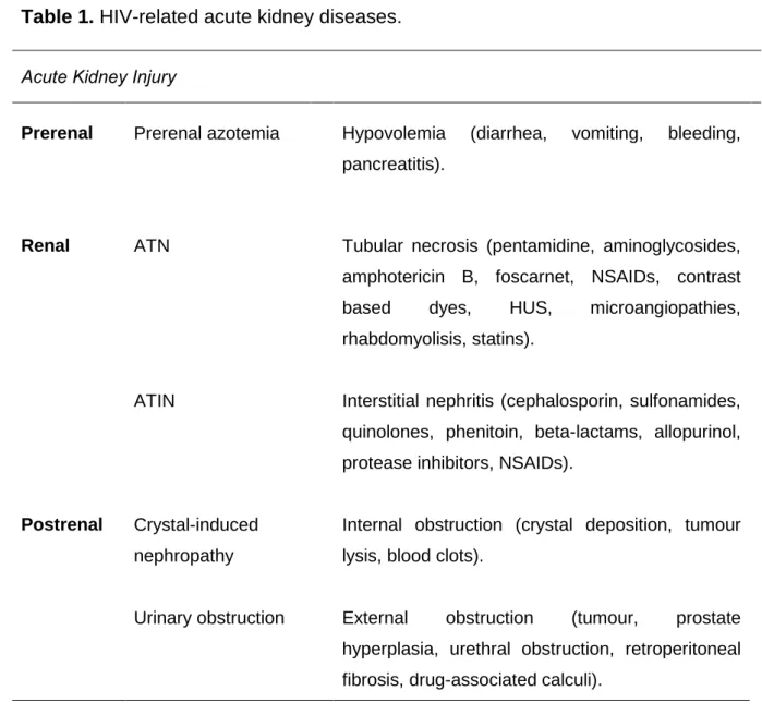
Table 1. HIV-related acute kidney diseases.
Acute Kidney Injury
Prerenal Prerenal azotemia Hypovolemia (diarrhea, vomiting, bleeding, pancreatitis).
Renal ATN Tubular necrosis (pentamidine, aminoglycosides, amphotericin B, foscarnet, NSAIDs, contrast based dyes, HUS, microangiopathies, rhabdomyolisis, statins).
ATIN Interstitial nephritis (cephalosporin, sulfonamides, quinolones, phenitoin, beta-lactams, allopurinol, protease inhibitors, NSAIDs).
Postrenal Crystal-induced nephropathy
Internal obstruction (crystal deposition, tumour lysis, blood clots).
Urinary obstruction External obstruction (tumour, prostate hyperplasia, urethral obstruction, retroperitoneal fibrosis, drug-associated calculi).
ATN- Acute tubular necrosis; ATIN- Acute tubulointerstitial nephritis; NSAIDs- nonsteroidal anti-inflammatory drugs; HUS- haemolytic uremic syndrome.
Acute tubular necrosis (ATN) is the death of tubular cells, when they do not get enough oxygen (ischemic ATN) or when they have been exposed to a toxic drug or molecule (nephrotoxic ATN). This can be due to hypovolemia, sepsis, shock and use of nephrotoxic agents for therapeutic and diagnosis purposes. However, medication-induced ATN is the most common cause of AKI in hospitalized HIV-infected patients
due to the variety of nephrotoxic drugs often required for treatment of opportunistic and nosocomial infections27. Commonly implicated drugs include: pentamidine, foscanet, amphotericin B, amynoglycoside antibiotics, non-steroid anti-inflammatory drugs (NSAIDs). Statin use for hyperlipidemia secondary to cART and concomitant use of gemfibrozil have also been found to cause rhabdomyolysis26.
Acute tubulointerstitial nephritis (ATIN) is an inflammation of the tubules that affect the interstitium. ATIN should also be considered as a possible cause of AKI when certain drugs are prescribed for HIV-infected patients. Common implicated drugs include: β-Lactam antibiotics, sulfonamides, quinolones, phenitoin, allopurinol, NSAIDs and indinavir25.
The deposition of insoluble crystals in the kidney can also cause AKI in HIV-infected patients. Severe AKI due to crystal-induced nephropathy can develop in the presence of risk factors that increase intra-tubular crystal precipitation. As an example, patients with AIDS-associated lymphoma which produce excessive amounts of uric acid during cell death24, that cannot be filtered and secreted by the kidney, which results in precipitation and deposit of uric acid crystals in the tubular lumens of the distal nephron causing intrarenal obstruction24. Indinavir, sulfadiazine, foscarnet and acyclovir have been implicated in crystalluria and intrarenal obstruction28. Intravascular volume depletion and low urinary flow rates increase the risk for crystal deposition of these substances, as well as uric acid28.
Abnormal results on urinalysis usually provide the only evidence of crystal-related kidney injury. Cristalluria, cylindruria, hematuria and proteinuria in varying degrees can be found on urinalysis24,28.
Obstruction of the urinary tract is a rare cause of AKI but gains importance in HIV-infected patients. Several unusual causes of obstruction have been described in these patients23,24, which are summarised in table 1. The most common cause of obstruction in HIV-infected patients is the development of drug-induced calculi. This has been noted with the antibiotic sulfadiazine and the protease inhibitor indinavir. Symptomatic nephrolithiasis, characterized by classic renal colic with hematuria and dysuria can also occur28.
Opportunistic infections and septicaemia are becoming less common causes of kidney injury in cART-treated patients. In opposition, drug toxicity is becoming an increasing cause of both AKI and chronic kidney disease in HIV-infected patients.
4. Chronic Kidney Disease
Chronic Kidney Disease (CKD) is a syndrome which results from progressive and irreversible destruction of the nephrons, regardless of the cause. It is defined by the maintenance of kidney injury or reduced kidney function for three months or longer (table 2). Proof of chronicity can be also provided by the demonstration of bilateral reduction of kidney size by diagnostic imaging techniques9. Other findings consist with long standing kidney disease, such as osteodystrophy, signs of uremia, anemia, hyperphosphatemia or hipercalcemia, however these are not specific. In contrast, the finding of broad casts in urinary sediments is specific for CKD. The wide diameter of
these casts reflects the compensatory function and hypertrophy of surviving nephrons9.
Recently, in a study with 1,239 HIV-infected patients, the CKD prevalence was 15.5% 29 and in another study it was reported that HIV-infected patients account for 1-2% of all the end-stage kidney failure population30.
In Portugal, the annual incidence of HIV-infected patients on dialysis grew from 0.5%, in 1997, to 0.9% in 2002. Approximately 75% of these patients were receiving treatment in the Lisbon metropolitan area31.
Table 2. Stages of chronic kidney disease.
Stage Description GFR (mL/min/1,73m2)
I Kidney injury with normal or elevated GFR ≥ 90
II Kidney injury with a mild GFR reduction 60 – 89
III Moderate GFR reduction 30 -59
IV Severe GFR reduction 15 – 29
V Kidney failure < 15
Chronic kidney disease is defined as kidney injury or GFR<60 mL/min/1.73m2 for more than three months. Injury is defined as a pathological alteration or existence of injury biomarkers, including changes presented in urinary or imaging exams.
In cases of CKD in HIV-infected patients, it is important to consider not only infection-associated etiologies, but also factors or comorbidity conditions which are frequent in the general population, such as diabetes mellitus or hypertension. In this group of patients, the most frequent cause of CKD is HIV-associated nephropathy (HIVAN). HIVAN was first reported in 1984 by Rao et al.32. This author described a focal and segmental glomerulosclerosis in nine patients with AIDS and nephritic syndrome. Before the cART era, the usual presentation of HIVAN was the onset of asymptomatic proteinuria, followed by a rapid decrease of kidney function and, in a matter of months, end-stage kidney failure33. It is considered that HIVAN pathogenesis involves a direct citotoxic effect, caused by the HIV infection and also by viral gene expression in kidney cells. It seems that a host genetic predisposition for occurrence of HIVAN might also be present34. Several studies have shown that HIVAN is predominant and more severe in black individuals. Nowadays in the USA, HIVAN corresponds to the third cause of CKD in the adult black population that initiates dialysis 35.
Non-HIVAN causes of CKD in these patients include: thrombotic microangiopathy, HIV immune complex-mediated kidney disease (HIVICK), diabetic nephropathy and hypertensive nephropathy (table 3).Thrombotic microangiopathy can occur due to an endothelial cell dysfunction, partially mediated by viral proteins36,37. This disease is characterized by five findings with variable expression: fever, neurologic dysfunction, thrombocytopenia, microangiopathic hemolytic anemia and kidney failure with hematuria. Presence of a high level of proteinuria is not frequent, which helps in the differential diagnosis with HIVAN38, 39.
Table 3. Causes and characteristics of CKD in HIV-infected patients.
Condition Characteristics
HIV-associated Nephropathy (HIVAN) Severe proteinuria, large echogenic kidneys,
absent of edema, urinalysis often bland.
HIV immune complex disease (HIVICK) Identical clinical presentation as HIVAN – varying
degrees of kidney dysfunction. Immune complex mediated glomerulonephritis
Ig A nephritis Proteinuria, hematuria, mild kidney dysfunction, elevated levels of serum Ig A.
Post-infectious glomerulonephritis Edema, proteinuria, hematuria, hypertension is frequent.
Membranous nephritis
Sub-epithelial immune deposits (thickened glomerular capillary walls), proteinuria, nephrotic urine sediment.
Membranoproliferative glomerulonephritis
Hematuria with dysmorphic RBCs and casts. Proteinuria (50% of patients have nephrotic proteinuria). Enlarged kidneys and hypercellular. Mesangial proliferative glomerulonephritis Edema, hematuria, proteinuria, hyperlipidemia.
Increase of mesangial cells.
Fibrillary or immunotactoid glomerulonephritis Nephrotic proteinuria, microhematuria, Ig G and C3 glomerular deposits.
Lupus-like nephritis
Microhematuria, proteinuria, relatively rapid progression to kidney failure. Large sub-epithelial deposits with a basement membrane reaction. Interstitial nephritis
Proteinuria, hematuria. Kidney biopsy shows mononuclear and often eosinophilic cellular infiltration of the kidney parenchyma.
Thrombotic microangiopathies
Schistocytes on a blood smear (fragmented RBCs), thrombocytopenia, lactate dehydrogenase level is extremely elevated.
Minimal change glomerulonephritis
Severe proteinuria, oval fat bodies can be observed, edema, hypoalbuminemia, hyperlipidemia, hyponatremia.
Diabetic nephropathy Persistent proteinuria, microalbuminuria.
Hypertensive nephropathy
Hyaline arteriolosclerosis in the kidneys. Nephrons with dilated tubules, often hyaline casts in the lumen.
HIVICK represents a variety of different presentations namely, glomerulonephritis lupus-like, proliferative, membranous, fibrillar, imunotactoid or post-infection40-43 (table 3). The pathogenesis of the different forms of HIVICK can have common etiologic features, including deposits of immune complexes containing HIV-1 antigens, specific cytokine expression profiles, genetic factors, inflammatory infiltrates, and the development of kidney scarring30.
Immunoglobulin A (IgA) nephropathy is characterized by mostly IgA deposition in the glomerular mesangium and, in HIV-infected patients, appears to be the result of immune complexes containing HIV antigens44. It is characterized by proteinuria, hematuria, and mild kidney impairment and is less severe than HIVAN and some other variants of HIVICK45. Elevated levels of serum IgA, as well as detectable serum IgA immune complexes and rheumatoid factor, may be present46.
IgA nephropathy seems to be more frequent in European men, being quite rare in Africa descendents47, as in the non-infected population48.
Diabetes is one of the leading causes of kidney disease in the general population. In Europe and USA, the prevalence of stage 3 CKD, among people with diabetes is 26.5%49. The pathogenesis of diabetic nephropathy is complex. Initially hyperglycaemia, oxidative stress and altered insulin seem to trigger kidney vasodilatation, leading to an increase of the glomerular filtration around 25-50% above the normal range10. This causes an increased blood pressure and volume flowing into the glomeruli, which results in glomerular capillary distension. The increased tension and inflammation causes changes to the kidney parenchyma, resulting in glomerular and tubular hypertrophy. This leads to a glomerular capillary compression, resulting in a decrease of glomerular filtration with microalbuminuria
and hypertension10. The progressive increase of cellular damage, including kidney podocytes apoptosis, leads to a further decrease on glomerular filtration and the occurrence of severe proteinuria (>3g/day). Without intervention, the patient eventually progresses to ESKD10.
Hypertension is another leading cause of CKD. High blood pressure leads to an increase of blood volume that passes though the glomeruli, which creates stress on the glomerular cells, including mesangial and epithelial cells and podocytes50. This leads to an overproduction of vasoactive substances that activate the production of inflammatory cytokines and growth factors, which promote cell damage and fibrosis, reducing glomerular filtration51. Patients with hypertensive nephropathy usually present severe proteinuria, edema and uremia, which can lead to death, frequently from cerebral hemorrhage10.
5. Mechanism of HIV infection of kidney cells
It is still an issue under discussion whether HIV can or cannot directly infect kidney cells. Although viral replication in kidney cells is probably restricted by the lack of CD4 and chemokine co-receptors required for viral entry, it was shown that HIV transfection allows kidney epithelial cells to produce viral particles52-54.
Another possibility is CCR5 transfer by microparticles released from peripheral blood mononuclear cells56.
The potential role of dendritic cells also needs to be considered. Dendritic cells are involved in binding, dissemination and transfer of HIV to lymphoid and non lymphoid tissues and, for that reason, may also be implicated in HIV infection of kidney cells55.
5. Combination Antiretroviral Therapy
The first treatment for HIV-infection was available in 1987 and consisted in the nucleoside analog reverse transcriptase inhibitor (NRTI) zidovudine (ZDV, Retrovir®, first manufactured by GlaxoSmithKline). At the time, most HIV-infected patients were dying within months after the diagnosis of AIDS, usually after a sequence of multiple episodes of Pneumocystis jiroveci pneumonia57.
ZDV was initially developed in 1964 as a candidate for cancer chemotherapy, but was never used before due to its toxicity57. In initial clinical trials, ZDV monotherapy was shown to decrease mortality, increase CD4+ T cell counts and lead to fewer opportunistic infections in patients with symptomatic HIV-infection or AIDS58, 59. Monotherapy limitations were later demonstrated, as studies showed that the duration of ZDV therapy benefits lasted for just one to two years60, 61. However, given the prognosis of a patient diagnosed with AIDS at the time, an apparent extension of a few months of life was regarded as worthwhile. Nevertheless, this opened a window of opportunities for the development of other antiretroviral drugs, with four
additional NRTI drugs licensed for this purpose: didanosine (ddI; Videx®, Bristol-Myers Squibb), zalcacitabine (ddC; Hivid®, Roche), stavudine (d4T; Zerit®, Bristol-Myers Squibb) and lamivudine (3TC; Epivir®, GlaxoSmithKline).
Combinations of two NRTI constituted the first cART. Compared to ZDV monotherapy, ZDV and ddI62,63, ZDV and 3TC62,63 or ZDV and ddC64 showed to be more effective in the increase of CD4+ count and decrease of viral load.
Two nonnucleoside reverse transcriptase inhibitors (NNRTI) became available around 1996, which were, delaviridine (Rescriptor®, Pfizer), which was never available in Portugal, and nevirapine (NVP, Viramune®, Boehringer Ingelheim). With these new antiretroviral drugs, it was possible to treat HIV-infected patients with three drugs. Combination of ZDV, ddI and NVP was found to outperform the efficacy of therapy with two nucleosides65, 66.
Protease inhibitors (PIs), the third class of antiretroviral drugs, were on the market by 199557. The introduction of PIs represented one of the biggest breakthroughs in HIV therapy. Highly active antiretroviral therapy (a term now being substituted by cART), that was a combination of a PI and two or three other antiretroviral drugs, help to transform HIV infection into a manageable chronic disease for the majority of patients with access to these drugs57.
Nowadays, the situation regarding HIV-infection is completely different from 23 years ago. Today there are more than 20 antiretroviral drugs available and life expectancy of HIV-infected patients is measured in decades. Because of that, today the weighing of antiretroviral drugs’ possible benefits of treatment against risk of drug toxicity is extremely important.
5.1. cART associated kidney disease
Studies have confirmed a kidney function improvement in HIV-infected patient, when cART is initiated67. However as previously mentioned, other studies have already associated kidney complications with administration of specific antiretroviral drugs68-95 (table 4).
Indinavir (IDV) (Crixivan®, Merch) seems to be the only PI with significant urinary excretion. Nephrolithiasis is known as the most important side effect68 and also pyuria associated with progressive loss of kidney function69. Ritonavir (RTV) utilization as IDV booster (IDV/r) elevates the risk of nephrolithiasis70 and of rhabdomyolysis with possible occurrence of kidney failure71.
Risk factors of IDV-associated kidney injury include: low body weight, IDV administration in doses equal or greater than 1000mg, administration twice a day, co-administration with cotrimoxazol72 and HCV or HBV co-infection73.
RTV (Norvir®, Abbot Laboratories) has also been associated with AKI74. Isolated cases of nephrolithiasis have also been referenced as associated with administration of saquinavir (Invirase® and Fortovase®, Roche)75 and nelfinavir (Viracept®, ViiV Healthcare)76. More recently, there was also a published case of AKI associated with atazanavir (ATV) (Reyataz®, Bristol-Myers Squibb) administration77.
Regarding NRTIs, dosages might have to be adjusted in patients with a GFR inferior to 60 ml/min/m3, with exception of abacavir that does not need dosage adjustments4. Tenofovir disoproxil fumarate (TDF) (Viread®, also present in Truvada®, Gilead sciences, and Atripla®, Bristol-Myers Squibb & Gilead Sciences) shares its molecular structure with adefovir which utilization is limited by its nephrotoxicity78
Table 4. Antiretroviral drug associated kidney injury
Antiretroviral drug Kidney injury Ref.
Nucleoside reverse transcriptase inhibitors Abacavir Didanosine Lamivudine Stavudine Zalcitabine Zidovudine
AKI (acute interstitial nephritis) in hypersensitivity reaction
Fanconi’s syndrome, AKI, lactic acidosis, nephrogenic diabetes insipidus
Kidney tubular acidosis and hypophosphataemia Kidney tubular acidosis and hypophosphataemia Not reported Not reported [88] [89,90] [91] [91]
Non-nucleoside reverse transcriptase inhibitors Nevirapine Delavirdine Efavirenz Etravirine Not reported Not reported
AKI (multiorgan hypersensitivity reaction) AKI
[94] [95] Nucleotide reverse transcriptase inhibitors
Tenofovir Proximal tubulopathy, nephrogenic diabetis insipidus, AKI [79-83] Protease inhibitors Amprenavir Indinavir Lopinavir Darunavir Fosamprenavir Tipranavir Nelfinavir Saquinavir Ritonavir Atazanavir Not reported
Intratubular precipitation, nephrolithiasis, kidney colic, AKI and CKD Not reported
AKI, nephrolithiasis (in association with ritonavir) Not reported
Not reported Nephrolithiasis
AKI (in association with ritonavir)
AKI, pancreatorenal syndrome, rhabdomyolysis Nephrolithiasis, AKI (interstitial nephritis)
[70,71] [96] [76] [75,93] [74,92,93] [77] Fusion inhibitors
Enfuvirtide Membranoproliferative glomerulonephritis in diabetic patients [97] CCR5 inhibitors Maraviroc Vicriviroc Not reported Not reported Integrase inhibitors
Raltegravir Rhabdomyolysis, mild impairment of a pre-existing kidney insufficiency, severe AKI
Figure 5 - Proximal tubular cell transport pathway for tenofovir.
Tenofovir is delivered to the basolateral membrane of proximal tubular cells and is transported into the cell by organic anion transporter-1 (OAT-1). Within the cell, tenofovir is transported via carrier proteins and is subsequently secreted into the kidney tubule by the apical efflux transporters multidrug resistance protein-2 (MRP-2) and MRP-4. Disturbances in this secretory pathway (due to increased OAT-1 activity or decreased MRP efflux transport activity) may lead to increased tenofovir concentrations within the cell, which can cause mitochondrial DNA depletion and dysfunction. Ultimately this can cause proximal tubulopathy (AKI or Fanconi syndrome).
NaDC- sodium–dicarboxylate symporter; OCT- organic cation transporter; OAT-1-organic anion transporter-1; MRP-multidrug resistance protein; Pgp- P-glycoprotein; TF- Tenofovir; AKI- acute kidney injury.
Although, initial studies did not show high rates of TDF associated with kidney failure78 there are already a number of published cases of Fanconi's syndrome post-TDF administration79-83. This suggests a possibility of specific risk factors in these patients. On the other hand, in most of these cases, patients were on TDF and RTV, including lopinavir/r, saquinavir/r, amprenavir/r and ATV/r79-83. In fact, pharmacokinetics studies demonstrated that an elevation of approximately 32% on TDF exposure occurs, when administrated with lopinavir/r and 37% when co-administrated with ATV/r, compared with TDF administration without these PIs83. The observation that around 93% of TDF-associated kidney impairment occurred in individuals on PIs, have led to the idea that RTV mediated inhibition of the multidrug resistance protein 2 (MRP-2) transporter, which mediates TDF excretion into urine in the proximal tubule84, may lead to intracellular accumulation of this antiretroviral drug (figure 5). For that reason, the higher TDF concentrations could cause proximal tubule necrosis.
Finally, one must not forget that antiretroviral drugs, besides having a direct toxicity, have been also associated with several metabolic complications that might lead to CKD, including insulin resistance, dislipidemia and hypertension85-87.
6. Kidney function assessment: traditional methods versus new
biomarkers
Accurate measurement of GFR is costly and time-consuming. Therefore, clinical routine monitoring of kidney function is based on urinalysis, proteinuria and/or albuminuria determination and, most importantly, it is based on serum creatinine (Cr) determination.
Cr is a by-product from the creatinine phosphate break-down of the muscle catabolism and usually is produced at a relatively constant rate. Because there is almost no tubular reabsortion of Cr, if the kidney filtration is affected, Cr blood levels rise. The normal serum Cr level varies with the technique used to measure it. For an adult, the normal range is between 0.6 to 1.2 mg/dl (53 to 106 μmol/L) by the kinetic or enzymatic method and 0.8 to 1.5 mg/dl (70 to 133 μmol/L) by the Jaffé reaction99. Cr is considered a reliable biomarker of kidney function. However its levels are influenced by age, gender, diet type, low body mass, and the presence of hepatic disease100-104.
To overcome some of these limitations, Cr levels are included in formulas that take into account other factors such as, age and/or weight, in order to obtain an estimated GFR (eGFR) or estimated creatinine clearance (eCrCl).
The two more commonly used formulae are the Cockcroft-Gault and the simplified MDRD (Modified Diet in Renal Disease) equations.
Nevertheless, these formulas are still not validated for HIV-infected patients. As the constant or slow production of Cr makes its rise too small and delayed for early detection of a kidney insult, these formulas also have serum Cr limitations. In addition, serum Cr concentration can remain relatively stable until approximately 50% of kidney function has already been lost105,106. In these cases, kidney function changes can be missed if only based on Cr levels.
Therefore, there remains the need for a reliable biomarker of kidney function that allows the detection of mild kidney dysfunction, in order to enable the timely diagnosis and intervention in the early stages of a kidney injury.
6.1 Cystatin C
Cyst C functions like a cysteine protease inhibitor and is present in several biologic fluids, such as serum or cerebrospinal fluid (CSF).
Cyst C is not secreted into the tubules and it is not reabsorbed back into the serum. Consequently, Cyst C concentration is almost completely dependent of GFR107, 108 Studies have demonstrated that Cyst C has a strong negative correlation with GFR109, 110 and seems to be an earlier87, 111 and superior marker than Cr, for GFR estimation112, 113. Cyst C also appears to be superior to serum Cr in paediatric114-117, elderly118 and diabetic patients119-121. On the other hand, the concept of Cyst C being independent of body mass has become controversial over the last years108, 122 and there are still no standardized methods for Cyst C measurement, nor validated formulas for eGFR123-125.
Figure 6-Ribbon diagram of human cystatin C structure. From: Janowski R et al. Nat Struct Biol 2001,8:316-20
Other factors mentioned as affecting Cyst C production are: thyroid dysfunction, causing a significant decrease in Cyst C levels, as well as high doses of corticosteroids that elevates Cyst C expression117, 126. Cyst C serum or plasma levels can be determined by two commercial available, fully automated assays: the particle-enhanced turbidimetric immunoassay and the particle-particle-enhanced nephelometric immunoassay.
6.2 Interleukin 18
Interleukin 18 (IL-18) is a pro-inflammatory cytokine, synthesized in the proximal tubules, as an inactive precursor form of 24 kDa, suffering a later cleavage by caspase-1 to originate its active form of 19 kDa127-129 (figure 7).
This protein is overexpressed in endogenous inflammatory processes, and seems to have an important role in host defence against several infections130.
Pre-clinical trials have indicated that IL-18 mediates acute tubule necrosis in mice131, 132 and a posterior study reported an IL-18 sensitivity superior to 99% for AKI diagnosis133. In another study, it was shown that IL-18 was an earlier marker of AKI in ICU patients, preceding serum Cr elevation in 1-2 days134. The elevation of IL-18 occurs approximately 12 hours post kidney injury and can be detected by enzyme-linked immunosorbent assay (ELISA) in urine.
6.3 Kidney injury molecule 1
Kidney Injury molecule 1 (KIM-1) is a 100 kDa transmembranar glycoprotein, with an immunoglobulin-like domain and a mucin domain on the extracellular fraction (figure 8)135.
Figure 8 – Ribbon diagram of kidney injury molecule 1 structure. From: Santiago C, et al. Immunity 2007:26,299–310.
The basal expression of this protein in the kidney is very low, in both rodents and humans136,137. Nevertheless, in animal models, KIM-1 is the main induced protein in the proximal tubule after a kidney injury, and can be detected in urine. Increase of KIM-1 occurs approximately 12 hours post kidney injury137.
In a recent study performed in mice with different mechanisms of injury in the proximal tubule, using histopathology as the reference method, a good performance of KIM-1 in nephrotoxicity identification was reported. Moreover, KIM-1 was the urinary biomarker with higher sensitivity and specificity from the 21 used in the study138. In research, KIM-1 is usually detected by ELISA or microbead. However there are still no commercial methods available.
6.4 Liver fatty acid binding protein
Fatty acid binding proteins (FABPs) are small cytoplasmatic proteins of 15 kDa which are expressed in all tissues with active metabolism of fatty acids139.
FABPs function as chaperons, facilitating free fatty acid chains transport from the cytoplasmatic membrane to oxidation sites such as the mitochondria or peroxissomes139,140.
Two types of FABPs were identified in the human kidney: a liver-type FABP (L-FABP) (figure 9), expressed in the proximal tubule and a heart-type FABP (H-(L-FABP), expressed in the distal tubule141, 142.
Pre-clinical and clinical trials reported that urinary L-FABP can be a potential biomarker for several pathologies, such as CKD, IgA nephropathy, contrast nephropathy and diabetic nephropathy142-145. Besides that, clinical studies demonstrated that L-FABP has a potential as biomarker for monitoring CKD progression. In another study from Kamijio et al. L-FABP increase was shown to be proportional to the kidney function deterioration in non-diabetic patients with CKD146. A preliminary study from Ferguson et al. suggested L-FABP as an earlier biomarker of AKI (compared with serum Cr), specifically in the case of aminoglicosides administration147.
A different study revealed that L-FABP elevation can be detected approximately four hours after a kidney injury148. Still, there seems to be some controversy around L-FABP specificity.
Figure 9 – Ribbon diagram of liver fatty acid binding protein structure. From: Protein Data Bank 2F72
6.5 Lipocalin-2 or Neutrophil gelatinase-associated lipocalin
Neutrophil gelatinase-associated lipocalin (NGAL) is a protein from the lipocalin family with a molecular weight of 25 kDa. It is composed of eight β-strands assembled in a β-barrel that encloses a calyx. This calyx binds and transports low molecular weight substances149 (figure 10).
NGAL is expressed by neutrophils and epithelial cells150. Distinct NGAL expression levels can be found in different tissues, such as uterus, prostate, lungs, salivary glands, trachea, stomach, colon and kidneys151.
It is known that NGAL binds to siderophores and possibly has an antibacterial action152-154. It is thought that this protein can also interact with specific cellular receptors, as the cationic transporter 24p3R, extracellular kinases, hepatocyte growth factors or the gelatinase B enzyme155-157.
This protein might also mediate kidney development158, 15 9 and act as a cellular growth factor160-162.
NGAL is one of the most over expressed proteins in the initial stages of AKI. In the kidney most of this protein is produced in the Loop of Henle and collecting duct163. Studies have shown that NGAL increase can be detected in plasma, serum or urine, just two to six hours after the occurrence of kidney injury164 and that this protein constitutes a sensitive and specific biomarker of AKI164-166. Moreover it was also demonstrated that it has a predictive ability for AKI167, 168 and CKD severity169-177. In a large prospective study by Wagener et al .178, it was shown that elevation of urinary NGAL (uNGAL) was correlated with cardio pulmonary bypass time and
aortic-a)
b)
Figure 10 - NGAL crystal structure with the bacterial enterochelin
ligand.
Bacterial enterochelin is composed of: a) a triserine lactone and b) three catecholate groups. These groups, between them, chelate an iron atom or ion. Enterochelin complex can be buried in the NGAL calyx, between three positively charged amino acids (R81, K125 and K134, where R is arginine and K is lysine).
Several methods have been used for NGAL measurement. Initial studies used Western blot assays182, while subsequent studies preferentially used immunoblotting and ELISA assays. There are automated commercial methods available, for instance Triage® NGAL (Biosite Incorporated, San Diego, USA) or ARCHITECT® (Abbott Diagnostics, Amadora, Portugal).
Beside the potential biomarkers mentioned here, there are several others currently in study. However, further studies are still necessary in this field, especially in the case of HIV-infected patients, where studies of these new biomarkers performances are still very limited.
7. Kidney dysfunction frequency in HIV patients of Hospital
Santa Maria
Hospital Santa Maria (HSM), Centro Hospitalar Lisboa Norte (CHLN) is one of the largest teaching hospitals in the country. It is a 1,300-bed referral tertiary center and serves a large proportion of the Lisbon population. Clínica Universitária de Doenças Infecciosas (Director: Professor Doutor Franciso Antunes) has a large HIV outpatient
In a previous study at Clínica Universitária de Doenças Infecciosas, during a 20 month period (between January 2007 and August 2008), it was shown that 6% (n=757) of 12,022 samples from 2,761 HIV-infected patients had a creatinine level greater than 1.2 mg/dL (range 1.2-13.5 mg/dL) (unpublished results) (figure 11). Using the CKD stages guidelines (Guidelines of HIV Medicine Association of the Infectious Diseases Society of America)4 and eGFR, it was found that 4.1% had moderate GFR reduction (30-59 mL/min/1.73m2), 0.6% had severe GFR reduction (GFR 15-29 mL/min/1,73m2) and 0.7% had kidney failure (GFR<15mL/min/1,73m2) (unpublished results).
Figure 11- Samples distribution per creatinine level.
Early identification of kidney injury is essential to promote initiation/adjustment of cART, suspension of nephrotoxic drugs and control of co-morbid diseases such as diabetes and hypertension. Still, to enable this, sensible (early stage detection) and specific biomarkers of kidney function that can be associated with disease prognosis, are necessary.
In spite of being influenced by several factors, serum Cr maintains a constant basal expression, which makes its rise slowly.
Furthermore, CKD can be present with minimal or no change in Cr, due to its basal reservoir and the raise of Cr tubular secretion. On the other hand, therapeutic interventions that would allow the prevention or treatment of AKI must be introduced in an early stage, before the rise of serum Cr can be detected181,182.
Early markers of kidney injury represent the only possibility for an in-time diagnostic and intervention, allowing kidney protection from further injuries and from other risk factors associated with HIV infection.
The HSM study mentioned above led us to study the kidney function obtained by serum Cr in comparison with the one obtained by an apparently promising potential biomarker, serum Cyst C, in HIV-infected patients attending the Infectious Diseases Department of HSM, CHLN, and evaluate whether this marker could be a better alternative than serum Cr for monitoring kidney function in HIV-infected patients undergoing antiretroviral therapy.
Introduction
Objectives
Material and Methods
Results
Discussion
Concluding remarks
Bibliography
Study objectives
I. Cross-sectional observational analysis of kidney function obtained through serum Cr and serum Cyst C determinations:
a) Analysis of kidney dysfunction associated risk factors for HIV-infected patients;
b) Determination of kidney dysfunction rate using Cr and eCrCl (patients on cART and without treatment);
c) Determination of kidney dysfunction rate based on serum Cyst C (same patient group);
d) Correlations of Cyst C with Cr, eCrCl and associations with risk factors of kidney injury.
II. Analysis of differences in kidney function between patients undergoing different antiretroviral regimens (with/without TDF, ATV/r).
a) Comparison of serum Cyst C, serum Cr and eCrCl levels between patients on cART with TDF, ATV/r and patients on cART that never were on these antiretroviral drugs.
Introduction
Objectives
Material and Methods
Results
Discussion
Concluding remarks
Bibliography
Material and Methods
Study settings
Cross-sectional study with HIV-infected patients who currently attend the HIV outpatient clinic, HSM, CHLN.
Inclusion criteria were: (a) HIV-1 infected patients; (b) age over 18 years; (c) capable of having an informed consent.
Exclusion criteria were: (a) HIV-infected patients with known kidney disease; (b) HIV-2 infection.
This study was approved by the Hospital’s Ethics Committee and Administration board.
Sample size
The sample size (N) was calculated using the following equation: N ≥ [Z 2*[P (1-P)/ (D2)], in which
Z-area under a normal curve corresponding to the desired confidence level (Z0.95); P-expected frequency of an event in the population
Assuming a kidney dysfunction frequency (CrCl<60 mL/min) of 5.4% in this population (frequency obtained from the previously mentioned HSM study), the inclusion of at least 218 patients as the sample size, will allow both the determination of kidney dysfunction frequency and associated risk factors with a confidence level of 95% (α= 0.05) and a precision of ± 3%.
The sampling method used in this study was the method of convenience sampling.
Study population
Information on age, gender, ethnicity, country of birth medical history and tobacco and alcohol use was retrieved.
Data on risk factors for HIV, duration and history of antiretroviral therapy were also assessed. The length of HIV infection was considered as the time since the first positive diagnosis.
CD4+ T cell nadir, current CD4+ T cell count and HIV RNA levels were also assessed.
HBV co-infection was considered positive when the presence of the hepatitis B surface antigen (HBsAg) was confirmed.
HCV co-infection was considered positive for RNA levels equal or higher than 43 IU/mL (COBAS® Ampliprep/TaqMan® HCV Test, Roche Diagnostics, Amadora, Portugal).
Measurements of alanine transaminase (ALT) and aspartate transaminase (AST) were collected in patients with HCV co-infection.
Height, weight, blood pressure and fasting glucose measurements were also collected.
Hypertension was defined as systolic blood pressure higher than 140 mmHg, and/or diastolic blood pressure greater than 90mmHg and/or use of antihypertensive medication.
Diabetes was defined as fasting glucose levels equal to or higher than 126 mg/dL and/or use of antidiabetic medication.
Kidney function markers measurements
Serum Cr level was determined using a modified Jaffé method (ADVIA® 2400 Chemistry System, Siemens, Portugal).
CrCl was estimated by the Cockroft-Gault equation:
eCrCl (mL/min) = [140 - age (years)] x weight (kg) x [0.85 if female] / 72 x serum Cr (mg/dL)
Increased Cr level was defined as a measurement greater than 1.2 mg/dL.
Serum Cyst C was determined with a particle-enhanced immunonephelomeric assay (N Latex Cystatin C, Dade Behring, Portugal), according to the manufacturer. Increased Cyst C was defined as levels greater than 0.96 mg/L.
Statistical analysis
Statistical analysis was performed using the software SPSS version 17.0 (SPSS Inc., Chicago, IL, USA).
Continuous data were summarized as mean (±SD, standard deviation) and median (IQR, inter-quartile range) for parametric and non-parametric data respectively, while categorical data were represented as proportions. Spearman’s and Pearson correlation coefficients were used to evaluate associations between non-parametric and parametric variables.
Pearson’s Chi Square (χ2) and Fisher’s exact test were used to compare categorical data, while Student’s t-test and Mann-Whitney U test were used to analyse parametric and non-parametric data respectively.
A multiple linear regression was also performed using age, gender, ethnicity, BMI as predictor variables and log transformed cystatin C as the dependent variable.
Differences, associations and regression coefficients between variables were considered significant for a p-value lower than 0.05.
Introduction
Objectives
Material and Methods
Results
Discussion
Concluding remarks
Bibliography
Annexes
Results
Demographic and laboratory description of the sample population
After the application of inclusion and exclusion criteria, 242 HIV-1 infected patients were enrolled in this study (table 5). Most of them were Portuguese (85%; n=205) and around a seventh of the patients (15%; n=37) were born in foreign countries, mainly Portuguese speaking countries (Angola, Cape Verde, Mozambique, Brazil, Guinea-Bissau), as well as in the United Kingdom, Switzerland and Zaire.
HIV infection was acquired, in most cases, by the sexual route (n=189), followed by intravenous drug use (n=52), and blood transfusion (n=1).
Seventy percent of patients were male (n=169), and 90% were Caucasian (n=218). The median age of the participants was 45 years old (range: 22-83 years; IQR=14). BMI was 24 Kg/m2 (median; range: 16-33 Kg/m2; IQR=5.9). Hypertension was present in 58 patients (24%) and diabetes in 27 patients (11%). Forty nine percent of the patients (n=119) were current smokers and 7% (n=17) had a documented history of alcohol abuse (table 5).
Overall, patients were infected for a median of 14 years (range 1-25 years, IQR=7). Median CD4+ T cell nadir was 216/mm3 (IQR=267), median CD4+ T cell count, at the time of the study, was 686/mm3 (IQR=509). From the patients on cART, most had viral-RNA levels lower than 40 copies/mL (95%; n=214).
Table 5. Demographic and laboratory data of the study population (n=242) Parameter Gender (n, %) Male Female 169 (70%) 73 (30%) Age (years) 45 (14) Ethnicity (n, %) Caucasian Black 218 (90%) 24 (10%) BMI (Kg/m2) 24 (6) Hypertension (n, %) 58 (24%) Diabetes (n, %) 27 (11%) Current smoking (n, %) 119 (49%) Alcohol abuse (n, %) 17 (7%) Years of infection 14 (7) Years on cART 12 (6) On cART (n, %) 225 (93%) HIV RNA>40copies/mL (n, %) Treatment failure Treatment naïve Treatment interruption 28 (12%) 11 (5%) 7 (3%) 10 (4%) CD4+ nadir (cell/mm3) 216 (267) CD4+ count (cell/mm3) 686 (509) HBV co-infection (n, %) 5 (2%) HCV co-infection (n, %) 63 (26%)
Data are presented as mean (standard deviation) and median (inter-quartile range) for parametric and non-parametric data and proportions (n, %) for categorical
Most patients were on antiretroviral treatment (n=225, 93%), of which 5% (n=11) had documented treatment failure. From those patients that were not on cART, seven (3%) were treatment naïve patients and 10 (4%) had interrupted treatment (due to patients’ own decision, a case of lupus and a case of lipodystrophy).
Among patients on antiretroviral treatment, the median duration of cART was 12 years (range: 5 months-24 years; IQR=6).
Table 6. Current cART regimens on 225 HIV-infected patients.
NRTI N(t)RTI (TDF) NNRTI PI/r II (RAL) N (%) X X 65 (29%) X X X 61 (27%) X X 36 (16%) X X X 55 (24%) X X X 2 (1%) X X X 2 (1%) X X X 4 (2%)
NRTI- nucleoside reverse transcriptase inhibitors; N(t)RTI- nucleotide reverse transcriptase inhibitor; NNRTI- non-nucleoside reverse transcriptase inhibitor; TDF-tenofovir disoproxil fumarate; ATV-atazanavir; PI/r- ritonavir boosted protease inhibitor; II- integrase inhibitor; RAL- raltegravir.