Selection of an antimicrobial culture to be
used in the prevention of neonatal listeriosis
Thesis submitted to the Universidade Católica Portuguesa to attain
the degree of PhD in Biotechnology - with specialization in Microbiology
By
Sandra Cristina Ferreira Borges
Selection of an antimicrobial culture to be
used in the prevention of neonatal listeriosis
Thesis submitted to the Universidade Católica Portuguesa to attain
the degree of PhD in Biotechnology - with specialization in Microbiology
By
Sandra Cristina Ferreira Borges
Under the supervision of Professor Paula Cristina Maia Teixeira
Under the co-supervision of Dr. Joana Gabriela Laranjeira Silva
Abstract
The capacity of lactic acid bacteria to produce acidic products and/or secrete antimicrobial compounds is important in the impairment of vaginal colonization by pathogens. Vaginal pH is normally acidic, varying between 3.5-4.5; an increase in vaginal pH (5.0 to 6.5) can be associated with colonization by pathogenic microorganisms.
The main goal of this study was to select an antimicrobial culture to be used in the prevention of vaginal colonization of Listeria monocytogenes during pregnancy, and consequently, prevent neonatal listeriosis.
The survival and biofilm formation of 20 isolates of L. monocytogenes in simulated vaginal fluid at normal vaginal pH (4.2) and at higher pH values (5.5 and 6.5) was investigated. This pathogen was inhibited by the normal vaginal pH but survives when pH increases. All isolates tested were biofilm producers at different pH values.
Streptococcus agalactiae is an important cause of neonatal infection and maternal
colonization. Therefore, its behavior in simulated vaginal fluid was also analyzed. As with L. monocytogenes, S. agalactiae (n=10) survived longer at higher pH values than at normal vaginal pH. All S. agalactiae isolates were also biofilm producers.
Therefore, since L. monocytogenes and S. agalactiae can survive at higher vaginal pHs, fetuses/neonates from women having increased vaginal pH values during pregnancy, may be at higher risk of neonatal infection. Biofilm production increases the probability of occurrence of neonatal infection.
The application of vaginal probiotics could have the potential for preventing vaginal
Listeria colonization in pregnant women and consequently reduce neonatal infections.
Thirty-five isolates of Pediococcus spp. showed antimicrobial activity against L.
monocytogenes, by production of a bacteriocin, but did not inhibit S. agalactiae isolates. Pediococcus spp. isolates demonstrated the ability to survive in simulated vaginal fluid
at pH 4.2.
Based on the higher bacteriocinogenic activity and survival in simulated vaginal fluid, one isolate of Pediococcus spp. was selected and characterized to evaluate its safety before use as vaginal probiotic. Pediococcus pentosaceus SB83 did not show the presence of virulence factors such as the production of gelatinase, lipase and DNase, hemolytic activity, nor the presence of virulence genes (i.e. surface adhesin, aggregation protein, cytolysin and extracellular metallo-endopeptidase). No relevant antibiotic
resistance traits were detected. Pediococcus pentosaceus SB83 produced biofilms at different pH values (4.2, 5.5 and 6.5) in simulated vaginal fluid, which could also serve as a protective layer against colonization by pathogenic bacteria.
The bacteriocin produced by P. pentosaceus SB83, designated as bacteriocin SB83, also showed inhibitory activity against Enterococcus faecalis and Enterococcus faecium, but did not inhibit vaginal lactic acid bacteria. Bacteriocin SB83 is resistant to several conditions, including conditions in the vaginal tract (pH and components of vaginal fluid).
The bacteriocinogenic activity of P. pentosaceus SB83 against L. monocytogenes was evaluated in simulated vaginal fluid at pH 6.5, since this is the ideal pH to the L.
monocytogenes survival and proliferation. There the inhibitory effect of the
bacteriocinogenic culture was assessed in suspension, as lyophilized powder and in tablets. Suspensions of P. pentosaceus SB83 (1010 CFU/mL) reduced the pathogen (10 CFU/mL)
5
after only 2 h of exposure to below the detection limit; the lyophilized bacteria after 24 h of contact and in tablet form, P. pentosaceus SB83 lost the antimicrobial activity. The pH of simulated vaginal fluid decreased in all the tested conditions.
Since P. pentosaceus SB83 lose its antimicrobial activity in tablet form, it could be therefore used in the form of lyophilized powder, which may be administered intra-vaginally, for instance as a washing solution. This formulation was selected to evaluate the anti-listerial activity during 12 months of storage. During storage at room temperature, lyophilized bacteria totally inhibited the pathogen (below the detection limit) only during one month; after this time, there was a decrease in the cell counts of
P.pentosaceus SB83 and, consequently, in antimicrobial potential. During storage at 4
ºC, P. pentosaceus SB83 showed antimicrobial activity throughout the time of storage investigated. The bacteriocin produced by P. pentosaceus SB83 after storage at 4 ºC, remained active at least during 12 months, however a slight decrease in antimicrobial activity occurred between 9 to 12 months. Therefore, the best formulation of P.
pentosaceus SB83 is as a lyophilized powder stored at 4 ºC.
These in vitro results prove a concept for the use of P. pentosaceus SB83 as a vaginal probiotic, to prevent vaginal colonization by L. monocytogenes in pregnant women.
Resumo
Os compostos antimicrobianos produzidos pelas bactérias do ácido láctico são importantes para a diminuição da colonização vaginal por agentes patogénicos. O pH vaginal é normalmente acídico, variando entre 3,5-4,5; um aumento do pH vaginal (5,0 a 6,5) pode ser associado à colonização por microrganismos patogénicos.
O objetivo principal deste estudo foi selecionar uma cultura com propriedades antimicrobianas com potencial para utilização na prevenção da colonização vaginal por
Listeria monocytogenes durante a gravidez e, consequentemente, evitar a listeriose
neonatal.
Foi investigada a sobrevivência e a capacidade de formação de biofilmes para 20 isolados de L. monocytogenes em fluido vaginal simulado a pH vaginal normal (4,2) e a valores de pH elevados (5,5 e 6,5). Este patogénico foi inibido em condições de pH vaginal normal, mas sobreviveu quando este aumentou. Todos os isolados testados formaram biofilme aos diferentes valores de pH.
Streptococcus agalactiae, é um importante agente de colonização maternal e causa de
infeção neonatal. Por este motivo, foi também analisado o seu comportamento em condições de simulação de fluido vaginal. Tal como L. monocytogenes, os isolados de S.
agalactiae (n=10) sobreviveram melhor a valores de pH elevados comparativamente ao
pH vaginal normal. Todos os isolados foram também produtores de biofilme.
O facto de L. monocytogenes e S. agalactiae sobreviverem a valores de pH vaginal elevados, sugere que as mulheres grávidas que tenham um aumento do pH vaginal, podem ter um maior risco de infeção neonatal. A produção de biofilmes aumenta a probabilidade de ocorrência destas infeções.
A aplicação vaginal de probióticos apresenta potencial para a prevenção de colonização vaginal por L. monocytogenes em mulheres grávidas e, consequentemente, reduzir as infeções neonatais.
Trinta e cinco isolados de Pediococcus spp., demonstraram atividade antimicrobiana contra L. monocytogenes, pela produção de uma bacteriocina, no entanto, não apresentou atividade contra os isolados de S. agalactiae. Estes isolados de Pediococcus spp. sobreviveram no fluido vaginal simulado a pH 4,2.
Com base nos resultados da avaliação da atividade bacteriocinogénica e sobrevivência em fluido vaginal simulado, Pediococcus pentosaceus SB83 foi selecionado para futura aplicação como probiótico vaginal. Pediococcus pentosaceus SB83 não demonstrou
possuir fatores de virulência tais como produção de gelatinase, lipase e DNase, atividade hemolítica, nem possuiu os genes de virulência estudados (adesina de superfície, proteína de agregação, citolisina e metalo-endopeptidase extracelular). Não foram detetadas resistências a antibióticos. Pediococcus pentosaceus SB83 demonstrou ser produtor de biofilme em fluido vaginal simulado a diferentes valores de pH (4,2, 5,5 e 6,5), que também pode atuar como camada protetora contra a colonização por bactérias patogénicas.
A bacteriocina produzida por P. pentosaceus SB83, designada por bacteriocina SB83, também demonstrou atividade inibitória contra Enterococcus faecalis e Enterococcus
faecium, mas não apresentou efeito inibitório para bactérias do ácido láctico autóctones
do trato vaginal. A bacteriocina SB83 demonstrou ser resistente a vários parâmetros, incluindo as condições do trato vaginal (pH e componentes do fluido vaginal).
A atividade bacteriocinogénica de P. pentosaceus SB83 contra L. monocytogenes foi avaliada em fluido vaginal simulado a pH 6,5, pois este demonstrou ser o pH ideal para a sobrevivência e proliferação de L. monocytogenes.Foi testado o efeito inibitório da cultura bacteriocinogénica em suspensão, na forma liofilizada e em comprimidos. A suspensão bacteriana de P. pentosaceus SB83 (1010 UFC/mL) reduziu o patogénico (10 UFC/mL) em
5
apenas 2 h (abaixo do limite de deteção), a bactéria liofilizada após 24 h de contacto e em comprimidos o P. pentosaceus SB83 perdeu a atividade antimicrobiana. O pH do fluido vaginal simulado diminuiu para todas as condições testadas.
Como P. pentosaceus SB83 perdeu a atividade antimicrobiana quando na forma de comprimido, poderá ser utilizado na forma liofilizada, a qual pode ser administrada intravaginalmente, por exemplo como uma solução de lavagem. Esta formulação foi a selecionada para avaliar a atividade anti-listerial durante 12 meses de armazenamento. Durante o armazenamento à temperatura ambiente, a bactéria liofilizada inibiu totalmente o patogénico (abaixo do limite de detecção) apenas durante um mês; após este período de tempo, houve uma diminuição SB83 e, consequentemente, o potencial antimicrobiano. Durante o armazenamento a 4 ºC, P. pentosaceus SB83 apresentou atividade antimicrobiana durante todo o tempo de armazenamento investigado. A bacteriocina produzida por P. pentosaceus SB83 após o armazenamento a 4 ºC, manteve-se ativa pelo menos durante 12 meses, no entanto, uma ligeira diminuição na atividade antimicrobiana ocorreu entre os 9 e 12 meses. Portanto, a melhor formulação
Estes resultados in vitro provam o conceito para a utilização de P. pentosaceus SB83 como probiótico vaginal, para prevenir a colonização vaginal por L. monocytogenes em mulheres grávidas.
Acknowledgments
I would like to thank to the Escola Superior de Biotecnologia of the Universidade Católica Portuguesa, for accepting me as PhD student and for providing the necessary conditions to carry out this work.
I am grateful to Fundação para a Ciência e Tecnologia for the financial support (SFRH/BD/45496/2008).
I am grateful to my supervisor, Professor Paula Teixeira, for accepting me as a PhD student, for all the dedication, support, encouraging, and for all the knowledge provided. To Professor Paul Gibbs, I am grateful for all the editions and for wise comments.
I would like to thank to Professor Paulo Costa to the Faculdade de Farmácia of the Universidade do Porto, for the help with pharmaceutical area, besides the provision of laboratory facilities.
I am thankful to Hospital S. Marcos (Braga), in the person of Dr. Alberta Faustino, for providing vaginal isolates of Group B Streptococcus and lactic acid bacteria.
My gratitude to all the people that work in CBQF for their help, companionship, availability and for all the support regarding my research.
I want to thank all my friends who are a constant presence in my life… you are part of me: Tanita, Mariana, Nesuxa, Óscar, Frank, Padrinho, Renato, Ni, Carla, Ju, Li.
To Helena, thank you for the friendship, the support and for all the help in scientific questions.
To the “babies of my PhD”, that bring me happiness and tears of emotion: Ariana, Clara, Matilde, Catarina and Samuel.
A special thank to three persons who were, are and will be present at all the times. Sarinha, thanks for your persistence and dedication. Thanks for the all the hours spent listening me, the advices and laughter shared… “I’ve got you under my skin”!
Joaninha, you made that all the hours of work seemed moments of "pleasure". Thanks for your help, for the laughter and for sharing all moments of my life. The PhD gave me a friend, a true friend!
Manu, thank you so much for your friendship and care. Thank you for sharing knowledge, for your precious help and for always being by my side.
To my family, “maninhos” Francisco and Nuno, Susana and Vânia, Francisquinho and Tiaguinho. Thanks to be present at all times, for strength and union. You are my equilibrium, my “porto seguro”. I am lucky to have you!
To my parents, one very special thanks, you are the person responsible for my life and you made it all possible. Thank you for letting me go on my way, but everytime I fall you always hold me in your arms. Thank you for showing me the real meaning of unconditional love!
A special thanks to all who were part of my life and will be forever in my heart… to my Grandparents!
Roadmap for the thesis
This thesis consists of six articles, five published in peer-reviewed scientific journals and one is in submission.
The concept of “normal microbiota” as a static and well-defined microbial population is in need of revision, particularly as better information (including that obtained through developed molecular methodologies that are not dependent on culture) is changing the present paradigm. It is recognized that a spectrum of microbial profiles can produce a stable vaginal ecosystem with the ability to maintain vaginal health without succumbing to disease (Farage et al., 2010).
The introductory section covers a general review of the vaginal microbiota and their role in maintaining vaginal health. The incidence of L. monocytogenes and its consequences in pregnant women and in fetuses/neonates, is also reported. The importance of the use of lactic acid bacteria as vaginal probiotics to control the colonization by pathogens, is also included.
Section 3, Survival and biofilm formation of Listeria monocytogenes in simulated
vaginal fluid: influence of pH and strain origin, describes the behavior of L.
monocytogenes in vaginal fluid in vitro, and the influence of the pH on survival of this
pathogen. As infection with L. monocytogenes in the neonate is very similar to Group B streptococci (Streptococcus agalactiae; vertical transmission and clinical manifestations), the behavior of this pathogen in simulated vaginal fluid was also analysed in Section 4, Survival and biofilm formation by Group B streptococci in
Section 5, entitled Evaluation of characteristics of Pediococcus spp. to be used as a
vaginal probiotic, describes the selection of isolates of lactic acid bacteria (Pediococcus
spp.) with antimicrobial activity against L. monocytogenes. In this section, the ability of these isolates to survive and show antimicrobial activity in simulated vaginal fluid, was analysed. The isolate which demonstrated the best results for use as a probiotic,
Pediococcus pentosaceus SB83, was characterized and was evaluated for its safety,
namely, the presence of virulence factors and antibiotic resistance traits.
As P. pentosaceus SB83 demonstrated anti-listerial activity by the production of a bacteriocin, in Section 6, Characterization of a bacteriocin of Pediococcus pentosaceus
SB83 and its potential for vaginal application, bacteriocin SB83 was characterized and
evaluated for its stability at different parameters (pH, temperature, detergents) and specifically in vaginal conditions.
Section 7, Effects of processing and storage on Pediococcus pentosaceus SB83 in
vaginal formulations: lyophilized powder and tablets, describes pharmaceutical
formulations for administration of P. pentosaceus SB83, namely lyophilized powder and tablets. In this section the stability of bacteria after manufacture and during storage was evaluated, including the viability and the bacteriocinogenic activity against L.
monocytogenes in simulated vaginal fluid.
The results of this research offer the potential for the use of P. pentosaceus SB83 to prevent vaginal colonization by L. monocytogenes and furthermore, neonatal listeriosis; these results are discussed in Section 8.
Publications
Borges, S., Silva, J., Teixeira, P. 2013. The role of lactobacilli and probiotics in maintaining vaginal health (submitted for publication).
Borges, S.F., Silva, J.G.L., Teixeira, P.C.M. 2011. Survival and biofilm formation of
Listeria monocytogenes in simulated vaginal fluid: influence of pH and strain origin.
FEMS Immunology and Medical Microbiology 62, 315-320.
Borges, S., Silva, J., Teixeira, P. 2012. Survival and biofilm formation by Group B streptococci in simulated vaginal fluid at different pHs. Antonie van Leeuwenhoeke 101, 677-682.
Borges, S., Barbosa, J., Silva, J., Teixeira, P. 2013. Evaluation of characteristics of
Pediococcus spp. to be used as a vaginal probiotic. Journal of Applied Microbiology,
doi: 10.1111/jam.12232.
Borges, S., Barbosa, J, Silva, J., Teixeira, P. 2013. Characterization of a bacteriocin of
Pediococcus pentosaceus SB83 and its potential for vaginal application. Anti-Infective
Agents 11(2) (in press).
Borges, S., Costa, P., Silva, J., Teixeira, P. 2013. Effects of processing and storage on
Pediococcus pentosaceus SB83 in vaginal formulations: lyophilized powder and tablets.
Keywords Listeria monocytogenes Neonatal listeriosis Pregnancy Genital tract Vaginal fluid Vaginal pH Vaginal probiotic Pediococcus spp. Antimicrobial activity Bacteriocin Vaginal administration Biopharmaceutical product
List of Abbreviations
agg - Aggregation substance gene
ANOVA - Analysis of variance
ATCC - American Type Culture Collection
AU - Arbitrary units
AV - Aerobic vaginitis
BLAST - Basic Local Alignment Search Tool
bp - Base pair(s)
BV - Bacterial vaginosis CFU - Colony forming unit
cyl - Cytolysin gene
DGGE – Denaturing gradient gel electrophoresis
DNA - Deoxyribonucleic Acid DNase - Deoxyribonuclease
dNTP - Deoxyribonucleotide triphosphate
DSMZ - German Collection of Microorganisms and Cell Cultures GmbH (Deutsche Sammlung von Mikroorganismen und Zellkulturen GmbH)
ECOFF - Epidemiological cut-off
efaAfm - Cell wall adhesins of Enterococcus faecium gene
efaAfs - Cell wall adhesins of Enterococcus faecalis gene
EFSA - European Food Safety Authority EPS - Extracellular polymeric substances esp - Enterococcal Surface Protein
FDA - Food and Drug Administration GBS - Group B Streptococcus
gel - Gelatinase
GRAS - Generally regarded as safe HIV - Human immunodeficiency virus
H2O2 - Hydrogen peroxide
HPMC - Hydroxypropylmethylcelluloses
LAB - Lactic acid bacteria MHA - Muller-Hinton agar
MIC - Minimum inhibitory concentration
MRS - de Man, Rogosa and Sharpe
NCCLS - National Committee for Clinical Laboratory Standards
OD - Optical density
PCR - Polymerase Chain Reaction
RAPD - Random amplified polymorphic DNA
rRNA - Ribosomal ribonucleic Acid
SDS - Sodium dodecyl sulphate
SDS-PAGE - Sodium Dodecyl Sulphate PolyAcrylamide Gel Electrophoresis
SVF - Simulated vaginal fluid
SXT - Trimethoprim/sulphamethoxazole
TAE - Tris, Acetic acid and EDTA TSA - Tryptone Soya agar
TSB - Tryptone Soya broth
UPGMA - Unweighted pairs group matching algorithm UTI - Urinary tract infection
WHO - World Health Organization YE - Yeast extract
List of Tables
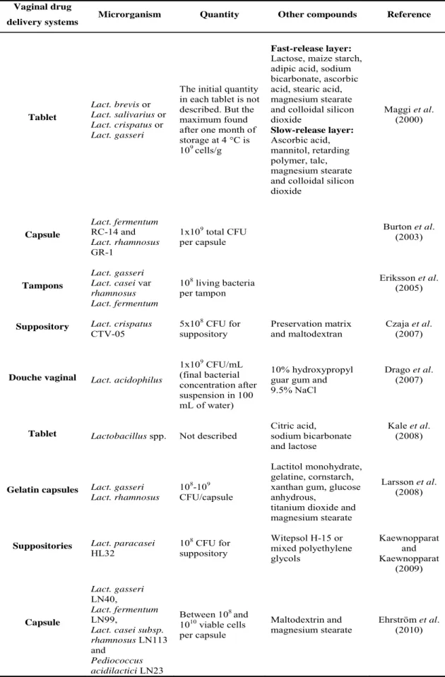
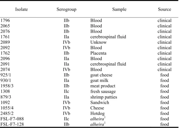
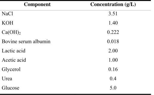
Table 1.1. Prevalence of Lactobacillus spp. in vaginal tract of different women (expressed in percentage values)………...4 Table 1.2. Several studies with different dosage forms for vaginal probiotics………..22 Table 3.1. Listeria monocytogenes strains used in this study, indicating their serogroups and sources………..33 Table 3.2. Percentage of L. monocytogenes strains that produce biofilms in respective media at different time intervals, based on the optical densities strains were classified as strong, moderate or weak biofilm producers………...38 Table 4.1. Composition of simulated vaginal fluid (SVF)....………..47
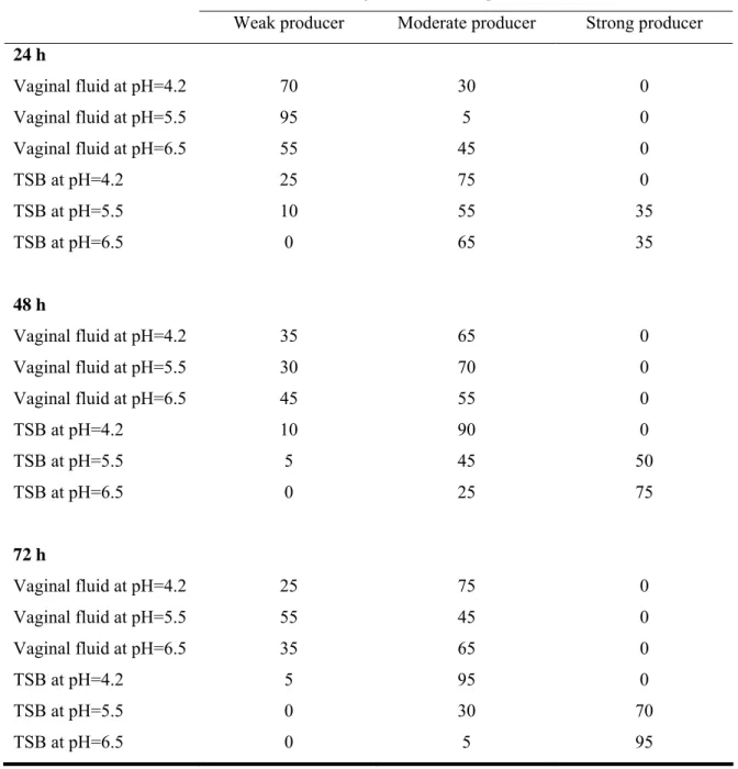
Table 4.2. Isolates of GBS that produce biofilm in respective media at different time intervals. Based on the optical densities strains were classified as strong, moderate or weak biofilm producers………...52 Table 5.1. Listeria monocytogenes isolates used in this study………....60 Table 5.2. Composition of SVF (Owen and Katz 1999)…...…...64
Table 5.3. Minimum Inhibitory Concentration (MIC; μg/mL) of seventeen antibiotics for P. pentosaceus SB83……….……….71
Table 6.1. Reduction of bacteriocin activity after incubation in different conditions (expressed in percentage values)……….90
List of Figures
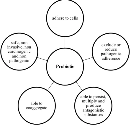
Figure 1.1. Requirements in the choice of a probiotic (Iannitti and Palmieri, 2010)…..20
Figure 3.1. Survival of L. monocytogenes in simulated vaginal fluid. Clinical strains at: (A) pH = 4.2; (C) pH = 5.5; (E) pH = 6.5. Food strains at: (B) pH = 4.2; (D) pH = 5.5; (F) pH = 6.5. All points are means ± standard deviations and for each strain origin (clinical or food), the symbols of the correspondent graphs are the same for each isolate………...36
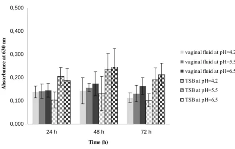
Figure 3.2. Production of biofilm by a representative isolate of L. monocytogenes (1761 IIa) at different time intervals, in SVF and TSB at different values of pH. Error bars represents mean ± standard deviation………...……….39
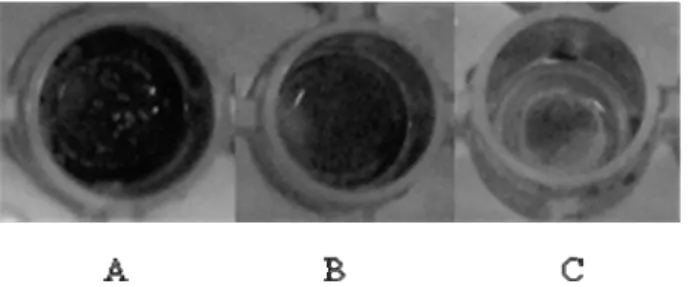
Figure 4.1. Biofilm stained with crystal violet. (A) strong producer; (B) moderate producer; (C) weak producer………...48
Figure 4.2. Survival of three representative GBS strains in SVF at different pH: ( ) strain more sensitive; ( ) strain with intermediate survival; ( ) strain more resistant. Data represented as the mean of the log (N/N0) ± standard deviation……….50
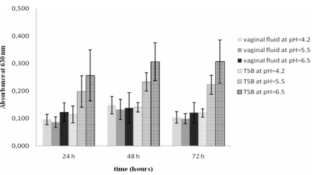
Figure 4.3. Production of biofilm by a representative isolate of GBS at different time intervals, in SVF and TSB at different values of pH. Error bars represent standard deviation of the mean………..51 Figure 5.1. Survival of P. pentosaceus SB83 in SVF at pH = 4.2. All points are means ± standard deviations………...…...…...70
Figure 5.2. Production of biofilm by P. pentosaceus SB83 at different time intervals, in SVF and MRS broth at different values of pH. Optical density was measured in SVF at
pH 4.2 (■), 5.5 (■) and 6.5 (■), and in MRS at pH 4.2 ( ), 5.5 ( ) and 6.5 ( ) Based on the OD, the strain was classified as a strong, moderate or weak biofilm producer. Error bars represent standard deviation of the mean……...…………..72 Figure 6.1. Production of bacteriocin by P. pentosaceus SB83. Antimicrobial activity is presented as AU/mL (bars), against L. monocytogenes ESB 2076 (■), L. monocytogenes ESB 2092 (■) and L. monocytogenes ESB 2096 (■). Changes in optical density (▲) and pH values are indicated (♦)………..88
Figure 6.2. Effect of bacteriocin SB83 on L. monocytogenes ESB 2076 (1/2b) (♦), L.
monocytogenes ESB 2092 (4b) (▲) and L. monocytogenes ESB 2096 (1/2a) (●). The
solid line indicates the effect of bacteriocin on L. monocytogenes and the dotted line represents the growth of L. monocytogenes without added bacteriocin (controls)………..91
Figure 6.3. Tricine-SDS-PAGE of bacteriocin SB83. Lane 1: the gel overlaid with the indicator strain and respective zone of the growth inhibition; lane 2: peptide band stained with Coomassie Blue R250 (40% ammonium sulphate saturated); lane M: molecular weight marker……….92
Figure 7.1. Release of P. pentosaceus SB83 from (A) tablets with retarding polymer and (B) tablets without retarding polymer. The release of bacteria was tested in SVF at pH = 4.2 (■); pH = 5.5 (▲); pH = 6.5 (●) and in Ringer’s solution (♦). All points are means ± standard deviations………...………….106
Figure 7.2. Viability of P. pentosaceus SB83 in preparations with (A) retarding polymer and (B) without retarding polymer during storage. The viability was examined in freeze-dried powder (♦), freeze freeze-dried powder with excipients (■), and tablets (▲) at room
temperature (black lines) and at 4 ºC (grey lines). All points are means ± standard deviations………...………..108
Figure 7.3. Antimicrobial activity of P. pentosaceus SB83 against L. monocytogenes 2092 4b in SVF at pH = 6.5. (A) Counts of viable cells of L. monocytogenes 2092 4b and (B) pH of SVF during 24 h of contact, are represented. The tested conditions were SVF inoculated with: bacterial suspension of P. pentosaceus SB83 and L.
monocytogenes 2092 4b (♦); lyophilized of P. pentosaceus SB83 and L. monocytogenes 2092 4b (■); tablet of P. pentosaceus SB83 with retarding polymer and L.
monocytogenes 2092 4b (▲) and tablet of P. pentosaceus SB83 without retarding polymer and L. monocytogenes 2092 4b (●). Controls were: SVF with 1 mg/mL of trypsin and inoculated with P. pentosaceus SB83 and L. monocytogenes 2092 4b ( ), SVF inoculated with a non- bacteriocinogenic LAB strain ESB67 and L. monocytogenes 2092 4b ( ), SVF inoculated with P. pentosaceus SB83 ( ), SVF inoculated with L.
monocytogenes 2092 4b ( ). All points are means ± standard
deviations………...……….109 Figure 7.4. Antimicrobial activity of lyophilized of P. pentosaceus SB83 against L.
monocytogenes 2092 4b in SVF at pH = 6.5, during storage at room temperature (black
lines) and at 4 ºC (grey lines). (A and D) Counts of viable cells of L. monocytogenes 2092 4b; (B and E) counts of viable cells of P. pentosaceus SB83; (C and F) pH of SVF overtime are represented. These parameters were evaluated at storage time of 0 (♦), 1 (■), 3 (▲), 6 (×), 9 (җ) and 12 (●) months. All points are means ± standard deviations………...…...112
Table of Contents
Abstract………. i
Resumo……….. iii
Acknowledgments………. vi
Roadmap for the thesis……….. viii
Publications………... x
Keywords.………. xi
List of Abbreviations……… xii
List of Tables……… xv
List of Figures………... xvi
1. Introduction………... 1
1.1. Physiology of vagina: the vaginal fluid………. 1
1.2. The microbiota of the vaginal tract……… 2
1.3. Vaginal microbiota and pregnancy……… 8
1.4. Listeria monocytogenes and pregnancy………. 10
1.5. Role of Lactobacillus spp. in maintaing vaginal health………. 13
1.5.1. Antimicrobial activity………. 13
1.5.1.1. pH………. 13
1.5.1.2. Hydrogen peroxide………... 14
1.5.1.3. Bacteriocins………... 16
1.5.2. Adherence……… 18
1.6. Potential of lactic acid bacteria for use as vaginal probiotics…………..…….. 19
1.6.1. Clinical trials………...… 23
1.7. Pediococcus spp. as bioprotective culture……….. 26
2. Objectives of this study………. 28 3. Survival and biofilm formation of Listeria monocytogenes in simulated vaginal fluid: influence of pH and strain origin………. 29 3.1. Abstract………... 30 3.2. Introduction……… 31 3.3. Material and methods………... 33 3.3.1. Microorganisms………... 33 3.3.2. Simulation of vaginal fluid……….. 33 3.3.3. Survival of Listeria monocytogenes in simulated vaginal fluid……….. 34 3.3.4. Biofilm assay………... 34 3.3.5. Statistical analysis………... 35 3.4. Results……… 35 3.4.1. Survival of Listeria monocytogenes in simulated vaginal fluid……….. 35 3.4.2. Biofilm assay………... 37 3.5. Discussion………... 39 4. Survival and biofilm formation of Group B streptococci in simulated vaginal
fluid at different pHs………. 43
4.1. Abstract………... 44
4.2. Introduction……… 45 4.3. Material and methods………. 46 4.3.1. Microorganisms and growth conditions……….. 46 4.3.2. Simulated vaginal fluid………... 46 4.3.3. Survival of Group B streptococci in simulated vaginal fluid…………..…… 47 4.3.4. Biofilm assay………... 48 4.3.5. Statistical analysis………... 49
4.4. Results……… 49 4.4.1. Survival of Group B streptococci in simulated vaginal fluid……… 49 4.4.2. Biofilm assay………... 50 4.5. Discussion……….. 53 5. Evaluation of characteristics of Pediococcus spp. to be used as a vaginal
probiotic………... 55 5.1. Abstract………... 56 5.2. Introduction……….... 57 5.3. Material and methods………. 59 5.3.1. Microorganisms………... 59 5.3.1.1. Lactic acid bacteria isolates……….. 59 5.3.1.2. Pathogenic strains………. 59 5.3.2. Screening for antimicrobial activity of lactic acid bacteria..………... 60 5.3.3. Quantification of antibacterial activity……… 61 5.3.4. Identification of bacteriocin-producing lactic acid bacteria……… 61 5.3.5. Random amplified polymorphic DNA (RAPD)………. 62 5.3.6. Survival of lactic acid bacteria in simulated vaginal fluid……….. 63 5.3.7. Characterization of Pediococcus pentosaceus SB83………... 64 5.3.7.1. Production of hydrolytic enzymes……… 64 5.3.7.2. Hemolytic activity……… 65 5.3.7.3. Virulence genes……… 65 5.3.7.4. Antibiotic susceptibility testing……… 66 5.3.7.5. Biofilm assay……… 67 5.3.8. Statistical analysis………... 68 5.4. Results………...…. 68
5.4.1. Screening for antimicrobial activy of lactic acid bacteria..………...……….. 68 5.4.2. Quantification of antibacterial activity……… 69 5.4.3. Identification of bacteriocin-producing lactic acid bacteria and RAPD-PCR 69 5.4.4. Survival of lactic acid bacteria in simulated vaginal fluid……..……… 69 5.4.5. Characterization of Pediococcus pentosaceus SB83………... 70 5.4.5.1. Virulence factors (production of gelatinase, lipase and DNase, hemolytic activity and virulence genes)………. 70 5.4.5.2. Antibiotic susceptibility testing……… 71 5.4.5.3. Biofilm assay……… 71 5.5. Discussion……….. 73 6. Characterization of a bacteriocin of Pediococcus pentosaceus SB83 and its
potential for vaginal application……… 79 6.1. Abstract……….. 80 6.2. Introduction……… 81 6.3. Material and methods………. 82 6.3.1. Lactic acid bacteria……….. 82 6.3.2. Bacteriocin activity spectrum……….. 83 6.3.3. Bacteriocin production during growth……… 83 6.3.4. Effect of various physical and chemical parameters on bacteriocin
activity... 84 6.3.4.1. Effect of enzymes………. 84 6.3.4.2. Effect of detergents……….. 85 6.3.4.3. Effect of pH………... 85 6.3.4.4. Effect of temperature……… 85 6.3.5. Mode of action………. 86
6.3.6. Adsorption of the bacteriocin to the producer cells………...…. 86 6.3.7. Determination of approximate molecular size of bacteriocin by SDS-PAGE 86 6.4. Results……… 87 6.4.1. Bacteriocin activy spectrum……… 87 6.4.2. Bacteriocin production during growth……… 87 6.4.3. Effect of various physical and chemical treatments on bacteriocin activity... 88 6.4.4. Mode of action (determination of the reduction of viable cells of test
microorganisms in the presence of bacteriocin)……… 91 6.4.5. Adsorption of the bacteriocin to the producer cells……….... 92 6.4.6. Determination of approximate molecular size of bacteriocin by SDS-PAGE 92 6.5. Discussion……….. 92 7. Effects of processing and storage on Pediococcus pentosaceus SB83 in vaginal formulations: lyophilized powder and tablets………... 97 7.1. Abstract……….. 98 7.2. Introduction……… 99 7.3. Material and methods………. 101 7.3.1. Microorganisms and culture conditions……….. 101 7.3.2. Preparation of freeze-dried cells………..……… 101 7.3.3. Preparation of vaginal tablets……….. 102 7.3.4. Release study of vaginal tablets……….. 102 7.3.5. Viability of Pediococcus pentosaceus SB83 in tablets and lyophilized
powder………... 103 7.3.6. Evaluation of antimicrobial activity of Pediococcus pentosaceus SB83 in
simulated vaginal fluid……….. 103 7.3.7. Statistical analysis………... 105
7.4. Results……….... 105 7.4.1. Enumeration of Pediococcus pentosaceus SB83 in lyophilized powder and
tablets………. 105
7.4.2. Release study of vaginal tablets……….. 105 7.4.3. Viability of Pediococcus pentosaceus SB83 in tablets and lyophilized
powder………... 107
7.4.4. Evaluation of antimicrobial activity of Pediococcus pentosaceus SB83 in
simulated vaginal fluid……….. 108 7.5. Discussion……….. 113 7.6. Conclusions……… 118 8. General discussion………. 119 9. Main conclusion……… 122 10. Proposals for future work……… 123 11. References………... 124
1. Introduction
1.1. Physiology of vagina: the vaginal fluid
The human vagina is often described as slightly S-shaped fibromuscular collapsible tubes between 6 and 10 cm long extending from the cervix of the uterus. The vaginal wall consists of three layers: the epithelial layer, the muscular coat and the tunica (Baloglu et al., 2009; Hussain and Ahsan, 2005). The surface of the vagina is composed of various folds, which are often called rugae. The rugae provide distensibility, support and an increased surface area of the vaginal wall. The vagina has an excellent elasticity because of the presence of smooth elastic fibers in the muscular coat. The loose connective tissue of the tunica adventia further increases the elasticity of this organ. The vaginal fluid is principally constituted for cervical secretion and transudation from the blood vessels with desquamated vaginal cells and leucocytes. Secretions from the endometrium and fallopian tubes also contribute to the vaginal fluid (Hussain and Ahsan, 2005).
Thus, vaginal fluid may include contributions from vaginal transudate, Bartholin’s and Skenes’s glands, exfoliated epithelial cells, residual urine, and fluids from the upper reproductive tract. The quantity and composition of human vaginal fluid have been studied by many researchers for a diversity of reasons. These include the diagnosis of pathological conditions such as bacterial vaginosis (BV), urinary tract infection (UTI), cancer, premature rupture of membranes during pregnancy, determination of the presence of semen for forensic analysis, determination of the time of ovulation, and the study of organic acids that may act as sexual attractants. Genital tract secretions contain a complex mixture of components, such as various salts, proteins, carbohydrates, low molecular weight organic compounds (Owen and Katz, 1999). Zegels et al. (2009)
collected cervical-vaginal fluid during colposcopy and identified 339 proteins, which included antimicrobial peptides such as human β-defensin 2 and cathelicidin. The enzymatic activity in the vagina is comparatively lower than in the gastrointestinal tract but there is still a wide range of enzymes present such as nucleases, lysozymes and esterases (Baloglu et al., 2009).
Previous studies suggest that the daily production of vaginal fluid is around 6 g/day, with approximately 0.5-0.75 g present in the vagina at any one time. The volume of vaginal fluid has been shown to increase substantially during periods of sexual stimulation (Owen and Katz, 1999). The discharge produced by postmenopausal women is reduced by 50% compared to that produced by women of reproductive age. At the time of ovulation, mucus secretion increases and it becomes clear, thin and alkaline (Baloglu et al., 2009).
1.2. The microbiota of the vaginal tract
The species that occupy sites in the human body can change based on intrinsic host factors such as stage of life cycle, hormone levels, immune responses, nutritional status and disease states. The normal microbiota can also be altered by external factors such as environmental exposures, microbial interspecies competition or commensalism, and hygiene behaviors (Bolton et al., 2008).
The first extensive study of human vaginal microbiota was published by Döderlein in 1892. Döderlein considered the vaginal microbiota to be homogenous, consisting only of gram-positive bacilli (Döderlein, 1892). These Döderlein’s bacilli are now known to be members of the genus Lactobacillus spp.. This concept has been modified by researchers who have found the microbiota of asymptomatic women to consist usually of a diversity of anaerobic and aerobic microorganisms. The understanding of
compositions and structure of the vaginal microbiome has significantly broadened as a result of using cultivation-independent methods based on the analysis of 16S ribosomal RNA (rRNA) gene sequences (Ma et al., 2012).
Lactobacilli are the most prevalent and often numerically dominant microorganisms, at 107 to 108 CFU/mL of vaginal fluid in healthy premenopausal women (Boris and
Barbés, 2000; Farage et al., 2010).
Among the lactobacilli species found in the vaginal microbiota, Lact. iners, Lact.
crispatus, Lact. gasseri, Lact. jensenii, followed by Lact. acidophilus, Lact. fermentum,
Lact. plantarum, Lact. brevis, Lact. casei, Lact. vaginalis, Lact. delbrueckii, Lact.
salivarius, Lact. reuteri, and Lact. rhamnosus are the most frequently isolated from
healthy women (Cribby et al., 2008). Individual differences in Lactobacillus spp. composition of the vaginal tract between women of different geographic locations, races and ethnicities have been noted across multiple studies (Table 1.1.). In a study by Ravel
et al. (2011), the vaginal bacterial communities of asymptomatic North American
women who represented four ethnic groups (white, black, Hispanic and Asian) were characterized by pyrosequencing of barcoded 16S rRNA genes and the vaginal microbiota varied among the four ethnic groups.
In addition to multiple species of Lactobacillus spp., other lactic acid bacteria (LAB) genera have been found in the vaginal tract, such as Pediococcus spp., Weisella spp.,
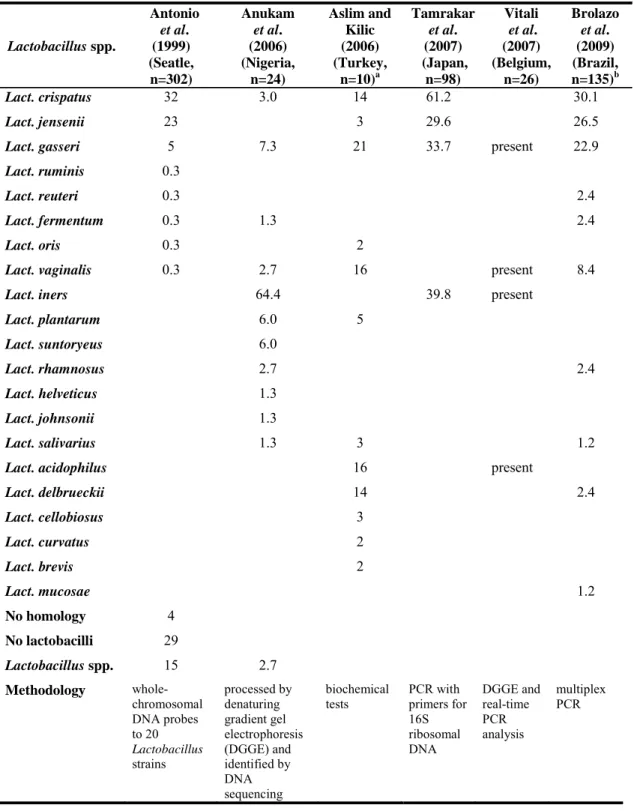
Table 1.1. Prevalence of Lactobacillus spp. in vaginal tract of different women (expressed in percentage values).
Lactobacillus spp. Antonio et al. (1999) (Seatle, n=302) Anukam et al. (2006) (Nigeria, n=24) Aslim and Kilic (2006) (Turkey, n=10)a Tamrakar et al. (2007) (Japan, n=98) Vitali et al. (2007) (Belgium, n=26) Brolazo et al. (2009) (Brazil, n=135)b Lact. crispatus 32 3.0 14 61.2 30.1 Lact. jensenii 23 3 29.6 26.5
Lact. gasseri 5 7.3 21 33.7 present 22.9
Lact. ruminis 0.3
Lact. reuteri 0.3 2.4
Lact. fermentum 0.3 1.3 2.4
Lact. oris 0.3 2
Lact. vaginalis 0.3 2.7 16 present 8.4
Lact. iners 64.4 39.8 present
Lact. plantarum 6.0 5 Lact. suntoryeus 6.0 Lact. rhamnosus 2.7 2.4 Lact. helveticus 1.3 Lact. johnsonii 1.3 Lact. salivarius 1.3 3 1.2
Lact. acidophilus 16 present
Lact. delbrueckii 14 2.4 Lact. cellobiosus 3 Lact. curvatus 2 Lact. brevis 2 Lact. mucosae 1.2 No homology 4 No lactobacilli 29 Lactobacillus spp. 15 2.7 Methodology whole-chromosomal DNA probes to 20 Lactobacillus strains processed by denaturing gradient gel electrophoresis (DGGE) and identified by DNA sequencing biochemical tests PCR with primers for 16S ribosomal DNA DGGE and real-time PCR analysis multiplex PCR
a) Percentage relative to the number of isolates (58 isolates of Lactobacillus spp.) b) Percentage relative to the number of isolates (83 isolates of Lactobacillus spp.)
The vaginal vault is colonized within 24 hours of a female child’s birth and remains colonized until death (Farage et al., 2010). Lactobacilli become the predominant inhabitants of the vagina at the time of puberty, presumably because of the effect of estrogens on the glycogen content of vaginal epithelial cells (Schwebke, 2001). Menopause is marked by a dramatic reduction in estrogen production, resulting in drying and atrophy of the vaginal epithelium. When estrogen levels drop, glycogen content in the vaginal epithelium drops as well, leading to depletion of lactobacilli. Diminishing numbers of lactobacilli result in a subsequent rise in vaginal pH, since glucose is not converted to lactic acid. High pH values promote growth of pathogenic bacteria, particularly colonization by enteric bacteria (Farage et al., 2010). Burton and Reid (2002) performed a study with 20 postmenopausal women (range 44-72 years) and 70% had either intermediate-grade bacterial colonization or BV.
Therefore, the vaginal microbial ecosystem suffers significant structural changes at various stages in a woman’s life that are directly associated to the level of estrogen in the body (Hickey et al., 2012).
The complex interactions of the various members of the vaginal microbiota are not well understood; however, it is recognized that the composition of the microbiota can vary from day to day, even in women without indication of infections. Studies using sequential vaginal cultures or vaginal smears have shown that in some women, there are significant, transient changes in the microbiota. The most unstable time appears to be around the time of menses, but the exact factors responsible for these changes are unknown (Schwebke, 2001).
The microorganisms that inhabit the vaginal environment, play a major role in preventing illnesses of the host, including BV, UTI, yeast vaginitis, and sexually
transmitted diseases including human immunodeficiency virus (HIV) (Reid and Bocking, 2003).
There seems to be an association between absence (or low concentrations) of vaginal lactobacilli and the development of BV. Compared with that of normal women, the vaginal microbiota of women with BV consists more commonly, and in higher numbers, of Gardnerella vaginalis, Mycoplasma hominis, Prevotella spp.,
Peptostreptococcus spp., Mobiluncus spp., Bacteroides spp., Atopium vaginae and
Megasphera spp. (Cribby et al., 2008, Falagas et al., 2007). Bacterial vaginosis is
characterized by the presence of three of five criteria (Amsel criteria): release of an amine fishy odor, release of amine odor after addition of 10% potassium hydroxide, vaginal pH values greater than 4.5, clue cells in the vaginal fluid, and milky homogenous vaginal discharge (Reid and Bocking, 2003). Nugent et al. (1991) suggested that BV should be diagnosed by vaginal smears examined following Gram’s stain. The examination consists of scoring the cell population as to being normal (0 to 3) and dominated by Lactobacillus spp., intermediate (4 to 6) with colonization by small gram-negative or gram-variable rods (Bacteroides spp. or G. vaginalis) and curved gram-variable rods (Mobiluncus spp.), and BV (7 to 10) with domination by pathogens and absence of lactobacilli.
Bacterial vaginosis is associated with high pH, a decrease in antimicrobial activity of the vaginal fluid compared to healthy women, and local impairment of the multiple innate immune pathways (Dover et al., 2008). A typical feature of BV is the absence of inflammation. In BV, there is only a slight increase in interleukin I and a low production of interleukin 8, preventing the attraction of inflammatory cells such as macrophages and neutrophils (Donati et al., 2010).
A second major abnormality of the vaginal microbiota, denominated as aerobic vaginitis (AV), has been described. In this disorder, the normally present Lactobacillus spp. are replaced with aerobic organisms, predominantly enteric commensals or pathogens. Such a community was most often classified as “intermediate”. Group B streptococci (GBS),
Escherichia coli, and Staphylococcus aureus are the organisms most frequently
associated with AV (Rampersaud et al., 2012).
Donders et al. (2002) proposed the following microscopic features to diagnose this disorder: (1) a paucity of lactobacilli; (2) an increased number of leukocytes; (3) the presence of parabasal cells (a sign of epithelial inflammation); and (4) the presence of cocci or coliform bacteria.
Despite the proximity of the vagina to the anus, the variety of microorganisms present in the vagina is much lower than in the gut. The reduced receptivity of the vagina, different nutrient availability and competition with indigenous organisms can be the reasons for this lower diversity in vaginal tract. Some microbes found in the gut, can also be found in the vagina, demonstrating the proper receptors, nutrients, and oxygen tension are present for these organisms to grow (Cribby et al., 2008).
Accessible data about types and numbers of bacteria present in the vagina have not correlated well with clinical symptoms, an observation which may well be the result of the incomplete analysis of the vaginal ecosystem available through traditional methods. Development of molecular techniques that can identify (without the need for laborious microscopy or prior culture) the organisms comprising the vaginal microbial community, are proving useful in defining organisms that have defied identification through traditional cultural methods (Farage et al., 2010).
1.3. Vaginal microbiota and pregnancy
Numerous types of microorganisms which can be found in association with preterm labor and delivery, such as Neisseria gonorrhoeae, Chlamydia trachomatis and
Trichomonas vaginalis, Haemophilus influenza only occasionally acts like a pathogen,
being associated with preterm labor. Group B streptococci can have devastating effects on the preterm or low birth weight infant. A number of genital microorganisms such as
E. coli, L. monocytogenes and viridans streptococci may be involved in
chorioamnionitis. Bacterial vaginosis is a polymicrobial condition increasingly associated with adverse perinatal sequelae (Donati et al., 2010).
The importance of LAB in life is perhaps best seen in relation to the health of women and babies (Reid, 2008). Lactobacilli are able to reduce the incidence of ascending infections of the uterus and the subsequent production of proinflammatory molecules (Wilks et al., 2004).
Once pregnant, the loss of lactobacilli from the vagina and subsequent development of BV is associated with serious complications include preterm delivery of low birth weight infants, spontaneous abortion, premature rupture of membranes, preterm birth, amniotic fluid infections, postpartum endometritis and endometritis following Caesarian section (Hickey et al., 2012). Toxins from BV-associated microorganisms (such as lipopolysaccharides) may cross the placenta and cause brain injuries in fetuses. The toxins may cause permanent neurological brain damage such as cerebral palsy, a risk of developing Parkinson’s disease and schizophrenia (Dover et al., 2008).
In terms of pregnancy, BV has been associated with a 40% increased risk of preterm labor and it occurs in approximately 16% of the untreated female population. Yet, the treatment of this common vaginal infection with antibiotics may or may not decrease the incidence of premature labor. Standard antibiotic therapy for BV with metronidazole
is quite ineffective in that more than 30% of women have yeast vaginitis after therapy and more than 50% get a recurrent BV infection within 3 to 6 months. The failure of antibiotic therapy for BV to prevent preterm delivery may reflect organisms already having ascended the uterus or antibiotics being unable to eradicate BV biofilms and negate their sialidase activity (Reid and Bocking, 2003).
Moreover, the healthy vaginal microbiota is disturbed by antibiotics and the risk of developing antimicrobial drug resistance increases dramatically with overall increased use of antimicrobial preparations (feminine hygiene and treatment). In vitro studies have shown that clindamycin and metronidazole inhibit Lactobacillus spp. at concentrations lower than doses topically applied for treatment. Therefore, there is an interest in developing alternative treatments against BV. Local treatment of BV with no systemic effects would be safer for pregnant women (Dover et al., 2008).
Prospective analyses have linked AV with several pregnancy-related complications including late miscarriage, chorioamnionitis, and preterm birth.The precise role of AV during pregnancy, the utility of screening, and potential therapies require further study (Rampersaud et al., 2012).
The placenta, fetal membranes, and cervical mucus have a collective function to defend the fetus from pathogenic microorganisms. Several adverse obstetric outcomes including miscarriage, chorioamnionitis, premature rupture of membranes and preterm birth have been associated with presence of bacteria in the intrauterine cavity. These consequences are supposed to be the direct result of the maternal and sometimes fetal inflammatory response to bacterial pathogens. It is difficult to establish whether bacteria may exist in these tissues without inducing a deleterious inflammatory response (i.e. colonization without infection). Because rupture of the fetal membranes and uterine contractions are independently associated with microbial invasion of the intrauterine
cavity, only studies using specimens obtained from women with intact membranes, prior to the onset of labor, can effectively address this question. Therefore, the majority of investigations are made in women delivering preterm secondary to maternal indications, such as pre-eclampsia, or in those presenting for elective cesarean section at term gestation. Variability in the specimen collected (placental tissues, fetal membranes or amniotic fluid) and methodologies employed for bacterial detection (culture-dependent and molecular-based techniques) contribute to the difficulty in interpreting reported data (Rampersaud et al., 2012).
1.4. Listeria monocytogenes and pregnancy
There are multiple species of Listeria including L. monocytogenes, L. innocua, L.
ivanovii, L. seeligeri, L. welshimeri, L. grayi and L. murrayi. Despite the range of
species, only L. monocytogenes can cause disease in humans. Listeria monocytogenes is a Gram-positive rod that is motile via flagella, catalase positive, oxidase-negative and a facultative anaerobe. It is distinguished from other species of Listeria. in that it forms a narrow zone of β-hemolysis. In some cases, L. monocytogenes can be Gram-variable and resemble cocci, diplococci, or diphtheroids, which may lead to a diagnosis of streptococcal or enterococcal infection (Delgado, 2008; DiMaio, 2000).
Infection with L. monocytogenes can be acquired from food, animals through direct contact, via vertical transmission from mother to fetus/neonate through the placenta or birth canal, and via nosocomial cross-infection (Delgado, 2008). The oral bacterial load required to cause infection has been estimated to be greater than 104. The vagina, cervix, and pharynx are other sites for potential carriage of the microorganism (DiMaio, 2000).
Listeria monocytogenes can cause systemic disease among immunocompromised
persons, the elderly, pregnant women and neonates. In United States, the annual incidence of listeriosis is 0.7 per 100,000 in the general population and 12 per 100,000 in pregnant women (a 17-fold increase). Listeriosis has an overall mortality rate of 20% to 23% (Delgado, 2008), but may be as high as 50% in the neonatal population. If a mother becomes infected with L. monocytogenes the fetus is affected in more than 90% of cases (DiMaio, 2000). In Portugal, listeriosis is not a notifiable infection and available data are scarce. In a study, data from 23 hospitals and a National Institute of Health delegation in Portugal, were recorded; 35 cases of listeriosis were identified for 1994-2003 and the mortality rate was greater than 17%. In 2003, the incidence of listeriosis in Portugal was at least 1.4 cases per million inhabitants (Almeida et al., 2006).
Generally, maternal illness is mild and sometimes even asymptomatic (Janakiraman, 2008). Common signs and symptoms include fever, muscle aches, nausea and vomiting, and diarrhoea (Delgado, 2008). However, listeriosis during pregnancy has serious consequences such as spontaneous abortion, stillbirth, premature delivery and neonatal infection. Neonatal disease can be divided into early or late onset listeriosis. In early onset, symptoms appear in 1.5 days and in late onset appear after several days to weeks. Early-onset disease is probably acquired in utero, due to the bacteremic phase of their mothers or by ascending infection. Early-onset disease often presents in the preterm infant with sepsis, respiratory distress, purulent conjunctivitis and skin lesions (Delgado, 2008; DiMaio, 2000). The highest concentrations of L. monocytogenes are found in the lung and gut, suggesting that infection can be acquired via inhalation and ingestion of infected amniotic fluid as well as via the hematogenous route (Posfay-Barbe and Wald, 2009).
Late onset disease is more likely in term infants who acquired L. monocytogenes from the vaginal tract of asymptomatic mothers, during passage through the birth canal (Delgado, 2008; DiMaio, 2000). The clinical manifestation in this group is more likely to be meningitis than sepsis, but it can be subtle, with fever, irritability, anorexia, diarrhea, and lethargy (Posfay-Barbe and Wald, 2009). Worldwide, L. monocytogenes is one of the three major causes of meningitis in neonates (Lecuit, 2007). Listeriosis in the neonate is very similar to GBS infection in both clinical manifestations and treatment (Delgado, 2008).
After birth, microscopic abscesses on the placenta or amnionitis in the pathology report can be indicative of L. monocytogenes. As some women have no symptoms or nonspecific, it is possible to miss cases of L. monocytogenes infection (Delgado, 2008). Physicians seldom take cultures from aborted tissue or fetus, which may also result in missing the diagnosis of perinatal infection, and many cases of neonatal listeriosis are not published. Thus, the incidence of perinatal infection may be underestimated (Chen et al., 2007).
The illness is usually self-limiting and mild in the mother, therefore treatment is aimed at preventing obstetric complications and improving neonatal outcomes. Prompt treatment during pregnancy can significantly decrease the rate of fetal infection and morbidity and mortality in the neonate. The antibiotic therapy of choice is a high dose of intravenous ampicillin, for neonatal listeriosis, treatment should be extended to 3-4 weeks. Trimethoprim-sulfamethoxazole is recommended in the pregnant patient with penicillin allergy. Cephalosporins and clindamycin, which usually work against Gram-positive pathogens, are not effective against L. monocytogenes. Aminoglycoside is unlikely to be effective and the recommendations may differ. Although L. monocytogenes is reported to be susceptible to meropenem in vitro, treatment failure of
meropenem in pediatric patients has been documented (Chen et al., 2007; Delgado, 2008; DiMaio, 2000). Prenatal vitamins or iron supplementation should be temporarily discontinued because iron appears to enhance the virulence of the microorganism (DiMaio, 2000).
Due to the nonspecific symptoms and the serious consequences in pregnancy, prevention of infections by L. monocytogenes should be the primary focus.
1.5. Role of Lactobacillus spp. in maintaining vaginal health
1.5.1. Antimicrobial activity
The role that the vaginal ecosystem plays in the maintenance of vaginal health is important in a functional equilibrium. This healthy equilibrium acts to supply a barrier to new colonization by pathogenic organisms and overgrowth of organisms that are otherwise commensal. The mechanisms by which lactobacilli stabilize the vaginal microbiota are the production of antimicrobial compounds (hydrogen peroxide, lactic acid, bacteriocin-like substances) and the capability to adhere and compete for adhesion sites in the vagina.
1.5.1.1. pH
The role that vaginal LAB play in inhibiting pathogens could be linked to their ability to acidify the vaginal tract (Charlier et al., 2009). The pH may become more alkaline as the microbiota shifts toward BV and may be affected by other factors, such as menses, douching, and semen (Schwebke, 2001). The vaginal pH varies from 6.6 (+/- 0.3) to 4.2 (+/- 0.2) between day 2 and day 14 of the menstrual cycle. The production of lactic acid by LAB can contribute to this acidification, nevertheless, this low pH is also maintained
by the secretion of organic acids by the vaginal epithelial cells themselves (Charlier et
al., 2009).
Acid production has long been known to be detrimental to some microorganisms, not only killing viruses such as HIV, rotavirus and even influenza virus, but also displacing some pathogens from surfaces. Some microorganisms in the vagina, such as yeast and enterococci, can tolerate acids and resist hydrogen peroxide action. This may be due to cell wall structures and biofilm formation. The end result is that very few probiotic strains are effective against Candida and enterococci in the vagina (Reid et al., 2006). There are evidences to suggest that the presence of Lactobacillus spp., the production of lactic acid, and the resulting low pH are important for preventing the colonization and proliferation of non-indigenous organisms in the vagina. However, these observations have been over-interpretedand have led to the statementthat Lactobacillus spp.must be present for maintained vaginal health. When women have a lack of Lactobacillus spp. in the vaginal community, this situation is considered abnormal. This fallacy is the premise of the Nugent criteria used for the diagnosis of BV in which the degree of ‘‘healthiness’’ is assessed by the abundance of Lactobacillus morphotypes, ignoring the possibility that their ecological function could be surpassed by bacteria with other morphotypes. It might be more logical to assume that an ecological function of vaginal communities, specifically the production of lactic acid, might be accomplished by a variety of taxa capable of homolactic and heterolactic fermentation of substrates (Hickey et al., 2012).
1.5.1.2. Hydrogen peroxide
Hydrogen peroxide (H2O2)is an oxidizing agent, toxic to catalase-negative bacteria such
prevent invasions since some vaginal species of Lactobacillus produce H2O2. Antonio et
al. (1999) found that H2O2 was produced by 95% of Lact. crispatus and 94% of Lact.
jensenii vaginal isolates. Aroutcheva et al. (2001) reported that approximately 80% of
the strains of vaginal origin produced H2O2.
There were striking differences between the amounts of H2O2 production, Aslim and
Kilic (2006) reported a range of 1.01 to 15.50 µg/mL H2O2.
Some studies in vitro suggest that women with H2O2-producing lactobacilli are less
likely to be infected with HIV-1, herpes simplex virus type 2 (Conti et al., 2009) and pathogens associated with BV (Matu et al., 2010). However, H2O2-producing strains,
deemed by some researchers to be critical for vaginal defense, can activate HIV-1 and increase production of intact virions (Klebanoff et al., 1999).
The H202-producing microorganisms that are present in the vagina of healthy women
have been suggested as the bacterial group that is responsible for the maintenance of the ecologic balance, mainly in pregnant women. Wasiela et al. (2008) demonstrated that the presence of H202 producing lactobacilli (Lact. acidophilus, Lact. fermentum, Lact.
minutus, Lact. jenseni and Lact. fermentum) seems to protect against the development of
BV during pregnancy. Pregnant women with BV lack lactobacilli, especially H202
-producing strains. Kim et al. (2006) observed that the distribution of H202-producing
lactobacilli in vaginal microbiota, as defense factors for infection, may have an important role in the pathophysiology of preterm labor.
Some studies have shown this is not likely to be the case in vivo, since dissolved oxygen levels in the vagina are exceptionally low and may limit the amount of H2O2 produced
by lactobacilli to sub-inhibitory levels (Hickey et al., 2012; Muench et al., 2009). Furthermore, O'Hanlon et al. (2010) measured the H202 concentration from women with
experiments, and this activity was blocked with cervicovaginal fluid and semen. According to this study, it is improbable that H2O2-production by vaginal lactobacilli is
a significant mechanism of protection in vivo.
Therefore, clinical studies concerning the role of H202-producing lactobacilli are
controversial.
1.5.1.3.Bacteriocins
Bacteriocins (antimicrobial peptides or proteins) are produced by almost all genera of LAB, and can be divided into different classes based on their biochemical properties. Class I bacteriocins are the lantibiotics, which are small and post-translationally modified to contain amino acids such as lanthionine and β-methyllanthionine, and several dehydrated amino acids. Class II includes unmodified heat-stable bacteriocins containing peptides with molecular masses of <10 kDa. Class II are subdivided into three subgroups, namely, class IIa (pediocin-like bacteriocins), class IIb (two-peptide bacteriocins), and IIc (other one-peptide bacteriocins). The class III peptides are thermo-sensitive proteins with molecular masses of >30 kDa (Drider et al., 2006; Riley and Wertz, 2002). Another group, known as class IV, is often included in classification. Bacteriocins of class IV are complex molecules with lipid and carbohydrate moieties (Papagianni and Anastasiadou, 2009).
Bacteriocins have a variety of killing mechanisms, including cytoplasmic membrane pore formation, interference with cellular enzymatic reactions (such as cell wall synthesis) and nuclease activity (Gillor et al., 2005).
Nisin (a lantibiotic produced by certain strains of Lactococcus lactis) is the only bacteriocin that has been approved by the World Health Organization (WHO) for use as
a food preservative. However, nisin may not be a good choice for vaginal application as it is strongly bactericidal for the healthy vaginal Lactobacillus spp. (Dover et al., 2008). Lactic acid bacteria strains have also been screened for bacteriocins production, in order to develop vaginal probiotics (Charlier et al., 2009).
A few bacteriocins from vaginal isolates of Lactobacillus spp. have been identified. Ocaña et al. (1999) found a vaginal Lact. salivarius subsp. salivarius CRL 1328 that produced a bacteriocin with activity against Ent. faecalis, Ent. faecium and N.
gonorrhoeae.
Aroutcheva et al. (2001) studied 22 vaginal isolates of Lactobacillus spp., of which approximately 73% exhibited bacteriocin activity against G. vaginalis. In that study, it was observed that not all strains of G. vaginalis responded identically to the bacteriocinogenic activity of lactobacilli. The authors believe that this difference in the ability of G. vaginalis to resist the action of bacteriocin may be responsible for the more resistant cases of BV.
Pascual et al. (2008a) studied a Lact. fermentum strain L23 that produced a small bacteriocin which displayed a wide inhibitory spectrum including both Gram-negative and Gram-positive pathogenic strains and two species of Candida spp..
Vera Pingitore et al. (2009) described a bacteriocin produced by vaginal Lact. salivarius CRL 1328 that exhibited activity against Ent. faecalis by dissipating membrane potential and trans-membrane proton gradient (both components of proton motive force).
However, some studies reported that the bacteriogenic activity of vaginal isolates, that inhibited the healthy vaginal microbiota, lead to the disruption of the ecology of the healthy vagina and paved the way for the establishment of the abnormal microbiota associated with BV. Dezwaan et al. (2007), characterized a bacteriocin produced by a
vaginal isolate of Ent. faecium strain 62-6; this bacteriocin had a wide spectrum of growth inhibition against Gram-positive bacteria (corynebacteria, streptococci, enterococci) of vaginal origin including lactobacilli. In a study performed by Karaoğlu
et al. (2002), 100 Lactobacillus spp. strains were isolated from vaginal samples of 75
reproductive women and 6 had bacteriocin activity against some common pathogenic bacteria colonized in the human intestine and vagina (G. vaginalis and Pseudomonas
aeruginosa) but also inhibited vaginal lactobacilli. Therefore, the natural inhibition of
vaginal lactobacilli by bacteriocins may be important in understanding the initiation of vaginal infections or BV associated with an unexplained decrease of vaginal lactobacilli.
1.5.2. Adherence
The capacity that lactobacilli have to adhere and compete for adhesion sites in the vaginal epithelium, can also be involved in the inhibition of colonization by a pathogen. The renewal of the superficial epithelium of the vagina can affect the equilibrium of the vaginal microbiota (Charlier et al., 2009). Factors such as hormonal changes (particularly estrogen), vaginal pH, and glycogen content can all affect the ability of lactobacilli to adhere to epithelial cells and colonize the vagina. The menstrual cycle can also cause changes in the vaginal microbiota, with high concentrations of estrogen increasing adherence of lactobacilli to vaginal epithelial cells (Cribby et al., 2008). In the healthy urogenital tract, it is believed that indigenous lactobacilli exclude the colonization of pathogenic bacteria by occupying or masking (by steric hindrance) their potential binding sites in the mucosa. However, in a depleted lactobacilli environment such as an infected urogenital tract, it should be supposed that exogenous probiotic lactobacilli have the capacity to compete for the same receptors and displace previously
attached pathogens. The blockage of urogenital pathogens adherence by lactobacilli may be by exclusion, competition for receptor sites and displacement of adhered pathogens. Several works have shown the ability of Lactobacillus spp. to adhere to epithelial vaginal cells in order to form a biological barrier against colonization by pathogenic bacteria (Coudeyras et al., 2008; Zárate and Nader-Macias, 2006). It has been considered that multiple components of the bacterial cell surface participate in this process. Boris et al. (1998) reported that the factors responsible for adherence to epithelial vaginal cells seemed to be glycoproteins and carbohydrates.
Self-aggregation may substantially increase the colonization potential of lactobacilli in environments with short residence times. Therefore, both self-aggregation and adhesion may favor the colonization of the vaginal epithelium through the formation of a bacterial film (Boris et al., 1998).
1.6. Potential of lactic acid bacteria for use as vaginal probiotics
The term probiotic derives from the Greek/Latin word “pro” and the Greek word “bios”, meaning for life. The concept of probiotic was possibly firstly introduced by the Russian Nobel laureate Elie Metchnikoff in 1907 (“The Prolongation of Life: Optimistic Studies”) where he proposed the idea that ingesting microbes could have beneficial effects for human beings, especially to treat digestive diseases. The term “probiotic” was first used in 1965, by Lilly and Stillwell, to describe substances secreted by one organism which stimulates the growth of another. The World Health Organization and the Food and Agriculture Organization of the United Nations have defined probiotics as “live microorganisms, which, when administered in adequate amounts, confer a health benefit on the host” (Iannitti and Palmieri, 2010).