Dissertação – Artigo de Revisão Bibliográfica
Mestrado Integrado em Medicina (MIM)
CARDIOVASCULAR RISK IN CHILDREN AND
ADOLESCENTS INFECTED WITH HUMAN
IMMUNODEFICIENCY VIRUS
Inês Fernandes Ferraz
Orientador: Dra. Laura Elvira Gonçalves Novo da Hora Marques
Assistente Graduada de Pediatria
Abstract
INTRODUCTION
:
The infection by HIV is a global pandemic. It is rapidly becoming a chronic disease whose course in the long term is very much unpredictable. In an era where disease progression is generally manageable, cardiovascular complications are an important cause of morbidity and mortality in HIV-positive children. The infection itself and the use of antiretroviral therapy are associated with increased risk of cardiovascular disease.OBJECTIVES
:
To present the state of the art in the various aspects of increased cardiovascular risk in HIV-infected paediatric community. The risk inflicted by both the infection itself and the currently used antiretroviral therapy on the cardiac and vascular systems will be presented.DISCUSSION
:
HIV infection has been shown to affect the heart and vascular system as early as during childhood. Infected children present with alterations of all layers of the heart. Structural and functional myocardial alterations have been identified as most prevalent, but pericardial effusion and endocarditis have also been reported. Vascular risk usually manifests later in life, with increased incidence of atherosclerotic disease. Biomarkers including increased intima-media thickness and decreased flow-mediated dilation are good indicators of increased risk even in earlier ages. The underlying mechanisms of increased cardiovascular risk are still under study and distinguishing between risk inflicted by the infection from risk inflicted by antiretroviral therapy is challenging.CONCLUSION
:
HIV infection in children may significantly affect the cardiovascular system. As HIV-positive children have added cardiovascular risk, control of traditional cardiovascular risk factors and monitoring of both cardiovascular and metabolic systems are of extreme importance. More in-depth paediatric research, which accounts for the interaction between normal growth and development, HIV infection and therapy, is fundamental.KEYWORDS:
human immunodeficiency virus, HIV, cardiovascular risk, heart, vasculature, metabolic syndrome, atherosclerosis, childrenResumo
INTRODUÇÃO: A infeção pelo HIV é uma pandemia global. Está rapidamente a tornar-se uma doença crónica cuja evolução a longo prazo é, em muitos aspetos imprevisível. Numa era em que a progressão da doença é em geral controlável, as complicações cardiovasculares são importante causa de morbilidade e mortalidade em crianças HIV-positivas. A infeção em si e o uso de terapia antirretroviral estão associados a risco cardiovascular aumentado.
OBJETIVOS: Apresentar o estado da arte nos vários aspetos de risco cardiovascular
aumentado na comunidade pediátrica HIV-infetada. O risco inferido nos sistemas cardíaco e vascular, pela infeção em si e pela terapia antirretroviral atualmente utilizada serão apresentados.
DESENVOLVIMENTO: Foi demonstrado que a infeção pelo HIV afeta o coração e o
sistema vascular tão precocemente como durante a infância. Crianças afetadas apresentam alterações de todas as camadas do coração. Alterações estruturais e funcionais miocárdicas foram identificadas como sendo as mais prevalentes, mas derrame pericárdico e endocardite foram também relatadas. Risco vascular habitualmente manifesta-se mais tardiamente por um aumento na incidência de doença aterosclerótica. Biomarcadores como a espessura intima-media aumentada e dilatação fluxo-mediada diminuída são bons indicadores de risco aumentado mesmo em idades mais precoces. Os mecanismos inerentes ao risco cardiovascular aumentado estão ainda a ser estudados e a distinção entre o risco relacionado com a infeção e o risco relacionado com a terapia antirretrovírica é desafiante.
CONCLUSÃO: A infeção pelo HIV nas crianças pode afetar significativamente o sistema
cardiovascular. Dado que as crianças HIV-positivas têm risco cardiovascular acrescido, o controlo dos fatores de risco cardiovascular tradicionais e a monitorização dos sistemas cardiovascular e metabólico são de extrema importância. Investigação na área pediátrica, tendo em conta a interação entre o crescimento e desenvolvimento normal, a infeção pelo HIV e a terapia é fundamental.
Table of Contents
1. Introduction . . . .8
2. Paediatric HIV Infection and the Heart . . . 10
2.1. Endocardium Injury . . . .10
2.2. Myocardium Injury . . . .10
2.2.1. Left Ventricular Systolic Dysfunction . . . 13
2.2.2. Left Ventricular Diastolic Dysfunction . . . .14
2.3. Pericardium Injury . . . .15
2.4. Pulmonary Hypertension . . . 16
2.5. Rhythm Disturbances . . . 17
2.6. In Utero Development . . . 18
3. Paediatric HIV Infection and the Vasculature . . . 20
4. Paediatric HIV Infection and Cardiovascular Malignancies . . . .23
5. HAART and Cardiovascular Risk . . . 24
6. Conclusion . . . 27 7. References . . . 28
List of Figures
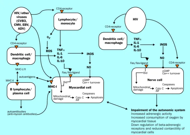
Figure 1: Mechanism of cardiac damage induced by HIV reservoir cells . . . 12 Figure 2: Mechanisms of HIV-associated atherosclerotic cardiovascular disease . .21
List of Tables
Table 1: Cardiac effects of antiretroviral drugs . . . .26
List of Abbreviations
ART – antiretroviral therapycIMT – carotid intima-media thickness CV – cardiovascular
ECG – electrocardiogram FMD – flow-mediated dilation
HAART – highly active antiretroviral therapy HIV – human immunodeficiency virus IMT – intima-media thickness
LV – left ventricle
MI – myocardial infarction
NRTIs – nucleoside reverse transcriptase inhibitors NYHA – New York Heart Association
PCR – polymerase chain reaction PIs – protease inhibitors
1. Introduction
The infection by the Human Immunodeficiency Virus (HIV) is a global pandemic. The first case was reported in the United States of America in 1981. Since then the epidemic has been on-going for the last three decades, affecting an increasing amount of individuals. As of 2015, it is estimated that a total of 36.7 million people are living with HIV, including 2.6 million children under the age of fifteen (UNAIDS, 2016). Whilst its incidence seems to be decreasing in the past years, HIV is rapidly becoming a chronic disease whose course in the long term is very much unpredictable. HIV infection in paediatric ages is mainly acquired through mother-to-child transmission, either during delivery or through breastfeeding. New infections in adolescents are mainly acquired horizontally by means of sexual intercourse, or intravenous drug use. The infected paediatric population seems to be decreasing, given that proper viral load control in pregnant women and the use of prophylactic measures have decreased the risk of mother-to-child transmission (NIH, 2016).
The discovery of Highly Active Retroviral Therapy (HAART) was a tremendous breakthrough in medical history and since its first introduction in 1996, HAART has been the main pillar in the treatment of HIV-positive patients. It has been largely responsible for a decline in morbidity and mortality in these patients. It is estimated that > 50% of HIV-positive persons will be > 50 years of age as of 2015, as a result of successful virological management (CDC, 2011). This changing epidemiological panorama presents a scenery of complications which were previously unknown and an added responsibility to evaluate the long-term effects of the disease on overall health.
One of the main complications of HIV infection currently acknowledged is cardiovascular (CV) disease. In an era where disease progression is generally manageable, cardiovascular complications are an important cause of morbidity and mortality in HIV-positive patients. The aging pattern of the HIV-infected population has led to an increased prevalence of traditional CV risk factors such as hypertension, diabetes, dyslipidaemia and smoking. HIV infection is also an independent risk factor for CV disease. The infection itself and the use of HAART are associated with increased risk of CV disease.
HIV can affect the heart and vasculature in multiple ways, both in short and long-term. Structural and functional disruptions can develop due to HIV infection and treatment and, to this day some manifestations are associated mainly to the disease itself, others to the use of HAART and some remain in a grey area where the mechanism of disease is yet to be determined or believed to be largely multifactorial. The effect of HIV and its therapy on
traditional CV risk factors is also relevant. A number of metabolic alterations are more prevalent in HIV-positive individuals, including insulin resistance, diabetes and dyslipidaemia. Smoking, interestingly enough, seems to have a higher prevalence in HIV-positive individuals when compared to HIV-negative individuals (Cerrato E, 2015).
It seems that, in adults, these alterations express themselves as a higher risk for atherosclerosis, cardiovascular events and heart failure. In children, the most immediate clinical presentations involve the heart whilst long term CV risk seems to be largely related to vasculature damage and its consequences. As HIV-positive children are expected to live longer with the disease than the an individual who is infected as an adult, measures of CV risk in this population become extremely relevant.
In this review article, I aim to assembly the current knowledge of CV risk in the paediatric population affected by HIV infection. An attempt at differentiating between infection-related and therapy-related alterations on the CV system will be made. All research was conducted using the PubMed database and only articles from the year 2000 onwards were accepted. A broad search on general cardiovascular risk was then refined to more specific areas of concern. Initially research was focused only on Paediatrics-related articles, but it was later on expanded to adults on topics where there was limited evidence for children. Given the relative difficulty in finding material on certain areas, bibliographies of retrieved articles were also considered to obtain additional information. A total of seventy articles were used as the final bibliography.
2. Paediatric HIV Infection and the Heart
HIV infection is a primary cause of acquired heart disease. All layers of the heart may be affected – endocardium, myocardium and pericardium – and manifestations seem to differ in a developed and in a developing heart. Structural and functional damage are, of course, many times correlated as one usually leads to the other. As disease of the pulmonary artery can correlate to heart dysfunction, this topic will be discussed in the current section. Before HAART was implemented, subclinical cardiac abnormalities in early childhood were extremely prevalent and these tended to progress to clinical disease within a few years (Orav EJ, 2002). Up to 25% of HIV-infected children presented with CV disease early in life, with serious cardiac events typically occurring between eight months and five years of age (Al-Attar I, 2003). In the P2C2 HIV longitudinal study of vertically HIV-infected children before HAART use,cardiac mortality was 35% (Al-Attar I, 2003). In children, as in adults with HIV infection, cardiac morbidity and mortality were associated with more advanced disease and with coinfections (Mas CM, 2012). The most common CV manifestations were myocardial disease, pericardial effusions, pulmonary hypertension and rhythm disturbances.
The panorama of heart manifestations has changed since the introduction of HAART. There is good evidence that HAART regimens significantly reduce the incidence of heart disease in HIV-infected children, even though some of the drugs used are by themselves cardiotoxic.
2.1. Endocardium Injury
Infective endocarditis and nonbacterial thrombotic endocarditis are some possible complications of HIV infection when it comes to the endocardium. These seem to be somewhat prevalent in the adult population but very rare in HIV-infected children (Fisher SD, 2011).
2.2. Myocardium Injury
The exact pathogenesis of heart structural and functional damage is still greatly unknown. Most studies on underlying mechanisms were done in adults but it is believed that these can be applied to the paediatric community as well. Although each type of cardiac pathology may have specific causes, general effects on
children’s hearts may occur through HIV-induced inflammatory mediators, such as cytokines and proteolytic enzymes, co-infection, nutritional deficiencies, autoimmune response to infecting pathogens, HAART cardiotoxicity and/or direct viral cell invasion (Merchant RH, 2012). Most researchers suggest a multifactorial aetiology (Brown SC, 2005). Studies of heart tissue have demonstrated that direct HIV infection of cardiomyocytes is rare, which suggests that indirect mechanisms predominate (Currie PF, 2003).
Dilated cardiomyopathy is more common in HIV-infected children and increases in frequency as the disease progresses. It carries a much worse prognosis than idiopathic dilated cardiomyopathy (Merchant RH, 2012).
Myocardial disease confers poor prognosis by itself as it results in symptomatic heart failure in up to 5-10% of HIV-infected children (Singh P, 2015).
Myocarditis is probably the most important cause of HIV-associated cardiomyopathy. A number of pathogenic routes have been discussed, including direct action of HIV on myocardial tissue, action of proteolytic enzymes or cytokine mediators induced by HIV alone or with co-infecting viruses (Pozzan G, 2009). Various pathogens have been found on HIV-infected patients, using endomyocardial biopsy. Some examples are cytomegalovirus, mycobacteria sp., toxoplasma, Candida sp., histoplasma and Staphylococcus aureus; however, the clinical significance of these findings is still unclear (Barbaro G, 2001). It is important to note that only scant and patchy inflammatory cell infiltrates and necrosis in the myocardium have been identified on autopsy and biopsy, suggesting that secondary infection is unlikely to cause myocarditis directly. The fact that, in adults, only PCR can identify these opportunistic pathogens indicates that pathogen load is low and supports the previous statement. A potentially important route of cardiomyocyte cell damage is through increased TNF-α production induced by HIV infection. Reservoir cells located in myocardial and brain interstitial tissue may harbour viruses for extended periods and chronically release cytotoxic cytokines, including TNF-α, IL-6, IL-1, TGF-β (Barbaro G, 2001). This cytokine release results in tissue damage. TNF-α alters intracellular calcium homeostasis and induces nitric oxide synthesis, which can injure myocardial cells and produce negative inotropic effects as a consequence. TNF-α induced apoptosis and myocardial diffuse regressive alterations have been observed in HIV-infected patients with biventricular dilation (Yearley JH, 2008).
Nutritional deficiencies are prevalent in HIV-infected individuals, particularly in the late stages and in young infants. Electrolyte imbalances and deficiencies in elemental nutrients usually result from poor intake, malabsorption and diarrhoea. Deficiency of trace elements, especially selenium, may be linked to cardiomyopathy in HIV uninfected children and this may be relevant in HIV-infected children (Longenecker C, 2012). Low levels of vitamin B12, carnitine, growth hormone and thyroid hormone may occur in HIV infection, and are also associated with left ventricle (LV) dysfunction (Miller TL, 2013). A lower body mass index is an independent risk factor for cardiomyopathy (Lemmer CE, 2011).
There are two types of HIV-associated cardiomyopathy: LV dilation with a reduced ratio of LV wall thickness to end-systolic dimension, and dilation with concentric hypertrophy, such that the ratio of wall thickness to end-systolic dimension is unchanged or increased (Easley KA, 2000). These may be manifested as LV systolic dysfunction, which can be clinically ranges from asymptomatic to symptomatic heart failure, and probably diastolic dysfunction (Miller TL, 2013).
2.2.1. Left Ventricular Systolic Dysfunction
Before HAART, the two-to-five year incidence of symptomatic heart failure ranged from 4 to 28% in HIV patients, suggesting a prevalence of symptomatic HIV-related heart failure of between 4 and 5 million cases worldwide (Easley KA, 2000).
As far as clinical presentation, patients can present with LV systolic dysfunction that is anywhere from asymptomatic to NYHA Class IV heart failure, with severe functional limitations (Mas CM, 2012). Echocardiography with strain measurements may be useful in assessing LV function, allowing for the diagnosis of LV dysfunction. Some of the possible findings are LV hypertrophy, dilation or low-to-normal wall thickness, as well as atrial dilation. Progressive LV dilation is quite prevalent among HIV-infected children and it may precede heart failure. This finding is associated with elevated LV afterload, LV hypertrophy and reduced LV function (Williams PL, 2013). Cardiomegaly could be identified in more than half of HIV-infected children and was associated with increased LV mass, symptomatic disease and pericardial effusion (Kearny DL, 2003). Echocardiographic evaluation should be performed at diagnosis and every 1-2 years thereafter. In selected patients, with higher CV risk or evidence of CV disease, these recommendations should be adjusted (Mondy KE, 2011). Electrocardiography (ECG) commonly shows nonspecific conduction defects or repolarization defects in HAART naïve patients. A number of studies have also shown an inverse correlation between blood natriuretic peptide concentrations and LV ejection fraction (Wilkinson JD, 2013).
In the pre-HAART era, median survival from diagnosis to an AIDS-related death was 101 days in patients with LV dysfunction. This data can be compared with an estimated time median survival of 472 days in patients without cardiac dysfunction and at a similar stage of HIV disease (Easley KA, 2000).
HAART seems to have a cardioprotective effect and to prevent LV dysfunction. Whether it does so directly, by reducing HIV-related cardiac damage, or indirectly, through improving overall health and nutritional status, is still not clarified (Williams PL, 2013). Patients receiving therapy still frequently show some asymptomatic abnormalities in cardiac structure. The hearts of children treated with multidrug antiretroviral therapy (ART) have less LV mass and smaller chambers but greater function. This increase in function may be a compensation of the smaller LV size. These children showed higher LV contractility than HAART-unexposed children, which resulted in such alterations being subclinical (Mas CM, 2012).
Echocardiographic studies have identified that around 18% of HAART treated patients have LV dysfunction, 6.5% have LV hypertrophy and 40% have left atrial dilation (Mondy KE, 2011). Left ventricular systolic dysfunction may be subtle with prolonged HAART duration (>10 years) and only detectable by strain echocardiography that measures myocardial deformation in circumferential, radial and longitudinal directions (Frank L, 2012). HIV-infected children and young adults may have impaired longitudinal strain and strain rate despite normal systolic function and ejection fraction (Al-Naami G, 2014). Among HAART-treated children, older age at treatment initiation, a regimen containing zidovudine, and a nadir CD4 %<15% were independent predictors of cardiomyopathy (Patel K, 2012) Although HAART exposure may be cardioprotective, this is only true for a finite period early in life. This effect decreases as the HIV-infected population ages into adolescence and early adulthood (Orav EJ, 2002). It is possible that HAART may block myocardial hypertrophy and hyperplasia and reduce myocardial energy production; increase cardiac apoptosis and block repair mechanisms as a result of mitochondrial toxicity (Mas CM, 2012).
Studies have suggested that any cardiac changes in the HAART era are generally subclinical in children. Further, in addition to characterizing lifetime HAART exposure, traditional non-HIV CV risk factors will be needed to best determine differences in global CV risk between perinatally HIV-infected children and the general population (Miller TL, 2013).
2.2.2. Left Ventricular Diastolic Dysfunction
Echocardiographic and clinical data have shown that diastolic cardiac dysfunction is relatively common in long-term survivors of HIV infection. In adults, an independent association between diastolic dysfunction and HIV infection was reported in a series of studies, even among asymptomatic HIV-infected patients in the early stages of the disease (Aggarwal P, 2009). Nearly 50% of HIV-infected, particularly older individuals with higher blood pressure and heart rate (both HAART naïve and exposed) have mild diastolic dysfunction. After adjustment for age and hypertension, HIV infected adults still present a 2.4-fold increased risk for diastolic dysfunction compared with uninfected subjects (Hsue PY, 2010).
LV diastolic dysfunction can be the first sign of underlying cardiac disease and it may precede LV systolic dysfunction (Reinsch N, 2010). LV compliance decreases as LV diastolic dysfunction worsens and left atrial pressure increases. Moderate to severe LV diastolic dysfunction independently predicts mortality, regardless of normal LV systolic function (O'Brien S, 2011). Whether LV diastolic dysfunction is associated with an increased risk of early coronary disease is unknown (Nayak G, 2009).
The physiopathology behind the diastolic dysfunction on these patients is not clear yet. It is postulated that it might result from HIV infection itself, HAART and/or other comorbidities. Although more studies are needed on this topic, it was noticeable that affected subjects tended to have lower CD4 count and/or a longer duration of treatment with nucleoside reverse transcriptase inhibitors (NRTIs) (Hsue PY, 2010). Data on the paediatric population is even scanter. Left or right ventricular diastolic dysfunctions, predominantly of the restrictive pattern, were found in roughly 40 and 30% respectively on clinically stable HIV-positive children (Silva ML, 2014). Other reports on left ventricular diastolic dysfunction in children are again limited, and the existing are almost always associated with systolic dysfunction, as these patients are usually symptomatic (Silva ML, 2014). The relationship between LV diastolic dysfunction and HIV treatment status has not been clearly demonstrated, but HAART may play a role as HIV-exposed but uninfected infants who have been exposed to prophylactic HAART have lower early diastolic annular velocity than controls (Cade WT, 2012). Future echocardiographic studies on the temporal occurrence of LV systolic versus diastolic changes may be important in determining the effects of HIV exposure and HAART exposure (Silva ML, 2014).
2.3. Pericardium Injury
Pericardial effusion was a major concern in the pre-HAART era, with up to 11% of HIV-positive patients presenting with this complication (Lindl A, 2011). Current data indicates that the prevalence in developed countries with easy access to ART has, in fact, very much decreased whilst it still accounts for around 12% of cardiac manifestations in African countries (Brown SC, 2005).
Generally, effusions are small and asymptomatic and drainage is rarely required but close monitoring is mandatory to detect possible opportunistic infections (Brown SC,
2005). Pericardiocentesis is indicated when there are clinical or echocardiographic signs of tamponade (Miller TL, 2013). Pericardial effusion is often part of a complex and generalized effusive process, usually involving pleural and peritoneal surfaces. It is believed that the pathogenesis behind it has to do with enhanced cytokine production associated with HIV infection, which leads to “capillary leakage” of serous fluid. Other related mechanisms include uraemia from HIV nephropathy or drug nephrotoxicity (Lindl A, 2011). There seems to be an association between pericardial effusion and tuberculosis infection in lower income countries, where it is highly prevalent (Brown SC, 2005). Children infected with HIV are at higher risk for tuberculosis (Matthews K, 2012).
HIV patients with pericardial effusions tend to have lower CD4 counts than those without effusions and the risk of death in these patients nearly triples (McLaughlin VV, 2009) (Lindl A, 2011).
2.4. Pulmonary Hypertension
Data on the prevalence of pulmonary hypertension associated with HIV infection is mainly available for the adult population. About 0,5% to 5% of the adult HIV-positive community has developed pulmonary hypertension, with an apparent female predominance (Bloomfield GS, 2014). It is believed that this could possibly be an underestimation as most patients accountable in this prevalence data are symptomatic, with asymptomatic going unaccounted for (Mirrakhimov AE, 2013). The introduction of HAART hasn’t altered the prevalence of this pathology (Opravil M, 2008).
On autopsy reports, findings of pulmonary arterial hypertension seem to be a consequence of the replacement of normal endothelial structure by arteriopathic remodelling with intimal fibrosis (Cicalini S, 2011). Although the pathogenic mechanisms behind these alterations are still largely in need of more studying, it is believed that it may involve HIV-related proteins (such as Gp-120 protein, Nef and Tat), HIV-induced inflammation, co-infections (particularly Human Herpes Virus 8, Hepatitis C and Hepatitis B) and heightened inflammation due to exposure of microbial products, thromboembolic events and genetic susceptibility through certain HLA subtypes (Cicalini S, 2011). It is also possible that HAART influences the
pathogenesis and severity of the pulmonary hypertension, however, there is not enough data to evaluate this correlation (Bloomfield GS, 2014).
The most common clinical presentation in these patients is dyspnoea and children may also present with signs of right-sided heart failure, such as facial oedema, hepatomegaly or oedema of the lower extremities. Upon evaluation, perfusion scans are usually normal (McLaughlin VV, 2009). Most adults show cardiomegaly, pulmonary arterial enlargement, dilated right ventricle and tricuspid regurgitation (Stewart D, 2011). In patients with HIV and pulmonary hypertension, pulmonary hypertension should be aggressively treated as it is life threatening. Most morbidity and mortality in these patients seem to be caused by pulmonary hypertension and, therefore, it should be clinically managed as a priority (McLaughlin VV, 2009).
2.5. Rhythm Disturbances
There seems to be a higher prevalence of electrocardiographic anomalies in HIV-infected children, with 50 to 93% of these patients presenting with voltage disturbances, conduction defects and dysrhythmias, between other alterations. Some of the most frequent dysrhythmias noted are second-degree atrioventricular block and supraventricular or ventricular tachycardia (Saidi AS, 2000). Most anomalies seem to be benign and without need to therapeutic action.
QT prolongation is also associated with HIV infection and seems to affect children with increased frequency as they age (Saidi AS, 2000). This alteration may predispose to torsades de pointes ventricular tachycardia (Miller TL, 2013). HAART may possibly be involved in the development of QT prolongation in HIV patients, particularly when in the use of protease inhibitors (PIs). The clinical significance of this finding is sill unclear (Hunt K, 2011).
Patients are at risk for sudden cardiac death late in HIV infection, and it is becoming increasingly common as the HIV-infected population ages (Miller TL, 2013). This is normally related to autonomic nervous system disturbances such as attenuation of the parasympathetic-mediated circadian rhythm heart variability and impairment of night-time QTc interval lengthening. Cardiac autonomic dysfunction correlates with adverse immunological status of HIV-infected children (Benchimol-Barbosa PR, 2009).
2.6. In Utero Development
Mother-to-child transmitted HIV infection was in the past one of the main concerns of the paediatric community. The use of HAART prophylaxis on HIV-positive pregnant women reduced this transmission path rates from more than 20% to approximately 8% (Mas CM, 2012). As of its implementation, the prevalence of HIV infection acquired through mother-to-child transmitted in developed countries has been reduced to less than 2% (Mofenson LM, 2000). However, where there are many positive aspects of the use of this prophylaxis, there also seems exist another side of the coin.
HIV can affect the developing heart both in utero, on a foetus of an HIV-positive mother, and after birth. Many approaches have come up on the issue of congenital anomalies of the heart related to HIV. Seemingly both HIV infection itself and HAART exposure are involved in this pathogenesis.
Children who are born to HIV-positive mothers present with LV dysfunction with reduced contractility indices that may persist over time, irrespective of the HIV infection status of the infant. This was suggested to be a consequence of the uterine environment provided by the HIV-positive mother, which gives rise to important cardiovascular effects on the foetus (Orav EJ, 2002). Apart from exposure to HIV itself, many children are exposed to HAART in utero to prevent transmission of the virus. There seems to be a higher rate of congenital anomalies in infants exposed to antiretroviral agents during pregnancy than in unexposed infants (Knapp KM, 2012). Foetal and early neonatal HAART exposure was associated with substantially lower LV wall thickness and mass, as well as with impaired myocardial growth (Shearer WT, 2011). Studies have been conducted on specific drugs used in HAART regimens and the their relation to cardiac congenital alterations. Abacavir, nelfinavir, nevirapine and zidovudine were some of the implicated drugs. Abacavir seems to be associated with decreased LV dimension; nelfinavir exposure was related to lower aortic valve diameter and reduced LV wall thickness. Nevirapine increased risk of increased LV wall thickness (Lipshultz SE, 2010). It is still unknown exactly what are the clinical implications of these HAART-associated cardiac alterations, however they confirm the potential of any adverse effects of HAART to be independent of the confounding effects of HIV infection (Mas CM, 2012). HAART is a greater good in reducing the rates of mother-to-child HIV transmission, however, children exposed to HAART in utero, regardless of their HIV status, should
have long term cardiac follow up because even mild LV dysfunction increases premature cardiac mortality over time (Mas CM, 2012).
3. Paediatric HIV Infection and the Vasculature
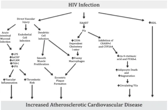
The relevance of the correlation between HIV infection and vascular disease is increasingly recognized. With the implementation of HAART, HIV-positive patients are living longer lives and the importance of chronic conditions such as atherosclerotic and metabolic diseases is growing as the population ages (Fitch K, 2011). A 1.5-1.7-fold increased risk for myocardial infarction (MI) has been noted in adults with HIV infection (Triant VA, 2007). Acute MI can be the primary presentation of atherosclerosis in these patients (Miller TL, 2013). All in all, HIV infection, its therapies and cardiometabolic consequences have marked effects on the vasculature.The pathogenesis behind the increased atherosclerotic disease in these patients is related to the state of constant low-grade inflammation produced by the infection, correlated to the level of viral control (Maniar A, 2013). Another related mechanism is the co-existence of a metabolic syndrome in HIV patients. The syndrome is characterized by lipodystrophy (redistribution of fat with reduction in subcutaneous and increase in visceral fat), dyslipidaemia, insulin resistance and abnormal glucose metabolism (Grinspoon S, 2005). Both the HIV infection and the use of HAART, particularly the exposure to PIs and certain NRTIs, can be in the genesis of the syndrome (Miller TL, 2013). This is a controversial topic as most data has been collected on the correlation between HAART and metabolic disease in HIV-positive patients, and not enough research has been done on the effect of HIV disease itself. In these studies, following exposure to HAART, there are increases in total, LDL, and HDL cholesterol in both adults and children (Miller TL, 2013). However, some data has shown that HIV-infection in adults may, in fact, independent of HAART exposure status be associated with dyslipidaemia, with high triglycerides and decreased HDL cholesterol (El-Sadr WM, 2005).
The pathophysiology of accelerated atherosclerosis in HIV-infected patients is therefore complex. The mechanisms are probably different in HAART-naïve and exposed patients. It is believed that in HAART-naïve patients, the immune activation and chronic inflammatory state predominates. In these patients, early endothelial dysfunction is promoted by immune mediators and is maintained by HIV viral persistence, microbial translocation associated with mucosal barrier damage, and co-infection (Baker JV, 2011). Additionally, normal vascular restoration after injury appears to be impaired in HIV infection. Although HAART controls many of the
pathogenic pathways of HIV when it comes to atherosclerosis, mainly by controlling viral load, it is well known that HAART-exposed patients may still have a higher cardiovascular risk than the general population. Chronic immune activation and inflammation may promote atherosclerosis and increased arterial stiffness in the absence of residual viral replication (Ross AC, 2009). In fact, HAART can have beneficial or detrimental effects on cardiovascular risk as the effect of the previously mentioned metabolic syndrome can be particularly deteriorating on the vasculature.
Atherosclerotic vasculopathy occurs earlier and to a greater extent of severity in HIV patients and it seems to affect both the coronary and the larger vessels (Mary-Krause M, 2003). Traditional risk factors such as family history, high LDL cholesterol, low HDL cholesterol, diabetes, hypertension and age > 55 also play a role in atherogenesis (Miller TL, 2013).
Evidence suggests that HIV infection also leads to poor atherosclerotic outcomes in children and it is particularly interesting to study the routes of accelerated atherosclerosis in this population, as they will both live longer with the disease and be exposed to HAART for a longer period as well. Whilst HAART improves growth and lean-body mass and improves overall health in children, it also associates with
Figure 2 - Mechanisms of HIV-associated atherosclerotic cardiovascular disease. Retrieved from (Lambert CT, 2016)
abnormalities in fat distribution in HIV-infected children. Both peripheral lipoatrophy and central obesity, otherwise known as lipodystrophy, occur in these children. Lipodystrophy is often, but not always, accompanied by metabolic disturbances, such as insulin resistance and dyslipidaemia. Up to 50% of HIV-infected children develop at some point lipodystrophy, especially with long-term exposure to NRTIs (European Paediatric Lipodystrophy Group, 2004). The mechanisms by which fat redistribution occurs in HIV-infected children are incompletely understood. The component of lipoatrophy is likely to be caused by mitochondrial DNA damage due to NRTI exposure, and possibly HIV infection itself, leading to respiratory chain dysfunction and decreased lipogenesis as well as increased fat apoptosis. An increase in fat circulation and failure of hepatocytes and endothelial cells in removing circulating lipids efficiently is the possible cause of lipohypertrophy and dyslipidaemia. In children, there is evidence to suggest that a disturbance in insulin sensitivity and lipid metabolism may be mediated by low levels of adiponectin and HAART-induced adipocyte dysfunction (Barlow-Mosha L, 2013). Lipodystrophy seems to increase cardiovascular risk significantly, specifically in terms of premature atherosclerosis, in HIV-infected adults (Behrens GM, 2003). The relationship in children is still unclear. It seems that older HIV-infected children with long-term exposure to HAART have increased intima-media thickness (IMT) and decreased flow-mediated dilation (FMD) on ultrasound. These two biomarkers are excellent and relatively recent measures being used in assessing vascular anomalies as a way of evaluating CV risk. The former is a marker for atherosclerosis and seems to be dynamic as variations can occur with duration of treatment. Carotid intima-media thickness (cIMT) may regress or increase with prolonged treatment. Diminished flow-mediated dilation is a marker of poor endothelial function (Bonnet D, 2004). The causes of structural and functional vascular changes in HIV-infected children are largely unknown and, as most data are from HAART-exposed children, it is not possible to differentiate between the effects of HIV infection and treatment. However, a relationship has been found between PI therapies and higher cIMT and lower FMD in children (Charakida M, 2005).
4. Paediatric HIV Infection and Cardiovascular
Malignancies
Certain cardiovascular malignancies appear to have a higher incidence within the HIV-‐ positive population. Whilst there is not much information on paediatric incidence of these particular tumours, it is believed that they generally affect individuals in the later stages of disease (Miller TL, 2013).
The main primary cardiac malignancy associated with HIV infection is cardiac lymphoma. Lymphoma has an increased incidence in HIV-‐infected populations and seems to be an AIDS-‐defining illness. Non-‐Hodgkin’s lymphoma is the most common type in this particular population and is believed to be 26-‐60 times more common in HIV-‐positive individuals when compared to HIV-‐negative controls. They can present as the first manifestation of AIDS, leading up to diagnosis in up to 4% of patients. The clinical picture is variable and some of the possible manifestations are chest pain, arrhythmias and signs heart failure on presentation. The prognosis is poor and cardiac lymphoma can rapidly progress to cardiac tamponade, myocardial infarction, conduction abnormalities and eventual heart failure (Miller TL, 2013). It seems that the implementation of HAART has not substantially affected the incidence of HIV-‐related non-‐Hodgkin’s lymphoma (Zoufaly A, 2009).
Kaposi’s sarcoma is a type of angiosarcoma that has been reported to affect primarily the cardiac tissue in HIV-‐positive patients. In the early years of HIV history, Kaposi’s sarcoma affected around 35% of HIV-‐positive persons. There is an association between this tumour and herpes virus 8. It affects endothelial cells and, in the heart it seems to have a preference for sub-‐pericardial fat around the coronary arteries. Contrary to what has been said about HIV-‐related Non Hodgkin’s lymphoma, HAART seems to have markedly decreased the incidence of Kaposi’s sarcoma (Fisher SD, 2011).
5. HAART and
Cardiovascular
Risk
HAART has improved immensely the longevity and quality of life of HIV-positive patients. Its relationship with CV risk in these patients is still in many parts unclear. Whilst in many ways it seems to control aspects of the disease that carry added CV risk, it has been also mentioned throughout this dissertation that in many cases HAART also seems to add risk on itself (Miller TL, 2013). Most the effects have been mentioned in-depth in the adequate sections. In this section a brief summarized presentation on the effect of HAART on CV risk will be made.
HAART may start adding cardiovascular risk in utero as it is associated with increased incidence in cardiac anomalies in foetuses of HAART exposed HIV-negative children who are born to HIV-positive mothers (Knapp KM, 2012). In these cases, HAART seems to substantially affect the LV. Children have been found to have decreased LV wall thickness and mass on echocardiographies performed early on in life (Shearer WT, 2011).
In HIV-positive children who are in use of HAART, certain aspects are also relevant. When it comes to heart anomalies, it seems that there is a higher incidence of low LV mass associated with overall smaller chambers. Drugs implicated on cardiac anomalies include abacavir, nelfinavir and nevirapine (Lipshultz SE, 2010). Changes occur during the first 10 years of exposure to therapy. In these children the effects are similar to the ones in foetuses exposed in utero as it seems that the consequences of HAART on the developing heart is transversal to both of these groups. Whilst some structural anomalies occur at an early age, it has been discussed that greater heart contractibility is found in the first years of life, in order to compensate of the smaller and structurally weaker heart. Mitochondrial toxicity has been found to block cardiac repair systems to damage and prevent adaptations such as hypertrophy and hyperplasia in the heart of an HIV-positive HAART treated child (Mas CM, 2012). Specifically, NRTIs have been associated with altered mitochondrial DNA replication and cardiac structure. It not yet clear whether altered mitochondrial DNA replication is the cause of cardiomyopathy, but in some patients, suspension of specific NRTI drugs may improve LV dysfunction (Fisher SD, 2011). Altered body composition and hyperlipidaemia are associated with some PIs, but theses lipid abnormalities vary with different drugs in the class (Dapena M, 2012). Ritonavir seems to have a stronger effect on lipid metabolism, with a mean increase
of total cholesterol concentration of 2.0 mmol/L and a mean increase in triglyceride concentration of 1.83 mmol/L. PIs also increased lipoprotein a in patients with elevated pre-treatment values. The overall effects of PIs on the lipid metabolism present an increased risk for atherosclerotic cardiovascular disease (Worm SW, 2010). NRTIs have also been discussed to confer metabolic alterations and increase CV risk (Dapena M, 2012). Control of lipid metabolic anomalies is important and measures such as low-level aerobic exercise and switching drugs may reverse both triglyceride concentrations and abnormal fat deposition (Fisher SD, 2011).
PIs may cause endothelial dysfunction through several mechanisms, such as reduced nitric oxide production, reduced expression of nitric oxide synthase and increased formation of reactive oxygen species (Mas CM, 2012). These effects don’t appear to be transversal to the whole class, as some specific drugs decrease endothelial function and others actually improve it (Dube MP, 2008).
An estimated 60% increase in hospitalizations due to strokes in HIV-infected persons has been attributed to HAART use. Several drugs appear to promote atherosclerosis. It is still unclear which exact mechanisms confer this increased risk but one of the hypotheses is the increase in life expectancy in HIV patients (Ovbiagele B, 2011). Whilst this hypothesis may contribute, it is quite clear that it doesn’t work on its own. HIV-infected children who are treated with HAART are at risk for accelerated atherosclerosis. A conjunction of metabolic anomalies and actual structural changes to the vasculature seem relevant. These structural changes are reflected by an increased cIMT and lower FMD (Charakida M, 2005). PIs have been associated with these changes both in children and adults (Lorenz MW, 2008). Heightened cIMT raises concern for premature atherosclerosis in HAART-treated patients (Ordovas K, 2012).
6. Conclusion
HIV infection has significant effects on the cardiovascular system. In children, in particular, given their significant longevity and longer expected exposure to HAART when compared to the infected adult population, the issue of long-term risk is a priority. In economically developed countries, where HAART is easily accessible, viral load control is nowadays not the main concern of physicians. Therapy is extremely effective in diminishing viral replication to levels almost undetectable. Due to this achievement, infectious complications became less prevalent and the mortality associated to these complications has reduced significantly. As such, HIV-infected children are living longer lives. Other complications arose from this new life expectancy. Cardiovascular risk has become one of the main areas of concern. Infected children are foreseen to suffer alterations in the heart, which manifest early in childhood, and on the vasculature. Studies have demonstrated that both the viral infection itself and the use of HAART cause added CV risk in these patients. When it comes to the underlying mechanisms, much is still poorly understood. Whilst some drugs included in HAART lead to alterations such as metabolic syndrome and accelerated atherosclerosis, the use of this therapy is still of great importance. Not only is HAART the main pillar in keeping these patients alive when it comes to viral control, but it also has a role when it comes to reducing certain cardiovascular complications. In no way does the CV risk added by HAART counteract all the benefits its use brings.
As HIV-positive children have added CV risk, control of traditional CV risk factors and monitoring of both cardiovascular and metabolic systems are of extreme importance. Lipid profile, ECG and echocardiogram can be used as a way of screening. Newer parameters used as biomarkers mainly in evaluating vascular risk, such as cIMT and FMD are promising as early markers of disease.
It is important to point out that current evidence on HIV infection and CV risk largely stems from research in adults. Modes of infection, immune maturity, growth and development and treatment are, however, in many ways different in children. As such, more in-depth paediatric research, which accounts for the interaction between normal growth and development, HIV infection and HAART is of great importance.
7. References
Aggarwal P, Sharma A, Bhardwaj R, Raina R (2009) Myocardial dysfunction in human immunodeficiency virus infection: an echocardiographic study. J Assoc Physicians India
57: 745-6.
Al-Attar I, Orav EJ, Exil V, et al (2003) Predictors of cardiac morbidity and related mortality in children with acquired immunodificiency syndrome. J Am College Cardiol 41: 1598-1605. Al-Naami G, Kiblawi F, Kest H, et al (2014) Cardiac mechanics in patients with Human
Immunodificiency Virus: A study of systolic myocardial deformation in children and young adults. Pediatric Cardiology 35: 1046-51.
Baker JV, Lundgren JD (2011) Cardiovascular implications from untreated human immunodeficiency virus infection. Eur Heart J 32: 945-51.
Barbaro G, Fisher SD, Lipshultz SE (2001) Pathogenesis of HIV-associated cardiovascular complications. Lancet Infectious Diseases 1:115-124.
Barlow-Mosha L, Eckard AR, McComsey GA, Musoke PM (2013) Metabolic complications and treatment of perinatally HIV-infected children and adolescents. J Int AIDS Soc 16: 18600.
Behrens GM, Meyer-Olson D, Stoll M, Schmidt RE. (2003) Clinical impact of HIV-related lipodystrophy and metabolic abnormalities on cardiovascular disease. AIDS 17: S149-54. Benchimol-Barbosa PR (2009) Circadian cardiac autonomic function in perinatally
HIV-infected preschool children. Braz J Med Biol Res 42: 722-30.
Bloomfield GS, Khazanie P, Morris A, et al (2014) HIV and noncommunicable cardiovascular and pulmonary diseases in low and middle income countries in ART era: What we know and best directions for future research. J Acquir Immune Defic Syndr 67 (Suppl. 1): S40-S53.
Bonnet D, Aggoun Y, Szezepanski I, et al (2004) Arterial stiffness and endothelial dysfunction in HIV-infected children. AIDS 18: 1037-41.
Brown SC, Schoeman CJ, Bester CJ (2005) Cardiac findings in children admitted to a hospital general yard in South Africa: a comparison of HIV-infected and uninfected children. Cardiovascular Journal of South Africa 16: 206-210.
Cade WT, Waggoner AD, Hubert S, et al (2012) Reduced diastolic function and left ventricular mass in HIV-negative preadolescent children exposed to antiretroviral therapy in utero. AIDS 26: 2053-58.
CDC. (2011). HIV testing among men who have sex with men: 21 cities, United States, 2008. CDC 60(21): 694-699.
Cerrato E, Calcagno A, D'Ascenzo F, et al (2015) Cardiovascular disease in HIV patients: from bench to bedside and backwards. Open Heart 2(1):e000174.
Charakida M, Donald AE, Green H, et al (2005) Early structural and functional changes of the vasculature in HIV-infected children: impact of disease and antiretroviral therapy.
Circulation 112: 103-09.
Cicalini S, Almodovar S, Grilli E, Flores S (2011) Pulmonary hypertension and human immunodeficiency virus infection: Epidemiology, pathogenesis, and clinical approach. Clin
Microbiol Infect 17: 25-33.
Currie PF, Boon NA (2003) Immunopathogenesis of HIV-related heart muscle disease: Current perspectives. AIDS 17: S21-S28.
Dapena M, Jiménez B, Noguera-Julian A, et al (2012) Metabolic disorders in vertically HIV-infected children: future adults at risk for cardiovascular disease. J Pediatr Endocrinol
Metab 25:
529-35.
Dube MP, Lipshultz SE, Fichtenbaum CJ, et al (2008) Effects of HIV infection and antiretroviral therapy on the heart and vasculature. Circulation 118: E36-40.
Easley KA, Orav EJ, Kaplan, et al (2000) Cardiac dysfunction and mortality in HIV-infected
children: the prospective P2C2 HIV multicenter study. Circulation 102(13):1542-8.
El-Sadr WM, Mullin CM, Carr A, et al (2005) Effects of HIV disease on lipid, glucose and insulin levels: Results from a large antiretroviral-naive cohort. HIV Med 6(2): 114-21. European Paediatric Lipodystrophy Group. (2004) Antiretroviral therapy, fat redistribution and
hyperlipidaemia in HIV-infected children in Europe. AIDS 18(10): 1443-51.
Fisher SD, Kanda BS, Miller TL, Lipshultz SE (2011) Cardiovascular disease and therapeutic drug-related cardiovascular consequences in HIV-infected patients. Am J Cardiovasc
Drugs 11(6): 383-94.
Fitch K, Grinspoon S (2011) Nutritional and metabolic correlates of cardiovascular and bone disease in HIV-infected patients. Am J Clin Nutr 94(6): 1721S-8S.
Frank L, Sims A, Cross R, et al (2012) Abnormal cardiac strain in children and young adults with HIV acquired in early life. J Am Soc Echo 25(7): 741-8.
Grinspoon S, Carr A (2005) Cardiovascular risk and body-fat abnormalities in HIV-infected adults. N Engl J Med 352(1): 48-62.
Hsue PY, Hunt PW, Ho JE, et al (2010) Impact of HIV infection on diastolic function and left ventricular mass. Circ Heart Fail 3(1): 132-39.
Hunt K, Hughes CA, Hills-Nieminen C (2011) Protease inhibitor- associated QT interval prolongation. Ann Pharmacother 45(12): 1544-50.
Kearny DL, Perez-Atayde AR, Easley KA, et al (2003) Postmortem cardiomegaly and echocardiographic measurements of left ventricular size and function in children infected with the human immunodeficiency virus: The prospective P2C2 HIV Multicenter Study.
Cardiovascular Pathology 12(3): 140-8.
Keesler MJ, Fisher SD, Lipshultz SE (2001) Cardiac manifestations of HIV infection in infants and children. Annals New York Academy of Sciences 946: 169-178.
Knapp KM, Brogly SB, Muenz DG, et al (2012) Prevalence of congenital anomalies in infants with in utero exposure to antiretrovirals. Pediatr Infect Dis J 31(2): 164-70.
Lambert CT, Sandesara PB, Hirsh B, et al (2016) HIV, highly active antiretroviral therapy and the heart: a cellular to epidemiological review. HIV Medicine 17(6): 411-24.
Lemmer CE, Badri M, Visser M (2011) A lower body mass index is associated with
cardiomyopathy in people with HIV infection: Envidence from a case comparison study.
South African Medical Journal 1(2): 119-21.
Lindl A, Reinsch N, Neuhaus K, et al (2011) Pericardial effusion of HIV-infected patients - results of a prospective multicenter cohort study in the era of antiretroviral therapy. Eur J
Med Res 16(11): 480-83.
Lipshultz SE (2010) Association of cardiac structure and function with in utero antiretroviral exposure among uninfected children born to HIV-infected mothers in the pediatric HIV/AIDS cohort study. Circulation 122: A16026.
Longenecker C, Hoit BD (2012) Imaging atherosclerosis in HIV: carotid-intima media thickness and beyond. J Lab Clin Med 159(3):127-39.
Lorenz MW, Stephan C, Harmjanz A, et al (2008) Both long-term HIV infection and highly active antiretroviral therapy are independent risk factors for early carotid atherosclerosis.
Atherosclerosis 196(2): 720-6.
Maniar A, Ellis C, Asmuth D, et al (2013) HIV infection and atherosclerosis: Evaluating the drivers of inflammation. Eur J Prev Cardiol 20(5): 720-28.
Mary-Krause M, Cotte L, Simon A, et al (2003) Increased risk of myocardial infarction with duration of protease inhibitor therapy in HIV-infected men. AIDS 17(17): 2479-86.
Mas CM, Lipshultz SE, Henkel JM, et al (2012) HAART to heart: highly active antiretroviral therapy and the risk of cardiovascular disease in HIV-infected or exposed children and adults. Expert Reviews Anti Infect Ther 10(6): 661-674.
Matthews K, Ntsekhe M, Syed F, et al (2012) HIV-1 infection alters CD4+ memory T-cell phenotype at the site of disease in extrapulmonary tuberculosis. Eur J Immunol 42(1): 147-57.
McLaughlin VV, Archer SL, Badesch DB, et al (2009) ACCF/AHA 2009 expert consensus document on pulmonary hypertension: a report of the American College of Cardiology
Foundation Task Force on Expert Consensus Documents and the American Heart Association. J Am Coll Cardiol 53(17): 1573-619.
Merchant RH, Lala MM (2012) Common clinical problems in children living with HIV/AIDS: systemic approach. Indian Journal of Pediatrics 79(11): 1506-13.
Miller TL, Lipshultz SE, Wilkinson JD, et al (2013). Cardiac effects in perinatally HIV-infected
and HIV-exposed but uninfected children and adolescents: a view from the United States of America. J Int AIDS Soc 16: 18597.
Mirrakhimov AE, Ali A, Barbaryan A,
Prueksaritanond S (2013) Human Immunodeficiency Virus and pulmonary arterial hypertension. ISRN Cardiol, vol. 2013.
Mofenson LM, McIntyre JA (2000) Advances and research directions in the prevention of mother-to-child HIV-1 transmission. Lancet 355
(9222): 2237-44.
Mondy KE, Gottdiener J, Overton ET, et al (2011) High prevalence of echocardiographic abnormalities among HIV-infected persons in the era of highly active antiretroviral therapy. Clin Infect Dis 52(3): 378-86.
Nayak G, Ferguson M, Tribble DR, et al (2009). Cardiac diastolic dysfunction is prevalent in HIV-infected patients. AIDS Patient Care STDs 23(4): 231-8.
NIH. (2016, 11 14). Preventing Mother-to-Child Transmission of HIV. Retrieved 03 22, 2017, from AIDSinfo: https://aidsinfo.nih.gov/education-materials/fact-sheets/24/50/preventing-mother-to-child-transmission-of-hiv#
O'Brien S, Sasaki N, Eidem BW, et al (2011) Left ventricular diastolic dysfunction in HIV-negative infants exposed in utero to antiretroviral therapy from HIV-positive mothers: the prospective NHLBI CHAART-I Study. Circulation 124: A10808.
Opravil M, Sereni D (2008) Natural history of HIV-associated pulmonary arterial hypertension: trends in the HAART era. AIDS 22(Suppl 3): S35-40.
Orav EJ, Lipshultz SE, Easley KA, et al (2002) Cardiovascular status of infants and children of women infected with HIV-1 (P2C2 HIV): A cohort study. Lencet 360(9330): 368-373. Ordovas K, Hsue PY, Lee T, et al (2012) Carotid intima-media thickness among human
immunodeficiency virus-infected patients without coronary calcium. Am K Cardiol 109(5): 742-47.
Ovbiagele B, Nath A (2011). Increasing incidence of ischemic stroke in patients with HIV infection. Neurology 76(5): 444-50.
Patel K, Van Dyke RB, Mittleman MA, et al (2012) The impact of HAART on cardiomyopathy
Pozzan G Pagliari C, Tuon FF, et al (2009) Diffuse-regressive alterations and apoptosis of myocytes: possible causes of myocardial dysfunction in HIV-related cardiomyopathy. Int J
Cardiol 132(1):90-5.
Reinsch N, Neuhaus K, Esser S, et al (2010) Prevalence of cardiac diastolic dysfunction in HIV-infected patients: results of the HIV-HEART study. HIV Clinical Trials 11(3): 156-62. Ross AC, Rizk N, O'Riordan MA, et al (2009) Relationship between inflammatory markers,
endothelial activation markers, and carotid intima-media thickness in HIV-infected patients receiving antiretroviral therapy. Clin Infect Dis 49(7): 1119-27.
Saidi AS, Moodie DS, Garson A Jr, et al (2000) Electrocardiography and 24-hour electrocardiographic ambulatory recording (Holter monitor) studies in children infected with human immunodeficiency virus type 1. Pediatr Cardiol 21(3): 189-96.
Shearer WT, Lipshultz SE, Thompson B, et al (2011) Cardiac effects of antiretroviral therapy in HIV-negative infants born to HIV-positive mothers: NHLBI CHAART-1 Cohort study. J
Am Coll Cardiol 57(1): 76-85.
Silva ML, Nassar SM2, Silva AP, et al (2014) Biventricular diastolic function assessed by Doppler echocardiogram in children vertically infected with human immunodeficiency virus. Jornal de Pediatria (Rio de Janeiro) 90(4): 403-07.
Sims A, H. C. (2011). Cardiovascular complications in children with HIV infection. Curr
HIV/AIDS Rep , 8, 209-214.
Singh P, Hemal A, Agarwal S, Kumar D (2015) Cardiac manifestations in HIV infected children. Indian Journal of Pediatrics 82(3): 230-4.
Stewart D, Mocumbi AO, Carrington MJ, et al (2011) A not-so-rare form o heart failure in urban black Africans: Pathways to right heart failure in the Heart of Soweto Study cohort.
Eur J Heart Fail 13(10): 1070-77.
Triant VA, Lee H, Hadigan C, Grinspoon SK (2007) Increased acute myocardial infarction rates and cardiovascular risk factors among patients with human immunodeficiency virus disease. J Clin Endocrinol Metabol 92(7): 2506-2512.
UNAIDS. (2016). Global AIDS Update - 2016. Joint United Nations Programme on HIV/AIDS. Wilkinson JD, Williams PL, Leister E, et al. (2013) Cardiac biomarkers in HIV-exposed
uninfected children: the pediatric HIV/AIDS cohort study (PHACS). AIDS 27(7): 1099-108. Williams PL, Lipshultz SE, Wilkinson JD, et al. (2013) Cardiac status of children infected with human immunodificiency virus who are receiving long-term combination antiretroviral therapy: results from the adolescent master protocol of the multicenter pediatric HIV/AIDS cohort study. JAMA Pediatrics 167(6): 520-527.
Worm SW, Sabin C, Weber R, et al (2010) Risk of myocardial infarction in patients with HIV infection exposed to specific individual antiretroviral drugs from the 3 major drug classes: the data collection on adverse effects of anti-HIV drugs. J Infect Dis 201(3): 318-30. Yearley JH, Mansfield KG, Carville AA, et al (2008) Antigenic stimulation in the simian model
of HIV infection yields dilated cardiomyopathy through effects of TNF-α. AIDS
22(5):585-94.
Zoufaly A, Stellbrink HJ, Heiden MA, et al (2009) Cumulative HIV viremia during highly active antiretroviral therapyis a strong predictor of AIDS-related lymphoma. J Infect Dis 200(1): 79-87.