ORIGINAL
ARTICLE
Risk of coronary artery disease in individuals infected
with human immunodeficiency virus
Authors
Felippe Dantas Vilela1 Andrea Rocha de Lorenzo2
Bernardo Rangel Tura3 Giovanna Ianini Ferraiuoli4 Marcelo Hadlich1 Marcelo Viana de Lima Barros5
Ana Beatriz Ribeiro Lima6 Vanderson Meirelles7
1Postgraduate in Cardiology, Instituto Nacional de Cardiologia, RJ, Brazil
2PhD in Cardiology, Universidade Federal do Rio de Janeiro (UFRJ), RJ, Brazil
3PhD in Biostatistics, UFRJ, RJ, Brazil
4Professor, Instituto Nacional de Cardiologia, RJ, Brazil
5Nutritionist, Instituto Nacional de Cardiologia, RJ, Brazil
6Social Worker, Instituto Nacional de Cardiologia, RJ, Brazil
7Physician, Instituto Nacional de Cardiologia, RJ, Brazil
Submitted on: 02/15/2011 Approved on: 06/27/2011
Correspondence to:
Felippe Dantas Vilela Departamento de Pesquisa Clínica Rua das Laranjeiras, 374/5o – Laranjeiras 22240-006 Rio de Janeiro, RJ Brazil
felippevilela@gmail.com
We declare no conflict of interest.
©2011 Elsevier Editora Ltda. All rights reserved.
ABSTRACT
Current treatment for human immunodeficiency virus (HIV) infection has improved survival and allowed infected patients to develop atherosclerotic coronary artery disease (CAD). Specific strate-gies to reduce cardiovascular risk in the infected population have not been developed. It is neces-sary to know the magnitude of cardiovascular risk in this population. Objectives: This study aimed to assess cardiovascular risk using a well-known clinical score and to investigate coronary artery calcium scoring (CACS) in this population. Methods: This was a cross-sectional study. Adults with HIV infection were studied. Demographic, clinical and anthropometric data, serum glucose and lipids were obtained. Cardiovascular risk was calculated through Framingham risk score (FRS) and CACS. Categorical variables were compared by Chi-square or Fisher’s exact test, and continuous variables were analyzed by Student t test or Mann-Whitney test. An analysis of concordance between FRS and CACS was performed using kappa statistic. Results: Forty patients, aged 45.9 ± 8.1 years, were studied. Age of risk for CAD were found in 30.0%, hypertension in 55.0%, diabetes in 10.0%, smoking in 35.0%, dyslipidemia in 67.5% and family history of CAD in 57.5%. Altered levels of total cholesterol, LDL-cholesterol, HDL-cholesterol and triglycerides were found in 30.0%, 25.0% and 82.5%, respectively. HDL-cholesterol and triglycerides were altered more frequently among protease inhibitors users. The FRS classified the risk as low for 72.5%, moderate for 25.0%, and high for 2.5%. CACS > 0 was found in 32.5% of the patients, in 67.5% the score was low, in 17.5% moderate, and in 15.0% high. Concordance between FRS and CACS showed a kappa = 0.435. Conclusions: There is a high prevalence of risk factors for CAD in the studied population, with dyslipidemia being the most frequent. HDL-cholesterol and triglycerides were the most frequently altered factors and were asso-ciated with the use of protease inhibitors. Risk assessed by the FRS was low in most cases. CACS > 0 was found in 32.5%, demonstrating the need to re-evaluate the strategies for assessing cardiovascular risk in the HIV-infected population.
Keywords: coronary artery disease; HIV; cardiovascular diseases; HIV protease inhibitors.
INTRODUCTION
Infection with human immunodeficiency vi-rus (HIV) is a public health problem world-wide. In 2009, the United Nations estimated that worldwide 33.3 million people were in-fected with HIV, being 1.4 million in South and Central Americas.1 In Brazil, updated data
up to June 2010 accounted for 592,914 cases of acquired immunodeficiency syndrome (AIDS) reported since 1980, with 38,538 new cases reported in 2009.2,3 Public
expendi-tures with hospitalizations and complications due to AIDS in Brazil amounted to about 8.2 million dollars in 2008 only, confirming the magnitude of the problem.3-5
Since 1996, with the advent of new antiretroviral therapies (ART), there have been significant gains in the fight against HIV infec-tion, with increased life expectancy of these patients.4-6 This allowed, on the other hand,
increased incidence of CAD in these individuals and over time, CAD has become a major cause of morbidity and mor-tality in this population.6-9
In the general population, the “IV Brazilian Guidelines on Dyslipidemia and Atherosclerosis Prevention” of the Brazilian Society of Cardiology recommends the use of stratification of cardiovascular risk and sets lipid targets for prevention and treatment of atherosclerosis.8 For that
purpose, the use of the Framingham Risk Score (FRS) is indicated. The coronary artery calcium scoring (CACS), obtained through computed tomography, is considered an aggravating risk factor for CAD when the individual’s per-centile adjusted for age and sex is > 75%.8,9
In these guidelines some recommendations are proposed for special groups, such as individuals with HIV infection, for whom the risk assessment for CAD through the FRS and lipid profile is suggested.8 The “Recommendations for Antiretroviral
Therapy in Adults Infected with HIV: 2008” also recommend that patients with HIV infection should be evaluated in order to identify the degree of cardiovascular risk. The approach is again based on the FRS, respecting some specific characteris-tics of this group, such as the use of protease inhibitors (PI).10
In this population, the use of complementary examinations is suggested, including imaging assessment such as CACS as an attempt to achieve better cardiovascular risk stratification.10
MATERIAL AND METHODS
We studied HIV positive individuals over 18 years of age re-ceiving care at specialized centers for treating HIV-infected patients, who presented no cardiovascular symptoms or his-tory of CAD. By that it is understood: (I) myocardial infarc-tion documented in hospital records or by clinical history associated with diagnostic methods; (II) stable or unstable angina, documented in hospital records, medical report or classical clinical history; (III) asymptomatic ischemic heart disease, but with coronary angiography showing a lesion > 50% in one or more coronary arteries or cardiac imaging assessment documenting ischemic alterations; (IV) previous CABG surgery; (V) prior coronary angioplasty; (VI) docu-mented cerebrovascular accident; (VII) aortic aneurysmal disease or aortic stenosis; (VIII) peripheral artery disease documented by imaging method.
All patients who agreed to participate were seen by a multidisciplinary team consisting of cardiologists, social worker and nutritionist. Demographic, clinical, and anthro-pometric data were obtained, including traditional risk fac-tors for cardiovascular disease11-13 [age (≥ 45 years for men
and ≥ 55 years for women), smoking (current smoking or stopped within the past 30 days), family history of early CAD (myocardial infarction or death from CAD of first-degree relatives, if male aged < 55 years and females aged < 65 years), systemic arterial hypertension (SAH with previous diagnosis and/or use of antihypertensive medication),
dys-lipidemia (previous diagnosis and/or medication to reduce lipid levels) and diabetes mellitus (DM, with prior diagno-sis and/or medication to reduce blood glucose)], current ART and PI use, body mass index (BMI, calculated as the ratio between the weight in kilograms and squared height in meters and considered normal from 18.5 to 24.9 kg/m2,
overweight 25.0 to 29.9 kg/m2 and obesity ≥ 30.0 kg/m2);14,15
abdominal circumference (AC, measured in cm, at the umbilicus and considered abnormal when > 102 cm in
men and > 88 cm in women15-17), systemic blood pressure
measurements at rest, with blood pressure considered to be altered when systolic blood pressure (SBP) levels were ≥ 140 mmHg or if diastolic blood pressure (DBP) levels were ≥ 90 mmHg.18
All patients underwent glucose, triglycerides, HDL-cholesterol, LDL-cholesterol and total cholesterol (TC) measurements after a 12-hour fasting period. For female patients of childbearing age, a qualitative beta-human cho-rionic gonadotropin (beta-HCG) was also performed, due to the subsequent performance of the CT scan, which in-volves radiation emission.
Serum glucose, TC, LDL-cholesterol and triglycerides were considered abnormal if, respectively, greater than 100 mg/dL,19 200 mg/dL, 100 mg/dL and 150 mg/dL11;
HDL-cholesterol was considered low when less than 40 mg/dL and altered, according to sex, when < 45 mg/dL in men and < 55 mg/dL in women.20
The CT scan was performed in a 64-detector CT scan-ner (Somatom Sensation 64, Siemens) within one week af-ter the collection of laboratory tests. The Agatston score21,22
was used in order to measure CACS,23-25 and percentiles
were generated according to age and sex.24,26 The risk was
categorized as “low” when there was no calcium in the cor-onary arteries (CACS = 0),27 “moderate” when the CACS
was less than or equal to the third quartile (75th
percen-tile)28 for age and sex, and “high” when CACS was greater
than the third quartile for age and sex.8,27,28
The FRS was classified as “low” risk when the rate ob-tained was < 10%, “moderate” when the percentage was ≥ 10% and ≤ 20%, and “high” when > 20%.8,29,30
RESULTS
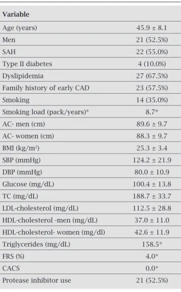
We studied 40 patients, aged 45.9 ± 8.1 years, ranging from 31 to 64 years. Only two (5%) were older than 60 years. Males ac-counted for 52.5% of the sample. The demographic and clin-ical characteristics of studied subjects are shown in Table 1. Two or more risk factors were found in 82.5% of patients and 5.0% had five risk factors. The age of risk for CAD was ob-served in 30.0% of individuals. A total of 27.5% individuals had altered blood pressure, and among individuals known to be hypertensive, 50% showed altered blood pressure. BMI was 25.3 ± 3.4 kg/m2, being classified as normal in 50.0% of
subjects, 42.5% as overweight and obesity in 7.5%. The AC was 89.6 ± 9.7 cm in men, and in females, 88.3 ± 9.7 cm. There were significantly more women than men with altered AC (52.6% and 4.8% respectively, p = 0.001).
Fasting blood glucose was 100.4 ± 13.8 mg/dL, and 42.5% of patients had altered glucose levels (≥ 100 mg/dL). Regarding the lipid profile, TC was 188.7 ± 33.7 mg/dL (127-268 mg/dL), and 30.0% of patients had altered TC lev-els (≥ 200 mg/dL). LDL-cholesterol was 112.5 ± 28.8 mg/dL (59-184 mg/dL) and was ≥ 130 mg/dL in 25.0% of the individuals. HDL-cholesterol was 39.7 ± 11.6 mg/dL (20-71 mg/dL), being 37.0 ± 11.0 mg/dL in men and 42.6 ± 11.9 mg/dL in women. HDL cholesterol was consid-ered abnormal for age and sex of individuals in 82.5% and < 40 mg/dL in 57.5%. Median triglyceride was 158.5 mg/dL (43-412 mg/dL, being the first quartile equal to 104.0 mg/dL and the third quartile, 274.5 mg/dL). Triglyc-erides were altered (≥ 150 mg/dL) in 52.5% of the sample.
All patients were on ART, and 52.5% on PI. Table 2 shows comparisons between patients who were on PI or not. No significant differences were detected, except for altered HDL-cholesterol adjusted for sex, which was more frequent in the group on PI (p = 0.040), and for altered triglycerides, which were more common in individuals who were on PI.
Assessment of cardiovascular risk through the FRS showed that the 10-year risk of MI or death due to CAD had a median of 4.0%, 2.0% in the first quartile and 10.0% in the third quartile. In this sample, 72.5% of patients were classi-fied as low risk, 25.0% as moderate risk and 2.5% (only one individual) as high risk.
The CACS had a median equal to zero, its value rang-ing from zero to 632 points. Coronary calcification was found (CACS > 0) in 32.5% of individuals. Patient charac-teristics according to the presence of coronary calcification are shown in Table 3. Subjects with coronary calcification were significantly older than patients without coronary cal-cification (p = 0.015). However, other significantly different variables were not found between subjects with and without coronary calcification.
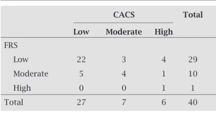
According to the percentiles of CACS, 67.5% of patients had low risk, 17.5% had moderate risk, and 15.0% had high risk. The assessment of agreement between the FRS and CACS showed that 22 subjects (75.8%) with low FRS were also considered low risk by CACS, 4 (40.0%) of those with moderate risk by the FRS, had moderate risk by CACS and 100% of those with high risk by the FRS had high risk by CACS, with a kappa value of 0.435 (Table 4).
DISCUSSION
Antiretroviral therapy has made HIV infection a chron-ic condition, and although it is not curable, it is treatable. This fact has allowed the infected population to age and be subject to chronic diseases such as cardiovascular disease, especially CAD. Thus, it is extremely important to know the cardiovascular risk of these patients in order to plan pos-sible interventions that might reduce this risk. The present study assessed patients with HIV infection in order to
Table 1. Population characteristics
Variable
Age (years) 45.9 ± 8.1
Men 21 (52.5%)
SAH 22 (55.0%)
Type II diabetes 4 (10.0%)
Dyslipidemia 27 (67.5%)
Family history of early CAD 23 (57.5%)
Smoking 14 (35.0%)
Smoking load (pack/years)* 8.7*
AC- men (cm) 89.6 ± 9.7
AC- women (cm) 88.3 ± 9.7
BMI (kg/m2) 25.3 ± 3.4
SBP (mmHg) 124.2 ± 21.9
DBP (mmHg) 80.0 ± 10.9
Glucose (mg/dL) 100.4 ± 13.8
TC (mg/dL) 188.7 ± 33.7
LDL-cholesterol (mg/dL) 112.5 ± 28.8
HDL-cholesterol -men (mg/dL) 37.0 ± 11.0
HDL-cholesterol- women (mg/dl) 42.6 ± 11.9
Triglycerides (mg/dL) 158.5*
FRS (%) 4.0*
CACS 0.0*
Protease inhibitor use 21 (52.5%)
Table 2. Comparisons according to the use of protease inhibitors
With use Without use p-value
n = 21 (52.5%) n = 19 (47.5%)
AC (cm) 90.4 ± 11.2 87.4 ± 7.5 0.336
BMI (kg/m2) 25.9 ± 3.9 24.6 ± 2.8 0.248
Glucose (mg/dL) 102.2 ± 12.7 98.5 ± 14.9 0.404
TC (mg/dL) 192.2 ± 27.9 184.7 ± 39.5 0.493
Altered TC 38.1% 21.1% 0.240
LDL-cholesterol (mg/dL) 112.1 ± 26.5 112.8 ± 31.8 0.936
LDL-cholesterol < 100 mg/dL 33.3% 26.3% 0.629
HDL-cholesterol (mg/dL) 38.7 ± 8.8 40.7 ± 14.3 0.581
HDL-cholesterol < 40 mg/dL 57.1% 57.9% 0.962
Altered HDL-cholesterol 95.2% 68.4% 0.040
Triglycerides (mg/dL) 219.0* 130.0* 0.040
Triglycerides ≥ 150 mg/dL 71.4% 31.6% 0.012
FRS (%) 4.0%* 3.0%* 0.375
CACS 0.0* 0.0* 0.782
Coronary calcification 33.3% 31.6% 0.906
Value expressed as percentages or mean ± standard deviation; *median; AC, abdominal circumference; IMC, body mass index; TC, total cholesterol; LDL, low-density lipoprotein; HDL, high-density lipoprotein; FRS, Framingham Risk Score; CACS, coronary artery calcium score.
Table 3. Comparison between patients with or without coronary calcification
With calcification Without calcification p-value
n = 13 (32.5%) n = 27 (67.5%)
Age (years) 50.3 ± 7.3 43.8 ± 7.7 0.015
Men 9 (42.8%) 12 (57.1%) 0.141
SAH 8 (36.3%) 14 (63.6%) 0.564
Type II diabetes 2 (50.0%) 2 (50.0%) 0.392
Dyslipidemia 9 (33.3%) 18 (66.6%) 0.584
Family history of early CAD 8 (34.7%) 15 (65.2%) 0.720
Smoking 4 (28.5%) 10 (71.4%) 1.000
Smoking load (pack/years) 5.0* 15.7* 0.229
AC (cm) 90.5 ± 10.3 88.3 ± 9.3 0.506
BMI (kg/m2) 24.9 ± 3.1 25.5 ± 3.6 0.627
SBP (mmHg) 117.6 ± 17.3 127.4 ± 23.5 0.194
DBP (mmHg) 78.4 ± 8.7 80.7 ± 11.9 0.543
Glucose (mg/dL) 100.1 ± 14.0 100.1 ± 13.9 0.833
TC (mg/dL) 196.6 ± 32.2 184.8 ± 34.3 0.309
LDL-cholesterol (mg/dL) 108.4 ± 29.5 114.4 ± 28.8 0.546
HDL-cholesterol (mg/dL) 39.6 ± 12.1 39.7 ± 11.7 0.975
Triglycerides (mg/dL) 193.0* 140.0* 0.525
FRS (% in 10 years) 8.0%* 3.0%* 0.142
Protease inhibitor use 33.3% 66.6% 0.906
determine the prevalence of traditional cardiovascular risk factors, assessing the risk of CAD through previ-ously validated clinical scores and investigate the use of CACS for the diagnosis of subclinical coronary athero-sclerosis and as an additional method for risk stratifica-tion in this populastratifica-tion.
In this sample, the mean age was 45.9 ± 8.1 years and only 30% were at the risk age. Therefore, it can be considered a young population for the development of CAD, but comparable to other studies of popula-tions with HIV.6,30-33 However, a high proportion of
patients (82.5%) had two or more traditional risk factors, highlighting the importance of proper stratification of cardiovascular risk in this group. SAH was present in 55% of patients, being more frequent than in other stud-ies, which showed prevalence rates of 35% to 41%34,35
and 27.5% of the patients had altered blood pressure levels. These findings are similar to those found in the general Brazilian population, in which the levels of hy-pertension control are still suboptimal,18,36 indicating
the need for greater attention to blood pressure control in the HIV-infected population.18
Prevalence of smoking among HIV-infected patients of 47% to 71% has been reported, much more elevated than in the general population.34,37 The prevalence
ob-served in our sample was 35% lower than the rates cited above, but still twice the 17.5% prevalence of the Brazil-ian population in 2008.38 Dyslipidemia was the most
fre-quent risk factor found in 67.5% of individuals. In the study by Cahn et al.,39 the prevalence of dyslipidemia in
the Brazilian population with HIV infection was 57.3%. The levels of TC and LDL-cholesterol were slight-ly higher than the percentage described in the study by Kingsley et al.40 HDL-cholesterol levels were altered in
the majority of the studied population (82.5%). The study by Kingsley et al.40 identified only 40% of individuals with
low HDL-cholesterol, whereas in our population, 57.5% were identified at this level. However, it is noteworthy that
these authors did not take into account the adjustment of HDL-cholesterol according to sex. As for triglycerides, our results are also similar to those previously described (38%).40
In this population, 52.5% of patients were using PI, which is similar to another Brazilian study.39 No
signifi-cant differences in levels of TC and LDL-cholesterol were found in our sample among individuals using PI or not, similarly to other studies, i.e., minor alterations in these lipid fractions.41-43 However, HDL-cholesterol, corrected
for age and sex, was significantly more frequent among patients using PI. This difference has been reported in the literature, associated with risk of MI.44 It was also possible
to demonstrate the association between the use of PI and increased levels of triglycerides, and the median in the group that used PI was 219 mg/dL, compared to 130 mg/dL in the group that did not use PI. This association has also been previously described.45
Risk assessment through the FRS showed that the pop-ulation of this sample had low risk (FRS median of 4.0% and the third quartile of 10.0%), which can be explained by their age, predominantly young. Notably, one fourth of individuals were classified according to the FRS as having moderate risk, and 2.5% as having high risk. A significant proportion of individuals with moderate risk underscores the importance of a refinement of risk stratification of this population in order to establish the best treatment and re-duce the incidence of adverse cardiovascular events.
Screening for subclinical atherosclerosis through CACS showed that about one third of patients had cor-onary calcifications, an indicator of the presence of atherosclerosis. Studies carried out in the general popu-lation have shown that at the age range of 40-45 years, there is a prevalence of coronary calcification of 13.3% to 21%.46,47 The frequency of calcification found in this
sample was similar to the study by Crum-Cianflone et al.48 among individuals with HIV infection, in which 34%
of patients showed calcification in the coronary arteries. Patients with calcification in the coronary arteries were significantly older than those without calcification, which has been shown in other studies of patients with and with-out HIV infection.32,40,47,49 Although other studies have
already reported an association between coronary calcifi-cation in individuals with HIV and other variables (SBP, triglycerides, TC, smoking, FH of CAD, dyslipidemia)30,40
these associations were not found in our population, pos-sibly due to the small sample size.
The agreement between the risk stratification by FRS and the CACS showed a kappa of 0.435. In this study, we did not have access to the clinical follow-up of patients, and therefore could not make any inference regarding the accu-racy of these two forms of risk stratification to predict the occurrence of events. Although it was not possible to estab-lish the best score for risk prediction in this population, it
Table 4. Agreement between risks assessed by FRS and CACS
CACS Total
Low Moderate High
FRS
Low 22 3 4 29
Moderate 5 4 1 10
High 0 0 1 1
Total 27 7 6 40
was possible to verify the frequency of modifications in risk classes defined by the FRS provided by CACS. The reclas-sification of risk occurred in 32.5% of individuals, and from the category of low risk by FRS, 24% had their risk increased (10.3% to moderate risk and 13.7% to high risk). From the category of moderate risk (the most widely described as the main indication for refinement of risk assessment), 10% had their risk increased and 50% had the risk decreased.
The effect of CACS on moderate-risk individuals by the FRS was consistent with published data, which showed a higher frequency of reclassifications exactly in this category.50-52 This capacity of CACS makes it
attrac-tive to refine risk stratification in the HIV-infected pop-ulation. For individuals considered high risk by CACS, the therapeutic approach would possibly be changed.8,11
CONCLUSIONS
One of the limitations of the study was the unavailabil-ity of data like the time of HAART, use of PI and HIV infection, which may be correlated with the occurrence of CAD. Larger studies with long-term clinical follow-up are necessary to confirm the findings and allow ob-taining a precise risk score, specific for the needs of this population.
REFERENCES
1. The Joint United Nations Programme on HIV/AIDS. UN-AIDS. AIDS epidemic update [Internet]. Geneva: [updated on 2010; cited 2011 Jan 11]. Available from: http://www.unaids. org/en/media/unaids/contentassets/documents/unaidspubli-cation/2010/20101123_globalreport_en[1].pdf
2. Ministério da Saúde. Aids.gov: Aids no Brasil [Internet]. Bra-sília: Ministério da Saúde. [cited 2011 Jan 11]. Available from: http://www.aids.gov.br/pagina/aids-no-brasil
3. Ministério da Saúde. Monitoraids [Internet]. Brasília: Ministé-rio da Saúde [cited 2011 Jan 10]. Available from: http://siste-mas.aids.gov.br/monitoraids/?keyWord=gasto&condicaoFich a=nomeIndicador&desagregacao=1
4. Lohse N, Hansen AB, Pedersen G, et al. Survival of persons with and without HIV infection in Denmark, 1995-2005. Ann Intern Med. 2007; 146(2):87-95.
5. Currier JS, Taylor A, Boyd F, et al. Coronary heart diseases in HIV- infected individuals. J Acquir Immune Defic Syndr. 2003; 33:506-12.
6. Triant VA, Lee H, Hadigan C, Grinspoon SK. Increased acute myocardial infarction rates and cardiovascular risk factors among patients with human immunodeficiency virus disease. J Clin Endocrinol Metab. 2007; 92;2506-12.
7. D’Arminio A, Sabin CA, Phillips AN, et al. Writing Committee of the D:A:D: Study Group. Cardio and cerebrovascular events in HIV-infected persons. AIDS. 2004; 18:1811-7.
8. Sposito AC, Caramelli B, Fonseca FA, et al. Sociedade Bra-sileira de Cardiologia. IV Brazilian Guideline for Dyslipidemia and Atherosclerosis prevention: Department of Atherosclero-sis of Brazilian Society of Cardiology. Arq Bras Cardiol. 2007; 88(Suppl 1):2-19.
9. Grupo de Estudo em Ressonância e Tomografia Cardiovascular (GERT) do Departamento de Cardiologia Clínica da Sociedade Brasileira de Cardiologia. Cardiovascular magnetic resonance and computed tomography imaging guidelines of the Brazilian Society of Cardiology. Arq Bras Cardiol. 2006; 87(3):e60-100. 10. Ministério da Saúde, Secretaria de Vigilância em Saúde,
Pro-grama Nacional de DST e Aids. Recomendações para terapia anti-retroviral em adultos infectados pelo HIV:2008. Brasília: Ministério da Saúde; 2008.
11. National Cholesterol Education Program (NCEP) Expert Panel on Detection, Evaluation, and Treatment of High Blood Cho-lesterol in Adults (Adult Treatment Panel III). Third Report of the National Cholesterol Education Program (NCEP) Expert Panel on Detection, Evaluation, and Treatment of High Blood Cholesterol in Adults (Adult Treatment Panel III) final report. Circulation. 2002; 106:3143-421.
12. Rodbard HW, Blonde L, Braithwaite SS, et al. AACE Diabetes Mellitus Clinical Practice Guidelines Task Force. American As-sociation of Clinical Endocrinologists medical guidelines for clinical practice for the management of diabetes mellitus. En-docr Pract. 2007; 13(Suppl 1):1-68.
13. American Diabetes Association. Standards of medical care in diabetes—2006. Diabetes Care. 2006; 29(Suppl 1):S4-S42. 14. Bailey KV, Ferro-Luzzi A. Use of body mass index of adults in
assessing individual and community nutritional status. Bull World Health Organ. 1995; 73:673-80.
15. National Institutes of Health. Clinical guidelines on the inden-tification, evaluation, and treatment of overweight and obesity in adults – the evidence report. Obesity Res. 1998; 6(suppl 2):51S-209S.
16. Janssen I, Katzmarzyk PT, Ross R. Waist circunference and not body mass index explains obesity-related health risk. Am J Clin Nutr. 2004; 79:379-84.
17. Lohman TG, Roche AF, Martorell R. Anthropometric stand-ardization reference manual. Champaign: Human Kinetics Book; 1998.
18. Sociedade Brasileira de Cardiologia, Sociedade Brasileira de Hipertensão, Sociedade Brasileira de Nefrologia. VI Diretriz Brasileira de Hipertensão Arterial. Arq Bras Cardiol. 2010; 95(1 suppl1):1-51.
19. American Diabetes Association. Screening for type 2 diabetes. Diabetes Care. 2004; 27(Suppl 1):S11-S4.
20. Castelli WP. Cardiovascular disease and multifactorial risk: challenge of the 1980s. Am Heart J. 1983; 106:1191-200. 21. Achenbach S, Daniel WG. Tomografia Computadorizada do
Coração. In: Braunwald E, Zipes DP, Libby P, Bonow RO. Braun-wald tratado de doenças cardiovasculares. 7ªed. Philadelphia: Elsevier Saunders; 2006. p. 355-371.
22. Agatston AS, Janowitz WR, Hildner FJ, et al. Quantification of coronary artery calcium using ultrafast computed tomography. J Am Coll Cardiol. 1990; 15(4):827-32.
23. Rumberger JÁ, Kaufman L. A rosetta stone calcium risk stratifi-cation: Agatston, volume, and mass score in 11,490 individuals. AJR Am J Roentgenol. 2003; 181:743-8.
24. Nasir K, Raggi P, Rumberger JA, et al. Coronary artery calcium volume scores on electron beam tomography in 12,936 asymp-tomatic adults. Am J Cardiol. 2004; 93:1146-9.
25. Hoff JA, Chomka EV, Krainik AJ, et al. Age and gender distri-butions of coronary artery calcium detected by electron beam tomography in 35,246 adults. Am J Cardiol. 2001; 87:1335-9. 26. Rumberger JA, Brundage BH, Rader DJ, et al. Electron beam
27. Sociedade Brasileira de Cardiologia. Departamento de Car-diologia Clinica. Grupo de Estudo de Ressonância e To-mografia Cardiovascular (GERT). Guideline of Sociedade Brasileira de Cardiologia for resonance and cardiovascular tomography. Exucutive summary. Arq Bras Cardiol. 2006; 87(Suppl 3):e1-e12.
28. O’Rourke RA, Brundage BH, Froelicher VF, et al; American College of Cardiology/American Heart Association Expert consensus document on electron-beam computed tomogra-phy for the diagnosis and prognosis of coronary artery dis-ease. Circulation. 2000; 102:126-40.
29. Truett J, Cornfield J, Kannel W. A multivariate analysis of the risk of coronary heart disease in Framingham. J Chronic Dis. 1967; 20:511-24.
30. D’Agostino RB Sr, Grundy S, Sullivan LM, et al. Validation of the Framingham coronary heart disease prediction scores: results of a multiple ethnic groups investigation. JAMA. 2001; 286:180-7.
31. R Development Core Team (2011). R: A language and envi-ronment for statistical computing. R Foundation for Statisti-cal Computing, Vienna, Austria. ISBN 3-900051-07-0, URL http://www.R-project.org/
32. Mangili A, Gerrior J, Tang AM, et al. Risk of cardiovascu-lar disease in a cohort of HIV-infected adults; a study using carotid intima-media thickness and coronary artery calcium score. Clin Infect Dis. 2006; 43:1482-9.
33. Mangili A, Gerrior J, Tang AM, et al. Metabolic syndrome and subclinical atherosclerosis in patients infected with HIV. Clin Infect Dis. 2007; 44:1368-74.
34. Steinn JH, Hadigan CM, Brown TT, et al. Prevention strate-gies for cardovascular disease in HIV-infected patients. Cir-culation. 2008; 118:e54-e60.
35. Kaplan RC, Kingsley LA, Sharrett AR, et al. Ten-year pre-dicted coronary heart disease risk in HIV-infected men and women. Clin Infect Dis. 2007; 45:1074-81.
36. Rosáorio TM, Scala LCNS, França GVA, et al. Prevalência, controle e tratamento da hipertensão arterial sistêmica em Nobres - MT. Arq Bras Cardiol. 2009; 93:672-8.
37. Gritz ER, Vidrine DJ, Lazev AB, et al. Smoking behavior in a low-income multiethnic HIV/AIDS population. Nicotine Tab Res. 2004; 6:71-7.
38. Ministério da Saúde. INCA. Publicações, e Pesquisa nacion-al por amostra de domicílio/tabagismo [internet]. Brasília: Ministério da saúde. [cited 2011 Jan 15]. Available from: http://www.inca.gov.br/inca/Arquivos/publicacoes/tabag-ismo.pdf
39. Cahn P, Leite O, Rosales A, et al. Metabolic profile and car-diovascular risk factors among Latin American HIV-infected patients receiving HAART. Braz J Infect Dis. 2010; 14:158-66. 40. Kingsley LA, Cuervo-Rojas J, Muñoz A, et al. Subclinical
coronary atherosclerosis, HIV infection and antiretrovi-ral therapy: Multicenter AIDS Cohort Study. AIDS. 2008; 22:1589-99.
41. Mulligan K, Grunfeld C, Tai VW, et al. Hyperlipidemia and insulin resistance are induced by protease inhibitors inde-pendent of changes in body composition in patients with HIV infection. J Acquir Immune Defic Syndr. 2000; 23:35-43. 42. Periard D, Telenti A, Sudre P, et al. Atherogenic dyslipidemia
in HIV-infected individuals treated with protease inhibitors: the Swiss HIV Cohort Study. Circulation. 1999; 100:700-5. 43. van der Valk M, Kastelein JJ, Murphy RL, et al.
Nevirapine-con-taining antiretroviral therapy in HIV-1 infected patients results in anti-atherogenic lipid profile. AIDS. 2001; 15:5186-92. 44. Duprez DA, Kuller LH, Tracy R, et al. Lipoproteina particle
subclasses, cardiovascular disease and HIV infection. Athero-sclerosis. 2009; 207:524-9.
45. Danner SA, Carr A, Leonard JM, et al. A short-term study of the safety, pharmacokinetics, and efficacy of ritonavir, na in-hibitor of HIV-1 protease. European-Australian Collaborative Ritonavir Study Group. N Engl J Med. 1995; 333:1528-33. 46. Davis PH, Dawson JD, Mahoney LT, et al. Increased carotid
intimal-medial thickness and coronary calcification are related in young and middle-aged adults: The uscatine study. Circula-tion. 1999; 100:838-42.
47. Loria CM, Liu K, Lewis CE, et al. Early adult risk factor lev-els and subsequent coronary artery calcification: the CARDIA study. J AM Coll Cardiol. 2007; 49:2013-20.
48. Crum-Cianflone N, Stepenosky J, Medina S, et al. Clinically significant incidental findings among human immunodefi-ciency virus-infected mem during computed tomography for determination of coronary artery calcium. Am J Cardiol. 2011; 107:633-7.
49. McClelland RL, Chung H, Detrano R, et al. Distribution of coronary artery calcium by race, gender, and age: results from Multi-Ethnic Study of Atherosclerosis (MESA). Circulation. 2006; 113:30-7.
50. Greenland P, Bonow RO, Brundage BH, et al. ACCF/AHA 2007 clinical expert consensus document on coronary artery calcium scoring by computed tomography in global cardiovas-cular risk assessment and in evaluation of patients with chest pain: a report of the American College of Cardiology Founda-tion Clinical Expert Consensus Task Force (ACCF/AHA Writ-ing Committee to Update the 2000 Expert Consensus Docu-ment on Electron Beam Computed Tomography) developed in collaboration with the Society of Atherosclerosis Imaging and Prevention and the Society of Cardiovascular Computed Tomography. J Am Coll Cardiol. 2007; 49:378-402.
51. Raggi P, Shaw LJ, Berman DS, et al. Prognostic value of coro-nary artery calcium screening in subjects with and without diabetes. J Am Coll Cardiol. 2004; 43:1663-9.