Bovine Besnoitiosis in Portugal
H. Cortes, DVM, MsC
Instituto de Ciências Agrárias Mediterrânicas (ICAM)
For correspondence with the author: Laboratorio de Parasitologia, Nucleo da Mitra, Universidade de Evora, Ap94, 7002-554, Portugal; E-mail: hcec@uevora.pt
M. J. Vila-Vicosa, DVM, PhD
Instituto de Ciências Agrárias Mediterrânicas (ICAM)
Laboratório de Reprodução, Polo da Mitra, University of Evora, Portugal.
A. Leitão, DVM, MsC, PhD
Centro de Investigação Interdisciplinar em Sanidade Animal (CIISA) Centro de Veterinária e Zootecnia (CVZ)
Instituto de Investigação Científica e Tropical, Lisboa, Portugal.
M. L. Ferreira, DVM, PhD
Centro de Investigação Interdisciplinar em Sanidade Animal (CIISA)
Faculdade de Medicina Veterinária, Universidade Técnica de Lisboa, Portugal
R. Vidal, PhD
Faculdade de Farmácia, Laboratório de Engenharia Genética, Universidade de Lisboa, Portugal
Emeritus Professor of Veterinary Medicine, University of California, Davis, USA
V. Caeiro, PhD
Laboratório de Parasitologia, Polo da Mitra, University of Evora, Portugal
Key words: bovine besnoitiosis; male infertility; orchitis; scleroderma; Besnoitia besnoiti.
Abstract
Besnoitia besnoiti is a coccidian parasite of cattle that is closely related to Toxoplasma gondii. B. besnoiti is endemic in many tropical and subtropical parts of the world. In these areas it causes both acute and chronic bovine besnoitiosis, and can be a significant economic constraint to commercial cattle production. However, most infections are mild or subclinical.
In the acute stage of disease, parasitic tachyzoites invade blood vessels of the skin, subcutaneous tissues, fascia and testes causing widespread vasculitis and thrombosis. The result is a severe generalized systemic reaction which is accompanied by edema of the skin and acute orchitis. During the chronic stages of disease, large numbers of tissue microcysts containing bradyzoites are formed. The most striking features of this stage are thickening, wrinkling and hair loss of the skin, accompanied by anorexia and severe weight loss. The case fatality rate is approximately 10%. Irreversible male sterility or impaired fertility is a common sequela in breeding bulls, and is one of the most negative aspects of the disease in animals that survive the acute and chronic stages of infection.
Prior to 1991, bovine besnoitiosis was seldom recognized in or reported from European countries. Since then, it has been receiving increasing attention. Here we report a single case in a newly purchased breeding bull that began showing clinical signs approximately 2 weeks following arrival on a southern Portuguese cattle farm. Because knowledge concerning the life cycle of B. besnoiti and the means of it's transmission to cattle is incomplete, and because no effective chemotherapeutic agents are available for treatment of clinical cases, a strong case can be made for expanded investment in the study of this disease.
Introduction
Bovine besnoitiosis is caused by Besnoitia besnoiti (Marotel 1912). This agent is a parasitic, tissue cyst-forming, coccidial protozoan, and is classified in the family Sarcocystidae within the Phylum Apicomplexa (Fayer 1981; Tadros and Laarman 1982). All breeds, both sexes, and animals of all ages are affected, except that clinical disease occurs rarely in calves less than 6 months of age (Bigalke 1968). The
occurrence is usually sporadic, and only a small proportion of infected animals develop clinical disease (Pols 1960; Bigalke 1968).
Bovine besnoitiosis is ubiquitous in Africa (Bigalke and others 1967; Shkap and others 1994) and Asia (Vsevolodov 1961; Peteshev and others 1974). In Europe, it has been reported only from France, (Besnoit and Robin 1912; Cadeac 1884) Spain, (Irigoien and others 2000) and Portugal. In the latter country, prior to 1991 (Malta and Silva, unpublished), the disease was seldom recognized or reported (Franco and Borges 1915; Leitao 1949). At present its prevalence may be increasing (Vidal and others 1999; Caeiro and others 2001; Cortes and others 2001; Cortes and others 2002; Cortes and others 2003). It has also been reported from Israel (Neuman 1972) and Venezuela (Vogelsang and Gallo 1941).
In the acute stages of disease, tachyzoites proliferate in macrophages, fibroblasts and endothelial cells within blood vessels. The result is vasculitis and thrombosis, especially of capillaries and small veins in the dermis, subcutis, fascia, testes and upper respiratory mucosae (Basson and others 1970). Clinical signs consist of fever, increased heart and respiratory rates, serous nasal and ocular discharges, anorexia, weight loss, generalized weakness, reluctance to move, swelling of superficial lymph nodes, generalized edema of the skin, acute orchitis with swollen, painful testes and, in some cases, anasarca (Schulz 1960). Inspiratory dyspnea may
result from inflammation of the upper respiratory mucosae (McCully and others 1966). Other less common manifestations are diarrhea and/or abortion (Pols 1960).
The subsequent, chronic stages of disease are characterized by formation of large numbers of tissue micro-cysts, up to 0.4 mm in diameter, containing
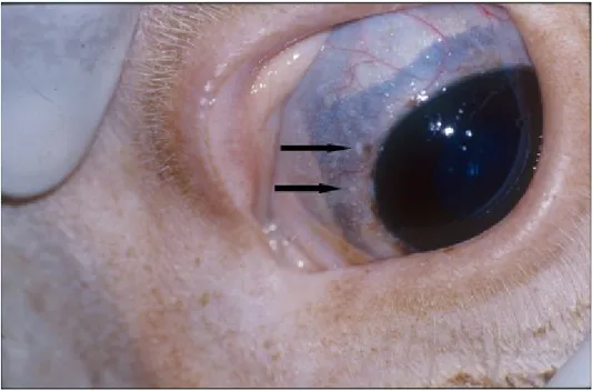
bradyzoites. A low-grade, intermittent febrile reaction may be observed, anorexia continues, and further weight loss results. The characteristic cysts form in the same tissues in which the tachyzoites were present during acute disease, especially in cutaneous and subcutaneous tissues, and in intermuscular fascia (McCully and others 1966). The skeletal muscles, tendons, tendon sheaths and periosteum of the limbs, the testicular parenchyma, and the upper respiratory mucosae may also be extensively involved (Basson and others 1970). Tiny cysts are usually apparent upon close visual inspection of the scleral conjunctiva, a feature which is of considerable value in clinical diagnosis (Sannusi 1991).
Dermal lesions are always present during chronic disease. These consist of rather dramatic thickening, hardening and folding or wrinkling of the skin, especially around the neck, shoulders and rump, always accompanied by hyperkeratosis,
hyperpigmentation and alopecia (Pols 1960). The thickening of the skin is caused by scleroderma. The latter is a sequela to granulomatous reactions provoked by the presence in tissues of dead bradyzoites (Basson and others 1970). Scleroderma and alopecia are permanent disfigurements in surviving animals (Bigalke 1960).
There may also be pronounced thickening of the limbs, and locomotion may be difficult and painful (Pols 1960). A mucopurulent nasal discharge may be
accompanied by inspiratory dyspnea (McCully and others 1966). Severely affected bulls often develop irreversible intratesticular lesions of vasculitis, focal necrosis,
sclerosis and atrophy, which usually result in permanent infertility (Ferreira and others 1982).
Few animals die during the acute stages of disease. The case fatality rate during the chronic stage is usually on the order of 10% (Pols 1960). Recovery is prolonged over a period of several months in severely affected individuals (Pols 1960).
A Case Report: Clinical, Pathological and Laboratory Findings:
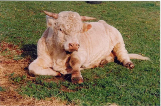
A 3-year-old, Charolais breeding bull was purchased on October 7, 2001. The farms of both buyer and seller were located not far apart from each other, in the Alentejo region of southern Portugal. Approximately 2 weeks after arriving at the buyer's farm, the bull developed clinical signs of fever and dyspnoea and, on October 22, was treated for respiratory disease by a veterinarian, using sulfadimethoxine and trimethoprim. Within a few days after treatment was administered, the dyspnea had significantly improved, but a subcutaneous edema of the extremities had developed (Fig. 1). Subsequently, the oedema progressively extended to involve the neck and lower abdomen, the skin became visibly thickened, generalised alopecia developed, and small nodules (less than 1mm in diameter) formed in the scleral conjunctival membrane (Fig. 2).
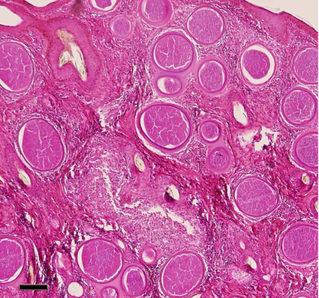
On November 12, approximately 22 days following initial appearance of clinical signs, a biopsy was obtained from the skin of the lateral side of the neck, and characteristic cysts of B. besnoiti were identified on histopathologic examination (Fig. 3). Following a clinical course of approximately 6 weeks, during which time
progressive loss of body condition occurred, the animal was euthanased and reproductive system pathology assessed.
At post mortem examination, the scrotal skin was severely fibrotic, causing the testes to be retracted up into the inguinal region. Both testes were markedly reduced in size, measuring 6 cm x 9 cm (as compared to expected normal measurements of 7 to 8 cm x 14 to 16 cm in animals of similar age and breed). Both testes were extensively fibrotic. A macroscopic area of necrosis was present in the central region of one testis (Fig. 4). Spermatozoa could not be demonstrated in the epididymus or vas deferens of either testis, as these structures were severely fibrotic and contained no fluid.
On October 20, 2001, 12 normal, asymptomatic Limousin bulls from this same farm, all between 18 and 24 months of age, were randomly selected from a total population of 32 bulls of similar age and breed. A biopsy was obtained from the cervical skin of each of these 12 animals. On histopathological examination of these specimens, B. besnoiti cysts were identified in 5 of these animals (42%).
Discussion
The life cycle of all 7 recognized species within the genus Besnoitia (for those species in which the life cycle has been elucidated) involves a definitive host and an intermediate host (Tenter and Johnson 1997). Cattle are believed to be the specific intermediate host for B. besnoiti (Bigalke and others 1967). The definitive host for B. besnoiti is unknown (Cortes and others 2003). Six other recognised Besnoitia species cause intermediate host-specific infections in (1) goats, (2) horses, burros and mules, and (3) several wildlife species (Dubey and others 2003). In naturally occurring disease of cattle, the usual means of infection is presumed to be by ingestion of mature oocysts of B. besnoiti that are shed in the feces of an unknown (but presumably carnivorous) definitive host or hosts. It is not clear how the definitive host(s) become(s) infected (Levine 1977).
Bovine besnoitiosis can also be experimentally transmitted by mechanical transfer of either tachyzoites or bradyzoites from an infected bovine to a susceptible one (Bigalke 1968). Epidemiological evidence suggests that biting flies, especially tabanids, are capable of mechanical transmission. Within endemic areas, outbreaks of bovine besnoitiosis always occur during seasons of the year when biting flies are active (Bigalke 1968; Cortes and others 2003). Since tachyzoites have been
demonstrated in lacrimal secretions (Bigalke 1968; Cortes and others 2003), there is a possibility that non-biting flies, such as Musca autumnalis and M. domestica, might also be capable of mechanical transmission.
The information developed from this on-farm study poses two questions and provides no unequivocal answers: (1) was this newly purchased bull incubating besnoitiosis at the time of arrival on the buyer’s farm, or (2) did the bull become infected following arrival on this farm?
The incubation period in cattle following experimental mechanical transmission of B. besnoiti has been reported to be 11 to 13 days (Bigalke 1968). Clinical signs in the present case were first observed approximately 2 weeks
following arrival at the buyer’s farm. Thus, mechanical transmission would have had to have occurred within the first day or two following arrival at the farm. Mitigating against this possibility is the fact that the weather in the Alentejo region during the month of October is usually cool, and populations of potential mechanical vectors, especially tabanids, are generally at low levels. Also mitigating against the case for mechanical transmission is the fact that, in experimental mechanical transmission studies, typical clinical cases (as are seen in the field) did not result. Instead, only very mild disease or subclinical infections resulted (Bigalke 1968).
The case history tends to support the hypothesis that transmission occurred by ingestion of mature oocysts of B. besnoiti, following arrival of the bull at the Alentejo farm. It should be noted that clinical besnoitiosis had been previously diagnosed in other cattle on this same farm, but not on the farm from which the bull was purchased.
Investigators in Kazakhstan reported an incubation period of 4 days following experimental exposure of cattle to mature oocysts of B. besnoiti. The infective oocysts were derived from the feces of a small, wild feline species (Felis lybica), which was thought to be a definitive host for B. besnoiti in that region (Peteshev and others 1974). However, other investigative teams have been unable to confirm these results (Busseiras and Chermette 1992; Dubey 1976; Ferreira 1985; Levine 1977; Rommel 1989; Soulsby 1982), and attempts to infect domestic cats by feeding the tissue cysts have failed (Ayroud and others 1995; Diesing and others 1988; Ng'ang'a and others 1994; Paperna and others 2001).
Infection by ingestion of mature oocysts would not be inconsistent with the finding that 5 of 12 clinically normal bulls in the herd of destination were infected with B. besnoiti, since it is well known that most naturally infected cattle are asymptomatic (Pols 1960).
Natural exposure of cattle to mature oocysts presupposes the presence in the immediate environment of (1) a definitive host, most likely a carnivore, and (2) an infective feed source (infected or contaminated with tachyzoites or bradyzoites of B. besnoiti) to which the definitive host has access (Tenter and Johnson 1997). On many farms on which bovine besnoitiosis is endemic (including the Portuguese farm in this study), dead cattle are conscientiously disposed of by rendering, incineration, deep burial, or other means that do not allow for recycling of infectious materials into potential definitive hosts. Therefore, it seems apparent that another intermediate host must exist in nature, that can serve as infective prey for the definitive host.
Two other transmission scenarios are also plausible: (1) the bull might have been (mechanically) infected immediately prior to being transported, and developed severe clinical disease after arrival at the Alentejo farm; (2) the bull might have been subclinically infected prior to being purchased, and an inapparent infection was converted to severe clinical infection, as a result of immune system suppression resulting from severe stresses associated with transportation and adaptation to changes in nutrition, housing, and management. It should be pointed out that this latter
occurrence has not been documented in the scientific literature (in connection with B. besnoiti infections).
In closing, the authors wish to emphasize five key points:
(1) Bovine besnoitiosis can have a significant adverse economic impact on cattle production in areas of the world where the disease is endemic, especially within
infected herds (Cortes and others 2003). Although the mortality rate may be relatively low, surviving animals experience severe weight loss, prolonged periods of
convalescence, adverse effects on male fertility, their hide is worthless for making leather, and their carcasses may require extensive trimming at slaughter, or even condemnation (Cortes and others 2003).
(2) No effective pharmaceutical agents are available for treatment of clinical cases (Pols 1960; Shkap and others 1985; Shkap and others 1987).
(3) A live, tissue culture vaccine has been developed and evaluated in South Africa. It has been shown to prevent clinical disease, but does not consistently prevent subclinical infections (Bigalke and others 1973).
(4) Bovine besnoitiosis is endemic in some EU countries (Besnoit and Robin 1912; Cortes and others 2003; Irigoien and others 2000).
(5) Responsible authorities should consider taking action to avoid introduction of bovine besnoitiosis into countries and regions of the EU where it does not now exist. Indirect immunofluorescence and ELISA tests have been developed to facilitate diagnosis (Shkap and others 1984), but have the disadvantage of lacking sensitivity (Janitschke and others 1984). At the present time, the most sensitive and specific test for detection of asymptomatic carriers is histopathological examination of skin biopsy material for the presence of characteristic cysts (Sannusi 1991).
Acknowledgements
This work was supported by Programa de Desenvolvimento Educativo para Portugal (PRODEP), ICAM and CIISA. We are thankful to Dr. Nuno Prates and Dr. José Miguel Leal da Costa for their collaboration in the study of this clinical case of
References
AYROUD, M., LEIGHTON, F. A., TESSARO, S. V. (1995) The morphology and pathology of Besnoitia sp. in reindeer (Rangifer tarandus tarandus), Journal of Wildlife Diseases 31, 319-326
BASSON, P. A., MCCULLY, R. M., BIGALKE, R. D. (1970) Observations on the pathogenesis of bovine and antelope strains of Besnoitia besnoiti (Marotel, 1912) infection in cattle and rabbits, Onderstepoort Journal of Veterinary Research 37, 105-126
BESNOIT, C., ROBIN, V. (1912) Sarcosporidioses cutanée chez une vache, Rec.Vet. 37, 649
BIGALKE, R. D. (1960) Preliminary observation on the mechanical transmission of cyst organisms of Besnoitia besnoiti (Marotel, 1912) from a chronically infected bull to rabbits by Glossina brevipalpis Newstead, 1910, Journal of the South African Veterinary Association 31, 37-44
BIGALKE, R. D. (1968) New concepts on the epidemiological features of bovine besnoitiosis as determined by laboratory and field investigations, The Onderstepoort journal of veterinary research 35, 3-138
BIGALKE, R. D., BASSON, P. A., MCCULLY, R. M., BOSMAN, P. P. (1973) Studies in cattle on the development of a live vaccine against bovine besnoitiosis. Conference Proceedings, Biennial Scientific Congress, South African Veterinary Association, Pretoria, 2-3.
and Besnoitia besnoiti of cattle, The Onderstepoort journal of veterinary research 34, 7-28
BUSSIÉRAS, J., CHERMETTE, R. (1992) Abrégé de Parasitologie Vétérinaire. Fascicule II: Protozoologie Vétérinaire, Service de Parasitologie - École Nationale de Vétérinaire d'Alfort, Maisons Alfort, pp 55-56, 131-132
CADÉAC, C. (1884) Identité de l'éléphantiasis et de l'anasarque du boeuf. Description de cette maladie, Revue Vétérinaire, 521
CAEIRO, V., FERREIRA, M. L., BRANCO, S. (2001) Besnoitiose bovina no Alentejo, Portugal, Acta Parasitológica Portuguesa 8, 143
CORTES, H., FERREIRA, M. L., SILVA, J. F., VIDAL, R., SERRA, P., CAEIRO, V. (2003) Contribuição para o estudo da besnoitiose bovina em Portugal, Revista Portuguesa de Ciências Veterinárias (545): 43-46
CORTES, H., NÓBREGA, F., VIDAL, R., FERREIRA, M. L., CAEIRO, V. (2002) Bovine besnoitiosis, its epidemiological aspects in Portugal and perspectives, Proceedings of the Veterinary Sciences Congress, 387-388
CORTES, H., VIDAL, R., CAEIRO, V., FERREIRA, M. L. (2001) Detecção, por PCR, de Besnoitia besnoiti que parasita o gado bovino da região de Évora assim como de um retrotransposão inserido no genoma cromossomal deste protozoário, Acta Parasitológica Portuguesa, 129
DIESING, L., HEYDORN, A. O., MATUSCHKA, F. R., BAUER, C., PIPANO, E., DE WAAL, D. T., POTGIETER, F. T. (1988) Besnoitia besnoiti: studies on the
definitive host and experimental infections in cattle, Parasitology Research 75, 114-117
DUBEY, J. P. (1976) A review of Sarcocystis of domestic animals and of other coccidia of cats and dogs, Journal of the American Veterinary Medical Association 169, 1061-1078
DUBEY, J. P., SREEKUMAR, C., LINDSAY, D.S., HILL, D., ROSENTHAL, B. M., VENTURINI, L., VENTURINI, M. C., GREINER, E. C. (2003) Besnoitia oryctofelisi n. sp. (Protozoa: Apicomplexa) from domestic rabbits, Parasitology 126, 521-539
FAYER, R. (1981) Coccidian Taxonomy and Nomeclature, Journal of Protozoology 28, 266-270
FERREIRA, M. L. (1985) Besnoitiose Bovina. Aspectos Anátomo-Clínicos., Tipografia Minerva Central: Maputo, R.P. Moçambique.
FERREIRA, M. L., NUNES PETISCA, J. L., DIAS, O. H. (1982) Alterações testiculares em touros de Moçambique assintomáticos e com sintomas clínicos de besnoitiose, Repositório de Trabalhos do Instituto Nacional de Veterinária 15, 97-108
FRANCO, E. E., BORGES, I. (1915) Nota sobre a sarcosporidiose bovina, Revista de Medicina Veterinária 165, 255-299
IRIGOIEN, M., DEL CACHO, E., GALLEGO, M., LOPEZ-BERNAD, F., QUILEZ, J., SANCHEZ-ACEDO, C. (2000) Immunohistochemical study of the cyst of
JANITSCHKE, K., DE VOS, A. J., BIGALKE, R. D. (1984) Serodiagnosis of bovine besnoitiosis by ELISA and immunofluorescence tests, The Onderstepoort journal of veterinary research 51, 239-243
LEITÃO, J. L. S. (1949), Globidiose bovina por Globidium besnoiti (Marotel 1912), Revista de Medicina Veterinária 330, 152-158
LEVINE, N. D. (1977) Nomenclature of Sarcocystis in the ox and sheep and on fecal Coccidia of the dog and cat, Journal of Parasitology 63, 36-51
MALTA, M., SILVA, M. (1991) Besnoitiose no Alentejo, XIV Jornadas de Medicina Veterinária "Bovinos de Carne". Unpublished.
MAROTEL, M. (1912) Discussion of paper by Besnoit and Robin, Bull.et Mém.de la Société de Sciences Veterinaires de Lyon et de la Société de Medicine Vetérinaire de Lyon et du Sud-Est 15, 196-217
MCCULLY, R. M., BASSON, P. A., VAN NIEKERK, J. W., BIGALKE, R. D. (1966) Observations on Besnoitia cysts in the cardiovascular system of some wild antelopes and domestic cattle, The Onderstepoort journal of veterinary research 33, 245-276
NEUMAN, M. (1972) Serological survey of Besnoitia besnoiti (Marotel 1912) infection in Israel by immunofluorescence, Zentralblatt Fur Veterinarmedizin.[B] 19, 391-396
NG'ANG'A, C. J., KASIGAZI, S. (1994) Caprine besnoitiosis: studies on the experimental intermediate hosts and the role of the domestic cat in transmission, Veterinary Parasitology 52, 207-210
PAPERNA, I., SON, R. L. (2001) Light microscopical structure and ultrastructure of a Besnoitia sp. in the naturally infected lizard Ameiva ameiva (Teiidae) from north Brazil, and in experimentally infected mice, Parasitology 123, 247-255
PETESHEV, V. M., GALUZO, I. G., POLOMOSHOV, A. P. (1974) Cats - definitive hosts Besnoitia (Besnoitia besnoiti), Izvestiae Akademii Nauk Kazakheskan SSR B, 33-38
POLS, J. W. (1960) Studies on Bovine besnoitiosis with special reference to the aetiology, The Onderstepoort journal of veterinary research 28, 265-356
ROMMEL, M. (1989) Recent advances in the knowledge of the biology of the cyst-forming coccidian, Angewandte Parasitologie 30, 173-183
SANNUSI, A. (1991) A simple field diagnostic smear test for bovine besnoitiosis, Veterinary Parasitology 39, 185-188
SCHULZ, K. C. A. (1960) A report on naturally acquired besnoitiosis in bovines with special reference to its pathology, Journal of the South African Veterinary Association 31, 21-35
SHKAP, V., DE WAAL, D. T., POTGIETER, F. T. (1985) Chemotherapy of experimental Besnoitia besnoiti infection in rabbits, The Onderstepoort journal of veterinary research 52, 289
SHKAP, V., PIPANO, E., UNGAR-WARON, H. (1987) Besnoitia besnoiti: chemotherapeutic trials in vivo and in vitro, Revue d Elevage et de Medecine Veterinaire des Pays Tropicaux 40, 259-264
SHKAP, V., UNGAR-WARON, H., PIPANO, E., GREENBLATT, C. (1984) Enzyme linked immunosorbent assay for detection of antibodies against Besnoitia besnoiti in cattle, Tropical Animal Health And Production 16, 233-238
SHKAP, V., PIPANO, E., MARCUS, S., KRIGEL, Y. (1994) Bovine besnoitiosis: transfer of colostral antibodies with observations possibly relating to natural transmission of the infection, The Onderstepoort journal of veterinary research 61, 273-275
SOULSBY, E. J. L. (1982) Helminths, Arthropods and Protozoa of Domesticated Animals, Baillière Tindall, London, p 686-687
TADROS, W., LAARMAN, J. J. (1982) Current concepts on the biology, evolution and taxonomy of tissue cyst- forming eimeriid coccidia, Advances in Parasitology 20, 293-468
TENTER, A. M., JOHNSON, A. M. (1997) Phylogeny of the tissue cyst-forming coccidian, Advances in Parasitology 39, 69-139
VIDAL, R., FERREIRA, M. L., CAEIRO, V., CABRAL, M. I., SILVESTRE, A. M., GARCIA E COSTA, F. J., JORGE VÍTOR, J. M. (1999) Caracterização genética preliminar da Besnoitia besnoiti isolada de tecidos fixados em formalina, VI Congresso (Is this correct?) Ibérico de Parasitologia, Córdoba, pp 107
VOGELSANG, E. G., GALLO, P. (1941), Globidium besnoiti (Marotel, 1912) y habronemosis cutanea en bovinos de Venezuela, Revista de la Facultad de Ciencias Veterinarias 3, 153-155
VSEVOLODOV, B. P. (1961) On besnoitiosis of cattle in Kazakhstan, in Natural focuses of disease and problems of parasitology, Academy of Sciences, Kazakh, SSR.
Fig. 1: Subcutaneous edema of the extremities of the bull with besnoitosis.
Fig. 2: Small nodules (Besnoitia cysts) on the limbal area of the scleral conjunctival membrane.
Fig. 3: Characteristic cysts of B. besnoiti, in a skin biopsy specimen from the infected bull. Scale bar = 200m.
Fig. 2: Small nodules (Besnoitia cysts) on the limbal area of the scleral conjunctival membrane.
Fig. 3: Characteristic cysts of B. besnoiti, in a skin biopsy specimen from the infected bull. Scale bar = 200m.


