UNIVERSIDADE DE TRÁS-OS-MONTES E ALTO DOURO
Emerging insights on canine hip dysplasia diagnosis:
clues from radiology and genetics
Dissertação de Mestrado em Medicina Veterinária
ANA RITA GONÇALVES GASPAR
Orientador: Professor Doutor Mário Manuel Dinis Ginja
Coorientadora: Professora Doutora Catarina Jorge Ginja
Composição do júri:
Presidente: Professor Doutor Nuno Francisco Fonte Santa Alegria
Vogais: Professor Doutor Luis Miguel Viana Maltez da Costa
Professor Doutor Mário Manuel Dinis Ginja
I hereby declare the authenticity of the present work in order to obtain the Integrated Master degree in Veterinary Medicine at the Universidade de Trás-os-Montes e Alto Douro in Vila Real, Portugal.
Ana Rita Gonçalves Gaspar
Portuguese national identification number 14167707
Official address: Avenida Dr. Renato Araújo, nº 369, 5ºesq, 3700-244, São João da Madeira, Portugal
i
Abstract
Canine hip dysplasia (CHD) is characterized by an abnormal hip and coxofemoral joint development with both hips usually affected, and is a common disorder that affects mainly large dog breeds. Hip joint laxity is the major risk factor leading to subluxation and poor congruence between the femoral head and acetabulum. Over time, degeneration of the joint occurs. Clinical sings include pain, decreased activity and lameness. Radiograph has been the gold standard to assess and quantify the joint changes associated but, with the development of genomic techniques, there is now the possibility to try to understand the genetic basis for CHD.
One of the most popular and ancient Portuguese native breeds of shepherd dogs is the “Cão da Serra da Estrela” (CSE) (Estrela Mountain Dog). This is a large, mastiff-type molossoid dog, with high prevalence of CHD. A large database of radiographic information for CHD in CSE animals already existed, thus further justifying the development of this genetic study to try to understand which regions of the genome are associated with this condition.
A review was published to describe and discuss the different diagnostic approaches concerning this condition, with special attention given to radiographic and molecular methods.
A retrospective radiographic study on 437 dogs of several breeds (33 of CSE breed) was completed to determine the ability of the Norberg angle at ≥105° to predict a non-dysplastic hip based on a distraction index cut-off of ≤0.3 or a dorsolateral subluxation score cut-off of ≥55%. It was possible to establish that at the commonly used cutpoint of 105°, the Norberg angle is not an accurate measurement of normal hip conformation. The specificity of a non-dysplastic diagnosis was maximized with a cut-off of 112º (when compared to the distract index) or 109º (dorsolateral subluxation score).
A study on 60 CSE animals, and for comparison purposes of an additional 10 Portuguese stray dogs and 10 Iberian wolves, was conducted to investigate within-breed genetic diversity and population structure using a whole-genome SNP approach. Y-chromosomal markers were also used to investigate phylogenetic relationships and within-breed haplotype composition. These analyses are key for the subsequent investigation of the genetic basis for specific CHD traits, e.g. hip joint laxity. CSE has high genetic diversity with negligible inbreeding. Weak genetic sub-structure was observed within CSE possibly
ii
explained by lineage sorting associated with different breeders in each geographic location where the samples were collected, i.e. the northern, central and southern regions of Portugal. All CSE animals shared the same Y-haplotype which belongs to the most prevalent H1-haplogroup found in European dogs.
The main purpose of the diagnosis and breeding schemes is to identify the individuals carrying hip dysplasia information in order to remove them from the breeding pool. This allows to eliminate carriers, to avoid the genetic inheritance associated with this condition and reduce its prevalence in the next generations. This study sets the basis for the future development of more comprehensive genomic studies to try to understand the genetic basis for CHD traits in this breed.
Key-words: ‘Cão da Serra da Estrela’, canine hip dysplasia, radiographic diagnosis, whole-genome SNP analyses
iii
Resumo
A displasia da anca no cão (DA) é caracterizada por um anormal desenvolvimento da anca e da articulação coxofemoral sendo usualmente ambos os lados afetados, e é uma condição frequente que afeta essencialmente cães de grande porte. A lassitude articular é o maior fator de risco levando a subluxação e a incongruência entre o acetábulo e a cabeça do fémur. Com o decorrer do tempo, a degeneração da articulação ocorre. Os sinais clínicos incluem dor, decréscimo de atividade e claudicação. A radiografia tem sido o método mais recorrente para avaliar a articulação e quantificar as modificações associadas mas, com o desenvolvimento de técnicas genómicas, existe agora a possibilidade de tentar descortinar a base genética para a DA.
Uma das raças nativas de cães pastores Portuguesas mais populares é o “Cão da Serra da Estrela” (CSE) onde se verifica uma elevada prevalência de DA. Uma vasta base de dados de informação radiográfica para DA nos CSE já estava disponível, justificando, assim, o desenvolvimento de estudos genéticos na tentativa de compreender que regiões do genoma podem estar associadas à DA.
Um artigo de revisão foi publicado com a descrição e discussão de diferentes abordagens de diagnóstico de DA, com especial atenção prestada aos métodos radiográficos e moleculares.
Um artigo original foi publicado que consistiu num estudo radiográfico retrospetivo em 437 cães de diversas raças (dos quais 33 CSE) com o objetivo de determinar a capacidade do ângulo de Norberg ≥105° de prever uma anca não displásica baseado no índice de distração ≤0.3 ou no índice de subluxação dorsolateral ≥55%. Foi possível estabelecer que com o cut-point recomendado de 105º, o ângulo de Norberg não foi uma medida precisa da conformação normal da anca. A especificidade do diagnóstico de uma anca não displásica foi maximizado com um cut-point de 112º (quando comparado com o índice de distração) ou de 109º (índice de subluxação dorsolateral).
Um estudo em 60 CSE e, para efeitos de comparação, em 10 cães de rua Portugueses e em 10 Lobos Ibéricos, foi conduzido com o objetivo de investigar a diversidade genética e a estrutura populacional na raça CSE usando polimorfismos de uma única base nucleotídica (SNPs). Utilizaram-se, ainda, marcadores específicos do cromossoma Y para aferir relações filogenéticas e a composição haplotípica. A raça CSE demonstrou elevados índices de variabilidade genética com consanguinidade negligenciável. Foi detetada sub-estrutura subtil
iv
nos CSE possivelmente explicada por diferentes linhagens familiares associadas a diferentes criadores em cada uma das zonas geográficas de amostragem, isto é, Norte, Centro e Sul de Portugal. Todos os CSE partilharam um único haplotipo do cromossoma Y pertencente ao haplogrupo H1 que é o mais prevalente em cães Europeus.
Os sistemas de diagnóstico têm como principal objectivo identificar os indivíduos portadores de informação genotípica e/ou fenotípica associada a DA para os remover dos programas de reprodução. Desta forma, conseguimos eliminar portadores, evitar a transmissão desta informação genética e reduzir a prevalência da DA nas gerações seguintes. Estas análises são a chave para uma investigação subsequente, nesta raça, da base genética de traços específicos associados a DA, como a lassitude articular.
Palavras-chave: Cão da Serra da Estrela, displasia da anca canina, diagnóstico radiográfico, análises genómicas com SNPs
v
Contents
Abstract ... i
Resumo ... iii
Contents ... v
List of figures ... vii
List of graphics ... iix
List of tables ... xi
List of publications ... xiii
Peer-reviewed international scientific journals ... xiii
Proceedings of scientific meetings ... xiii
List of abbreviations ... xv
1.Introduction ... 1
2.Canine Hip Dysplasia ... 3
2.1. Pathogenesis ... 3
2.2. Clinical signs ... 3
2.3. Diagnosis ... 4
2.3.1. Physical examination ... 4
2.3.2. Radiographic examination ... 5
2.3.3. Other diagnostic techniques ... 13
2.4. The Portuguese native dog breed ‘Cão da Serra da Estrela’ ... 14
3.Emerging insights into the genetic basis of canine hip dysplasia ... 17
3.1. Abstract ... 17
3.2. Introduction ... 18
3.3. Epidemiology, physical signs, and outcomes ... 19
3.4. Diagnosis – physical examination and imaging ... 19
3.4.1. Physical examination ... 20
3.4.2. Diagnostic imaging ... 21
3.5. Prevention and treatment ... 24
3.6. Genetic and environmental factors in the pathogenesis of canine hip dysplasia ... 25
3.7. Selection of breeding stock with low hip scores – progress toward reducing incidences of hip dysplasia ... 26
3.8. Genome-wide association studies and genetic analyses in the identification of susceptibility alleles ... 28
3.9. Prospects for the development of commercially available DNA tests for screening and diagnosis ... 33
vi
3.11. Acknowledgments ... 34
4.The Norberg Angle is not an Accurate Predictor of Canine Hip Conformation Based on the Distraction Index and the Dorsolateral Subluxation Score ... 35
4.1. Abstract ... 35
4.2. Introduction ... 36
4.3. Materials and methods ... 37
4.3.1. Animals ... 37
4.3.2. Sedation ... 38
4.3.3. Radiographic projections and measurement ... 38
4.3.4. Statistical Analysis ... 39 4.4. Results ... 40 4.4.1. Prediction analysis ... 42 4.5. Discussion ... 44 4.6. Conclusion ... 46 4.7. Acknowledgments ... 47
5.Genetic diversity and structure of the Portuguese ‘Cão da Serra da Estrela’ dog breed using whole-genome SNPs ... 49
5.1. Abstract ... 49
5.2. Introduction ... 50
5.3. Materials and Methods ... 52
5.3.1. Sampling and SNP genotyping ... 52
5.3.2. Genetic diversity and population structure ... 53
5.3.3. Y chromosome analysis ... 53
5.4. Results ... 54
5.4.1. Genetic diversity and population relationships ... 54
5.4.2. Population structure ... 59
5.4.3. Y chromosome analysis ... 62
5.5. Discussion ... 63
5.6. Conclusion ... 65
5.7. Acknowledgements ... 66
6.Concluding remarks and future perspectives ... 67
References ... 69
vii
List of figures
Figure 1: Standard ventrodorsal hip extended view and Norberg angle calculation. ... 6
Figure 2: Animal placement for the PennHIP distraction view. ... 12
Figure 3: PennHIP distraction view and distraction index calculation. ... 12
Figure 4: Animal placement for the dorsolateral subluxation test radiograph. ... 12
Figure 5: Dorsolateral subluxation score ilustration. ... 13
Figure 6: ‘Cão da Serra da Estrela’ individuals - long-haired variety. ... 15
Figure 7: ‘Cão da Serra da Estrela’ individual - short-haired variety. ... 15
Figure 8: Ortolani test performed with the dog in lateral recumbency. ... 21
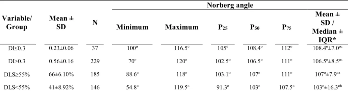
Figure 9: Hip views of an 8 months old dog. (A) Ventrodorsal hip extended projection with a Norberg angle >105°. (B) PennHIP distraction projection showing a distraction index >0.30. (C) Dorsolateral subluxation projection showing a femoral head coverage score <55%. ...41
Figure 10: Neighbor-joining dendogram of allele sharing distances depicting genetic relationships between individuals of the ‘Cão da Serra da Estrela’ breed, Iberian wolf and stray dogs populations. ... 58
Figure 11: Representation of individual genotype membership coefficients obtained with faststructure in each canid population for k=2 (top), k=3 (center) and k=4 (bottom). ... 60
Figure 12: Representation of individual genotype membership coefficients obtained with faststructure for the within-breed analysis of ‘Cão da Serra da Estrela’ for k=2 (top) and k=3 (bottom)... 62
ix
List of graphics
Graphic 1: Area under receiver operating characteristic for norberg angle ≥105º as a predictor of distract index ≤ 0.3 (a) and dorsolateral subluxation score≥55% (b). ... 43 Graphic 2: Sensitivity and specificity curves for norberg angle ≥105º as a predictor of distract index ≤ 0.3 (a) and dorsolateral subluxation score≥55% (b). ... 43 Graphic 3: Principal components analyses of ‘Cão da Serra da Estrela’, Iberian wolf and stray dogs populations. ... 56 Graphic 4: Principal components analyses showing within-breed sub-structure of ‘Cão da Serra da Estrela’.. ... 57
xi
List of tables
Table 1: Fédération Cynologique Internationale grading scheme for canine hip dysplasia. .... 6 Table 2: British Veterinary Association/Kennel Club scoring scheme for canine hip dysplasia.
... 8 Table 3: Orthopedic Foundation for Animals grading system for canine hip dysplasia. ... 11 Table 4: Comparison of four canine hip dysplasia scoring systems: Fédération Cynologique Internationale, Orthopaedic Foundation for Animals, British Veterinary Association/Kennel Club and Flückiger method. ... 23 Table 5: Summary of the single nucleotide polymorphism and nearby candidate genes identified in six genome-wide association studies of canine hip dysplasia and other related traits in reference breeds. ... 30 Table 6: Norberg angles scores for distraction indices and dorsolateral subluxation scores
groups. ..………... 40 Table 7: Genetic diversity parameters calculated in each canine population from whole-genome SNPs data: observed and expected heterozygosities, inbreeding coefficient and confidence intervals. ... 54 Table 8: Frequencies of Y-chromosome haplotypes observed in 90 males of the following populations: 32 ‘Cão da Serra da Estrela’; 46 dogs from other Portuguese native breeds; 12 stray dogs; and 32 Iberian wolves. ... 63
xiii
List of publications
Peer-reviewed international scientific journals
Ginja M., Gaspar A.R., Ginja C., 2015. Emerging insights into the genetic basis of canine hip dysplasia - review. Veterinary Medicine: Research and Reports, 6, 193–202.
Gaspar A.R, Hayes G., Ginja C., Ginja M. M., Todhunter R. J., 2016. The Norberg Angle is not an Accurate Predictor of Canine Hip Conformation Based on the Distraction Index and the Dorsolateral Subluxation Score. Preventive Veterinary Medicine, 135, 47-52.
Gaspar A.R., Muñoz-Mérida A., Pires A.E., Godinho R., Hayward J., Todhunter R.J., Ginja M.M., Ginja C., 2016. Genetic analysis of the Portuguese ‘Cão da Serra da Estrela’ dog breed using whole-genome SNPs. (manuscript in preparation to submit to Animal Genetics, ISNN: 1365-2052).
Proceedings of scientific meetings
Gaspar A.R., Godinho R., Muñoz-Mérida A., Hayward J., Todhunter R.J., Ginja M.M., Ginja C., 2016. Genetic analysis of the Portuguese Serra da Estrela dog breed using whole-genome SNPs. ConGenomics 2016 - Conference on Conservation Genomics, 3-6 May 2016, ESF – CIBIO-InBIO – Universidade do Porto, Campus de Vairão, Vairão, Portugal (poster presentation)
xv
List of abbreviations
AE – Acetabular edge AF – Acetabular fossa
BVA/KC – British Veterinary Association/ Kennel Club bp – Position in base pair
CHD – Canine hip dysplasia CaAE – Caudal acetabular ridge CFA – Canis familiaris autosome CI – Confidence interval
CrAE – Cranial acetabular edge
CSE – “Cão da Serra da Estrela” breed DA – Displasia da anca
DAE – Dorsal acetabular edge DI – Distraction index
DJD – Degenerative joint disease DLS – Dorsolateral subluxation score DNA – Deoxyribonucleic acid
EBV – Estimated breeding value
FCI – Fédération Cynologique Internationale Fis – Inbreeding coefficient
Fst – Coefficient of genetic differentiation GBV – Genomic breeding value
H – Haplotype diversity He – Expected heterozygosity HJL – Hip joint laxity
Ho – Observed heterozygosity
ICC – Intraclass correlation coefficient IW – Iberian wolf
JPS – Juvenile pubic symphysiodesis
k – Partition of the dataset in the Bayesian cluster analyses Mb – Megabase
xvi NA – Norberg angle
nt – Nucleotide
NPV – Negative predictive value OA – Osteoarthritis
OFA – Orthopedic Foundation for Animals PCA – Principal components analysis PPV – Positive predictive value
Q – Mean genotype membership coefficient q – Individual genotype membership coefficient QTL – Quantitative trait loci
ROC – Receiver operator characteristic SD – Stray dogs
SNPs – Single nucleotide polymorphisms STRs – Short tandem repeats
SVDV – Standard ventrodorsal hip-extended view UTR – Untranslated region
1
1. Introduction
The present study was accomplished to obtain the Integrated Master degree in Veterinary Medicine and was accomplished based on collaborative research between teams from Hospital Veterinário da Universidade de Trás-os-Montes e Alto Douro (Vila Real, Portugal), Cornell University Hospital for Animals (Ithaca NY, USA) and CIBIO/InBio - Centro de Investigação em Biodiversidade e Recursos Genéticos (Vairão, Portugal). The general aims were to investigate radiographic methods for the diagnosis of canine hip dysplasia (CHD), and to try to understand the genetic basis of traits associated to this condition in the Portuguese native dog breed ‘Cão da Serra da Estrela’ (CSE).
This dissertation begins with a short revision of the literature regarding CHD in the second chapter. Special attention is given to the CSE breed, which is one of the oldest populations of Portuguese native dogs with high prevalence of CHD.
The third chapter consists of a review article, entitled “Emerging insights into the genetic basis of canine hip dysplasia – review”, which is coauthored by M Ginja, AR Gaspar and C Ginja and published in a peer reviewed scientific journal - Veterinary Medicine: Research and Reports 2015;6:193-202. The main purpose of this review was to present and discuss medical aspects of CHD for which knowledge is incomplete and have therefore merited major current research efforts.
The fourth chapter is an original study which was designed in straight collaboration with the team of Cornell University College of Veterinary Medicine, entitled “The Norberg Angle is not an Accurate Predictor of Canine Hip Conformation Based on the Distraction Index and the Dorsolateral Subluxation Score”. The purpose of this study was to determine the ability of the Norberg angle (NA) at ≥105° to predict a non-dysplastic hip based on a Distraction Index (DI) cut-off of ≤0.3 or a Dorsolateral Subluxation Score (DLS) cut-off of ≥55%. The manuscript was coauthored by AR Gaspar, G Hayes, C Ginja, M Ginja, R Todhunter and published in the specialty journal Preventive Veterinary Medicine 2016;135:47-52.
The fifth chapter, is an original study designed in collaboration with research teams from CIBIO/InBio - Centro de Investigação em Biodiversidade e Recursos Genéticos and Cornell University College of Veterinary Medicine, entitled “Genetic analysis of the Portuguese ‘Cão da Serra da Estrela’ dog breed using whole-genome SNPs”. In this study, we aimed to carry out a comprehensive analysis of the within-breed genetic variation and
2
structure of CSE using whole-genome markers (i.e. single nucleotide polymorphisms, SNPs). Namely, the levels and patterns of genetic diversity and the putative population structure caused by the different breed uses (work vs pet) were investigated. Y-chromosomal markers were also used to investigate phylogenetic relationships and within-breed haplotype composition. Individuals of the distinct Iberian wolf population and Portuguese stray dogs were included in the analyses for comparison purposes. Preliminary results of these analyses were presented as a poster in the scientific meeting “CONGENOMICS 2016 – Conference on conservation genomics”, in May 3-6, 2016, at CIBIO/InBIO (Vairão, Portugal). A manuscript coauthored by AR Gaspar, A Muñoz-Mérida, AE Pires, R Godinho, J Hayward, R Todhunter, M Ginja and C Ginja, is being prepared for publication in the specialty journal Animal Genetics. It is imperative to first genetically characterize the CSE population to be able to pursuit an association genetic study for CHD traits in this breed, in which this condition has high prevalence.
The last chapter of this dissertation includes a brief discussion of the overall results, final considerations, and future perspectives concerning CHD diagnosis in the CSE breed.
3
2. Canine Hip Dysplasia
Canine hip dysplasia (CHD) is a relatively common skeletal disorder in which occurs an abnormal development of the hip and coxofemoral joint. This defect can affect mainly the health of large to giant breed dogs but it can also be seen in small breed dogs and cats (Janutta and Distl, 2006; Lopez and Schachner, 2015). Hip joint laxity (HJL) is the major risk factor for hip dysplasia leading to subluxation and poor congruence between the femoral head and acetabulum. Over time, degeneration of the joint occurs. Usually both hips are affected (Demko and McLaughlin, 2005; Ginja et al., 2010). Numerous factors influence the development and progression of hip dysplasia: genetics is considered determinant, and others aspects of environmental nature, such as rapid weight gain in growing animals can influence the disease severity (Kealy et al., 2000; Smith et al., 2001, 2006; Lopez and Schachner, 2015).
2.1. Pathogenesis
The hip capsule and chondro-osseous conformations, with two articulating surfaces – the acetabulum and the femoral head who grow in synchrony - are both major contributors to stability of the coxofemoral joint under load. For the normal development of the acetabular concavity it is fundamental the presence and pressing of the femoral head and its integrity. When these structures are affected, the joint becomes unstable (Alexander, 1992; Fries and Remedios, 1995; Todhunter and Lust, 2003).
The primary cartilage lesion results from focal stress in that area secondary to the abnormal magnitude and direction of load. Joint inflammation, pain, articular cartilage degeneration, and bone remodeling may occur. Abnormal weight bearing continues to cause excess wear on the articular cartilage and damages the underlying bone causing microfractures and sclerosis. The hip laxity eventually decreases as the capsule fibroses and the synovial effusion resolves (Alexander, 1992; Morgan, 1992; Fries and Remedios, 1995; Todhunter and Lust, 2003).
2.2. Clinical signs
The main clinical signs are hip pain, resulting in decreased activity, difficulty in rising, reluctance in running or climbing stairs, lameness, atrophy of the thigh muscles, hypertrophy of the shoulder muscles, crepitus, reduced hip joint motion and HJL. However, a number of stoic animals don’t show any clinical sign and this condition goes undetected to the owners (Fry and Clark, 1992; McLaughlin, 2001; Demko and McLaughlin, 2005).
4
The age of clinical detection varies depending on the trait’s severity and the owner’s awareness. It is assumed that dysplastic dogs during growth, around 8-12 months, often develop lameness or gain abnormalities. However, sometimes, the young dog will often spontaneously improve to normal function due to fibrosis that contains and stabilizes the joint improving clinical signs. By the time it becomes adult, and osteoarthritis is already severe, clinical signs resurge and the dog is presented for veterinary care in a late stage of the disease (Todhunter and Lust, 2003; Demko and McLaughlin, 2005).
2.3. Diagnosis
2.3.1. Physical examination
Clinical examination of an animal with a CHD suspicious should include a thorough retrieve of phenotypic information as well of the relevant history. A detailed orthopedic and neurologic examination is recommended to rule out other causes of pelvic limb lameness. Palpation and manipulation of the hip joint is an important step, both with the animal conscious and under anesthesia (McLaughlin, 2001; Ginja et al., 2010). Synovitis, joint capsule thickening, and articular injury are uniformly present (McLaughlin, 2001; Todhunter and Lust, 2003).
The Ortolani (Ortolani, 1976), Barlow (Barlow, 1962), and Bardens (Bardens and Hardwick, 1968) tests are a subjective evaluation of HJL. The first two were originally designed for diagnosis of human congenital hip dysplasia and then adapted for veterinary medicine. In most instances, general anesthesia is necessary to demonstrate these tests adequately (Ortolani, 1976; Fry and Clark, 1992). The Ortolani test will be clarified in detailed ahead in the review article (third chapter).
For the Barlow’s test, while in lateral recumbency, the application of an axial pressure down the femoral shaft towards the coxofemoral joint produces subluxation. If the femoral head slips forward into the acetabulum the hip has been dislocated. Barlow’s test is essentially a modification of the Ortolani test to be used in younger puppies, since, occasionally, when the hips are abducted the dislocated femoral head slides so smoothly over the low rim of the acetabulum that it does not make a click, and therefore appears to be normal (Barlow, 1962; Fry and Clark, 1992).
For the Bardens test, the dog is similarly positioned in lateral recumbency and the proximal femur is elevated laterally from the body. Simultaneously, a thumb or forefinger on one hand is placed over the greater trochanter of the femur while the other hand firmly grasps
5
the thigh, lifting it laterally without abduction. This displacement is measured and a positive sign is considered when the distance is greater than 2 mm (Bardens and Hardwick, 1968; Fry and Clark, 1992). These palpation techniques should be considered with other clinical data when attempting to diagnose CHD.
2.3.2. Radiographic examination
Radiograph has been the gold standard to assess and quantify joint changes associated with CHD and is the diagnostic method evaluated in the present study. It is possible to assess canine radiographic coxofemoral joint conformation and degenerative changes in adult animals or to estimate HJL in young dogs. Presently, for the first scenario, worldwide, there are 3 popular standardized scoring systems: Fédération Cynologique Internationale (FCI), Bristish Veterinary Association/ Kennel Club (BVA/KC) and Orthopedic Foundation for Animals (OFA). These systems are similar to each other and it is possible to establish correlations between them (detailed ahead in the review article, third chapter). To quantify HJL 2 techniques are commonly used: the PennHIP method and the dorsolateral subluxation (DLS) test.
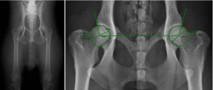
The FCI grading scheme is majorly used in Europe, South America, Africa and Asia. Limb radiographs in standard ventrodorsal hip-extended view (SVDV) (Figure 1), performed at 1 year of age (18 months for large breed dogs), are scored by a radiologist approved by breed-specific kennel clubs using FCI scoring system. The Norberg angle (NA), the degree of subluxation, acetabular shape and depth and signs of secondary joint disease are taken in consideration. The hip joint is scored from A (normal) to E (severe dysplasia), being the final grade based on the worst hip (Table 1). The first two categories are considered non-dysplastic while the last three are dysplastic (Flückiger, 2007; Ginja et al., 2010; Verhoeven et al., 2012; Lopez and Schachner, 2015; Fédération Cynologique Internationale website, 2016a).
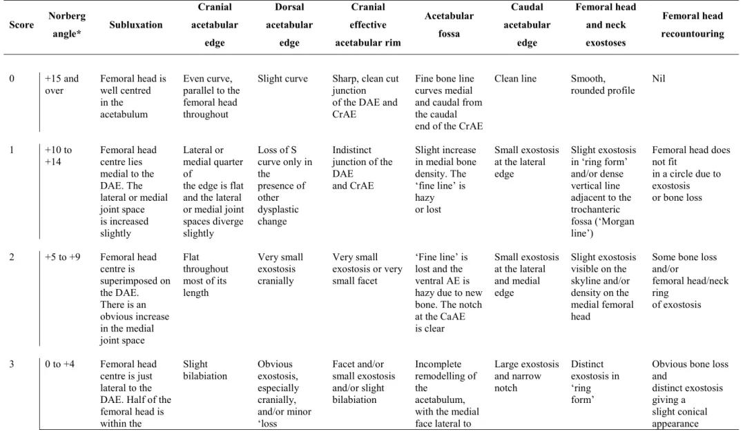
The BVA/KC scoring scheme is popular in Britain, Ireland, Australia and New Zealand. For scoring, the dogs must be at least 1 year of age and the criteria used are the NA, subluxation, acetabular shape and depth and femoral head and neck’s shape and with eventual signs of degenerative joint disease (DJD) (Table 2). In a SVDV (Figure 1) both hips are individually classified with a score, 0-53 (the sum being 0-106), with higher values implicating worse hip status (Gibbs, 1997; Flückiger, 2007; Ginja et al., 2010; Dennis, 2012; Verhoeven et al., 2012; Lopez and Schachner, 2015; Kennel Club website, 2016).
6
Table 1: Fédération Cynologique Internationale grading scheme for canine hip dysplasia (Flückiger, 2007).
A No signs of
CHD
The femoral head and the acetabulum are congruent. The craniolateral acetabular rim appears sharp and slightly rounded. The joint space is narrow and even. The NA ≈ 105°. In excellent hip joints the craniolateral rim encircles the femoral head somewhat more in caudolateral direction.
B Near normal
hip joints
The femoral head and the acetabulum are slightly incongruent and the NA ≈ 105° or the femoral head and the acetabulum are congruent and the NA<105°.
C Mild CHD
The femoral head and the acetabulum are incongruent, the NA ≈ 100° and/or there is slight flattening of the craniolateral acetabular rim. No more than slight signs of osteoarthrosis on the cranial, caudal, or dorsal acetabular edge or on the femoral head and neck may be present.
D Moderate CHD
There is obvious incongruity between the femoral head and the acetabulum with subluxation. The NA>90° (only as a reference). Flattening of the craniolateral rim and/or osteoarthritic signs are present.
E Severe CHD
Marked dysplastic changes of the hip joints, such as luxation or distinct subluxation are present. The NA<90°. Obvious flattenting of the cranial acetabular edge, deformation of the femoral head (mushroom shaped, flattening) or other signs of osteoarthrosis are noted.
CHD, canine hip dysplasia; NA, Norberg angle.
Figure 1: Standard ventrodorsal hip extended view (left) and Norberg angle calculation. Hospital Veterinário da Universidade de Trás-os-Montes e Alto Douro.
7
The OFA grading system is mainly represented in the United States of America and Canada. Standard ventrodorsal hip-extended views (Figure 1) performed at 2 years of age are assessed by three independent board-certified radiologists in excellent, good, fair, borderline, mild, moderate, or severe (Table 3). The first three categories are considered non-dysplastic while the last three are dysplastic. Hip conformation and evidence of DJD are considered (Flückiger, 2007; Ginja et al., 2010; Verhoeven et al., 2012; Lopez and Schachner, 2015).
The PennHIP method measures HJL which reveals the susceptibility for a dog to develop DJD at 16 weeks of age. Three different radiographic views are taken: a SVDV for evidence of DJD, a compression view for congruity between the femoral head and the acetabulum, and a distraction view to measure HJL (Figures 2 and 3). Joint laxity is estimated measuring the distraction index (DI), based on quantification of the relative displacement of the femoral head center from the acetabular center and it ranges from 0-1. This way passive HJL is measured in addition to subjection radiographic information. Dogs with looser hips (DI≥0.7) are more likely to develop hip dysplasia than dogs with tighter hips (DI<0.3) (Smith et al., 1990, 1993; Smith, 1997).
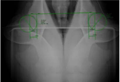
The DLS test reveals HJL in a weight-bearing position at 4-8 months of age (Figures 4 and 5). With the hip joints in a normal standing orientation, the DLS measures the femoral head coverage by the lateral aspect of the cranial acetabular rim. It is assumed that DLS scores equal to or above 55% are related to low susceptibility to develop osteoarthritis while joints with less than 45% coverage of the femoral head have a higher probability of developing osteoarthritis (Farese et al., 1998; Todhunter et al., 2003a).
8
Table 2: British Veterinary Association/ Kennel Club scoring scheme for canine hip dysplasia (Flückiger, 2007; Dennis, 2012; Verhoeven, 2012).
Score Norberg angle* Subluxation Cranial acetabular edge Dorsal acetabular edge Cranial effective acetabular rim Acetabular fossa Caudal acetabular edge Femoral head and neck exostoses Femoral head recountouring 0 +15 and
over Femoral head is well centred
in the acetabulum Even curve, parallel to the femoral head throughout
Slight curve Sharp, clean cut
junction of the DAE and CrAE
Fine bone line curves medial and caudal from the caudal end of the CrAE
Clean line Smooth,
rounded profile Nil
1 +10 to +14 Femoral head centre lies medial to the DAE. The lateral or medial joint space is increased slightly Lateral or medial quarter of
the edge is flat and the lateral or medial joint spaces diverge slightly Loss of S curve only in the presence of other dysplastic change Indistinct junction of the DAE and CrAE Slight increase in medial bone density. The ‘fine line’ is hazy or lost Small exostosis at the lateral edge Slight exostosis in ‘ring form’ and/or dense vertical line adjacent to the trochanteric fossa (‘Morgan line’)
Femoral head does not fit in a circle due to exostosis or bone loss 2 +5 to +9 Femoral head centre is superimposed on the DAE. There is an obvious increase in the medial joint space Flat throughout most of its length Very small exostosis cranially Very small exostosis or very small facet ‘Fine line’ is lost and the ventral AE is hazy due to new bone. The notch at the CaAE is clear Small exostosis at the lateral and medial edge Slight exostosis visible on the skyline and/or density on the medial femoral head
Some bone loss and/or femoral head/neck ring of exostosis 3 0 to +4 Femoral head centre is just lateral to the DAE. Half of the femoral head is within the
Slight
bilabiation Obvious exostosis,
especially cranially, and/or minor ‘loss Facet and/or small exostosis and/or slight bilabiation Incomplete remodelling of the acetabulum, with the medial face lateral to Large exostosis and narrow notch Distinct exostosis in ‘ring form’
Obvious bone loss and
distinct exostosis giving a
slight conical appearance
9 Score Norberg angle* Subluxation Cranial acetabular edge Dorsal acetabular edge Cranial effective acetabular rim Acetabular fossa Caudal acetabular edge Femoral head and neck exostoses Femoral head recountouring
acetabulum of edge’ the AF. The
ventral AE is lost, the AF is hazy and the notch is irregular 4 -1 to -5 Femoral head centre is clearly lateral to the DAE. A quarter of the femoral head is within the acetabulum Moderate
bilabiation Exostosis well lateral to the
edge and/or moderate ‘loss of edge’ Obvious facet and/or obvious exostosis and/or moderate bilabiation Marked remodelling. The medial face of the
acetabulum is clearly lateral to the AF. The ventral AE is lost
and the notch is partly closed
Marked exostosis and ‘hooking’ of the lateral end
Obvious complete collar of exostosis Gross remodelling. There is obvious bone
loss and exostosis gives a
mushroom-like appearance
5 -6 to -10 Femoral head
centre is well lateral to, and just touches, the DAE
Gross
bilabiation Marked exostosis all
along the edge and/or gross ‘loss of edge’ Gross exostosis and/or facet and/or gross bilabiation Gross remodelling, with dense new bone throughout the acetabulum. The CaAE notch is lost and the AF is obscured Gross distortion due to mass of new bone in the acetabulum. The notch is lost completely Massive exostosis giving a mushroom-like appearance Very gross remodelling with marked bone loss and much new bone
6 -11 and
10 Score Norberg angle* Subluxation Cranial acetabular edge Dorsal acetabular edge Cranial effective acetabular rim Acetabular fossa Caudal acetabular edge Femoral head and neck exostoses Femoral head recountouring
dislocation cranially the
cranial to caudal edge Massive exostosis and/or gross facet new articular surface, well lateral to the AF. The notch is lost infill of the trochanteric fossa and below the femoral head due to maldevelopment of the femoral head centre
11
Table 3: Orthopedic Foundation for animals grading system for canine hip dysplasia (CHD) (Flückiger, 2007; Verhoeven, 2012).
Excellent
Superior hip conformation in comparison to other animals of the same age and breed. There is a deeply seated femoral head which fits tightly into a well-formed acetabulum with minimal joint space. There is almost complete coverage of the acetabulum over the femoral head.
Good Slightly less than superior but well-formed congruent hip joint. The femoral head fits
well into the acetabulum and good coverage is present.
Fair
Minor irregularities are present. The hip joint is wider than a good hip phenotype due to slight subluxation causing a minor degree of joint incongruence. There may also be slight receding of the weight-bearing surface of the dorsal acetabular rim causing the acetabulum to appear slightly shallow
Mild CHD
The femoral head is partially subluxated causing an incongruent and widened joint space. The acetabulum is usually shallow only partially covering the femoral head. There are usually no arthritic changes present and if the dog is young (24 to 30 months of age), a second radiograph may be submitted for re-evaluation when the dog is older. Most dogs will remain dysplastic showing progressive degenerative joint disease.
Moderate CHD
There is significant subluxation present with the femoral head barely seated into a shallow acetabulum. There are secondary arthritic changes usually along the femoral neck and head (remodelling), acetabular osteophytes and various degrees of trabecular bone pattern changes (sclerosis).
Severe CHD
Radiographic evidence of marked CHD. There is significant subluxation with the femoral head partly or completely out of a shallow acetabulum. There are massive secondary arthritic bone changes along the femoral neck and head, acetabular rim changes and large amounts of abnormal bone pattern changes.
12
Figure 3: PennHIP distraction view and distraction index calculation. Hospital Veterinário da Universidade de Trás-os-Montes e Alto Douro. Figure 2: Animal placement for the PennHIP distraction view. Hospital Veterinário da Universidade de Trás-os-Montes e Alto
Douro.
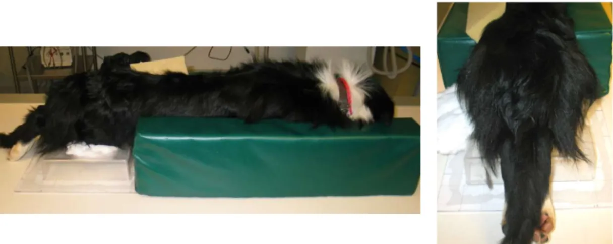
Figure 4: Animal placement for the dorsolateral subluxation test radiograph. Cornell University Hospital for Animals.
13
Figure 5: Dorsolateral subluxation score ilustration. Cornell University Hospital for Animals.
2.3.3. Other diagnostic techniques
There are other tests available for CHD diagnosis such as computed tomography imaging. This technique is becoming more popular, it has been used in several studies and it is more suitable to evaluate bone structure (Fujiki et al., 2007; Ginja et al., 2009; Andronescu et al., 2015). Magnetic resonance imaging can also be used, nevertheless its cost is more prohibitive and it is more appropriate to evaluate the structure of articular soft tissues (Ginja et al., 2009). The use of ultrasonography in dogs is not supported due to the non-congenital nature of CHD and early femoral head ossification preventing the correct visualization of the acetabulum (Greshake and Ackerman, 1993; Adams et al., 2000; Ginja et al., 2009). Examination of arterial blood supply to the hip joint with Doppler sonography was also investigated but not associated to HJL or hip dysplasia (Rademacher et al., 2005).
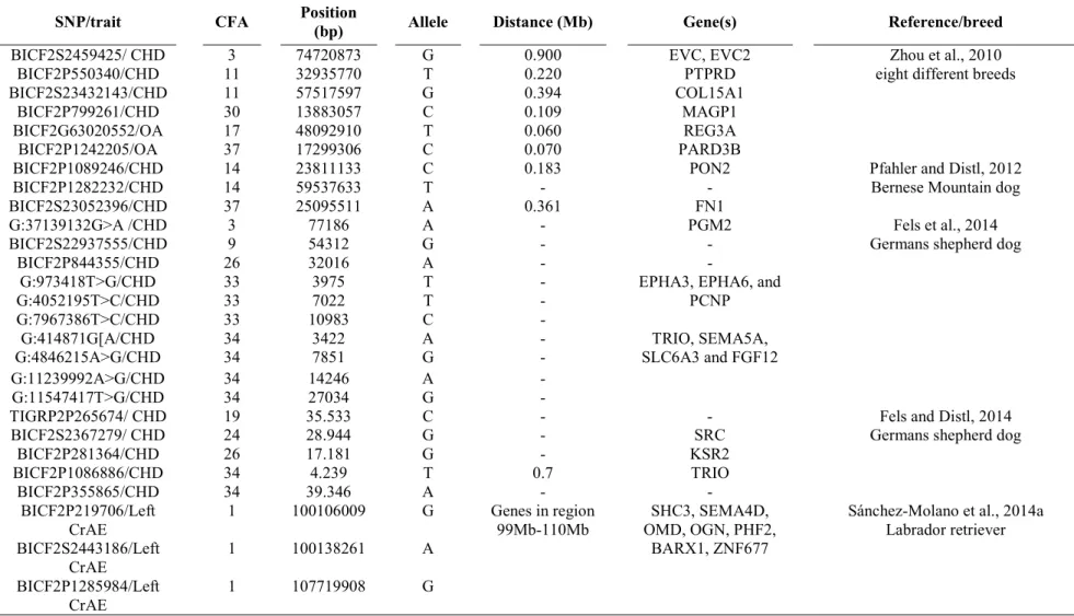
Molecular studies have been developed for CHD diagnosis searching genetic markers linked to loci responsible for different CHD phenotypes (Todhunter et al., 1999; Chase et al., 2004, 2005; Todhunter et al., 2005; Marschall and Distl, 2007; Phavaphutanon et al., 2009; Sánchez-Molano et al., 2014a; Fels and Distl, 2014; Fels et al., 2014). Genome-wide association studies, considering the joint effect of multiple SNPs, have been published by several researchers (Zhou et al., 2010; Pfahler and Distl, 2012; Lavrijsen et al., 2014; Fels et al., 2014; Hayward et al., 2016). Presently, there is a commercial DNA-based test for the Labrador Retriever breed that predicts the risk for the dog to develop CHD (Sanchez et al.,
14
2013). However, the applicability of such test to diagnose CHD in dogs from other breeds remains limited.
The cost of genomic techniques is becoming much more accessible. The availability of complete genomes from several breeds of dogs will increase. Also, the inexpensive genotyping by sequencing of genomic regions of interest will allow to characterize genetic variability in a wide range of breeds and to investigate more efficiently the regions of the genome that influence CHD traits in dogs.
The main purpose of the diagnosis and breeding schemes is to identify the individuals carrying CHD information in order to remove them from the breeding pool. This allows to eliminate carriers, to avoid the genetic inheritance associated with this disease and reduce its prevalence in future generations.
2.4. The Portuguese native dog breed ‘Cão da Serra da Estrela’
One of the most popular Portuguese native dog breeds is “Cão da Serra da Estrela” (Estrela Mountain Dog), a large, mastiff-type molossoid dog recognized by FCI - number 173 (Fédération Cynologique Internationale website, 2016b) with approximately 500 to 600 registrations annually in the Portuguese Kennel Club (Clube Português de Canicultura website, 2016a).
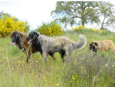
Considered one of the oldest breeds in Portugal, this dog breed has been protecting flocks of sheep for many centuries. Shepherds depend on their ability to identify and scare off wolves and other predators. This breed has been developed over a period of hundreds of years. Shepherds would have chosen to breed the dogs that had the necessary characteristics to survive in their mountain environment and to do their job: large size, strength, endurance, agility, a deep chest, ability to tolerate a marginal diet, a powerful set of the legs and mouth, a warm coat, and a watchful, mistrustful, yet loyal temperament. The first CSE dog entered a show ring in 1908, and the official breed standard was established by 1933. In 1972, the United States of America became the first foreign country to receive CSE individuals outside of Portugal. Animals of this breed can now be found in several countries around the world. We can distinguish two different varieties – long-haired (Figure 6) mainly related to show and pet dogs and short-haired (Figure 7) being used mostly as work dogs (Pye, 2002).
In the CSE breed, which is characterized by large sized dogs, CHD is a frequent problem breeders have to face. A comprehensive radiology study of the CSE breed reported 65% of the animals affected (Ginja et al., 2009). For this reason, and for being one of the most
15
important Portuguese native breeds of shepherd dogs, it was decided to carry out this study to investigate the genetic basis for CHD traits.
Figure 6: ‘Cão da Serra da Estrela’ individuals - long-haired variety.
Figure 7: ‘Cão da Serra da Estrela’ individual - short-haired variety. © R aq ue l S im õe s | G ru po L ob o
17
3. Emerging insights into the genetic basis of canine hip
dysplasia
Mário M. Ginja1, Ana Rita Gaspar1, Catarina Ginja2,3
1Department of Veterinary Sciences - CITAB, University of Trás-os-Montes and Alto Douro, Vila Real,
Portugal;
2Ce3C – Centro de Ecologia, Evolução e Alterações Ambientais, Faculdade de Ciências, Universidade
de Lisboa, Lisboa, Portugal;
3CIBIO-InBIO – Centro de Investigação em Biodiversidade e Recursos Genéticos, Universidade do
Porto, Vairão, Portugal.
3.1. Abstract
Canine hip dysplasia (CHD) is the most common inherited polygenic orthopedic trait in dogs with the phenotype influenced also by environmental factors. This trait was described in the dog in 1935 and leads to a debilitating secondary hip osteoarthritis. The diagnosis is confirmed radiographically by evaluating signs of degenerative joint disease, incongruence, and/or passive hip joint laxity. There is no ideal medical or surgical treatment so prevention based on controlled breeding is the optimal approach. The definitive CHD diagnosis based on radiographic examination involves the exposure to ionizing radiation under general anesthesia or heavy sedation but the image does not reveal the underlying genetic quality of the dog. Phenotypic expression of CHD is modified by environmental factors and dogs with a normal phenotype can be carriers of some mutations and transmit these genes to their offspring. Programs based on selection of dogs with better individual phenotypes for breeding are effective when strictly applied but remain inferior to the selection of dogs based on estimation of breeding values. Molecular studies for dissecting the genetic basis of CHD are ongoing, but progress has been slow. In the future, the recommended method to improve hip quality in controlled breeding schemes, which will allow higher selection pressure, would be based on the estimation of the genomic breeding value. Since 2012, a commercial DNA (deoxyribonucleic acid) test has been available for Labrador Retrievers using a blood sample and provides a probability for development of CHD but we await evidence that this test reduces the incidence or severity of CHD.
Keywords – canine hip dysplasia, phenotype, breeding stock, genome-wide association studies, screening
18
3.2. Introduction
Canine hip dysplasia (CHD) is the most common inherited polygenic orthopedic trait with the phenotype influenced by environmental factors (Ginja et al., 2010). This trait was described in the dog in 1935, in the USA, and leads to a debilitating secondary hip osteoarthritis (Ginja et al., 2009). Heritability estimates for CHD vary from 0.1 to 0.83 (Ginja et al., 2008; Zhu et al., 2009), due to different pedigrees, methods used to calculate the heritability, and the hip phenotypes analyzed (Silvestre et al., 2007). Canine hip dysplasia is more prevalent in large and giant breeds of dogs often resulting in mild or no clinical signs (Barr et al., 1987; Ginja et al., 2010). However, for some dogs, clinical signs can be severe and resistant to medical management needing aggressive and expensive surgical treatments (Manley et al., 2007; Ginja et al., 2010). The definitive diagnosis of CHD is made if characteristic radiographic signs are evident on a standard or stressed ventrodorsal view of the pelvis, occurring along a gradual scale from nearly normal to severely affected (Ginja et al., 2010). This is a crucial aspect of CHD as the radiographic diagnosis has been essential for the selection of breeding stock (Ginja et al., 2010). Studies attempting to find genetic markers for CHD diagnosis are now frequent (Chase et al., 2004; Todhunter et al., 2005; Fels and Distl, 2014; Sánchez-Molano et al., 2014a). The sequencing and annotation of the canine genome has resulted in renewed interest in research of the genetic underpinnings of canine orthopedic disorders, particularly those of a multifactorial etiology, such as CHD (Breur et al., 2012). Recently, the first commercial CHD diagnostic genetic test for Labrador Retrievers appeared (Sanchez et al., 2013), but, the imaging diagnosis continues to be of major importance for disease screening and treatment. Humans are also affected by hip dysplasia and both conditions have phenotypic similarities of joint subluxation and the development of osteoarthritis (Zhou et al., 2010). However, the main medical approach in humans is different, being mainly based on the preventive management and with good results (Wenger and Bomar, 2003; Ginja et al., 2010). Currently, molecular CHD studies are considered useful for the understanding of the genetic basis of analogous conditions in humans, mainly because the heterogeneity in human populations and the complexity of this disorder makes the genetic dissection of human hip osteoarthritis more difficult (Breur et al., 2012). The main purpose of this review is to present and discuss medical aspects of CHD for which knowledge is incomplete and have therefore merited major current research efforts.
19
3.3. Epidemiology, physical signs, and outcomes
Canine hip dysplasia continues to be a common trait mainly in large and giant breeds, in pet and working dogs, with prevalence higher than 50% in some breeds (Ginja et al., 2009). The clinical presentation of the disease is not correlated with the radiographic changes (Barr et al., 1987; Ginja et al., 2009). Clinical signs of CHD are more evident in dogs younger than 1 year of age due to hip instability, or in adult dogs with chronic pain from osteoarthritis (Riser, 1975; Manley et al., 2007). Chronic hip alterations, such as fibrosis and thickening of the joint capsule, result in joint stability and improvement of limb function, masking the clinical signs and functional limitations in middle-aged animals (Barr et al., 1987; Ginja et al., 2009). Clinical signs warrant medical and/or surgical treatment (Farrell et al., 2007; Vezzoni et al., 2008). Preventive conservative or surgical management could be indicated in puppies at risk of developing CHD (Lust et al., 1992; Vezzoni et al., 2008), but early intervention is hampered because there are no pathognomonic clinical signs for CHD. Common clinical signs are: slight to moderate lameness; gait and running abnormalities, such as shortened stride length and bunny hopping; difficulty in rising and reluctance to climb stairs (Fry and Clark, 1992).
3.4. Diagnosis – physical examination and imaging
Information about the conformation of the hip joint can be obtained using clinical or diagnostic imaging tests (Fry and Clark, 1992; Vezzoni et al., 2005). These medical tests are usually performed on sedated or anesthetized animals and are separated into two main categories: to evaluate hip joint laxity (HJL), mainly used on young animals; to detect clinical or radiographic signs of osteoarthritis, as crepitation and reduced range of motion on joint palpation or degenerative joint disease (DJD) signs on radiographs (Fry and Clark, 1992; Ginja et al., 2010). However, it would be very helpful to develop medical CHD screening techniques for fully conscious young animals, similar to hip dysplasia screening examination of human neonates (Ginja et al., 2010). The imaging diagnosis of CHD has been the main area of research of CHD in the last 50 years, for the purposes of reproductive control. Clearly, in terms of human medicine the main focus has been different, trying to refine diagnostic accuracy and preventive management (Wenger and Bomar, 2003; Ginja et al., 2010).
20 3.4.1. Physical examination
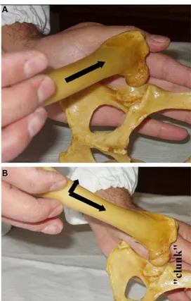
The Ortolani test is the most common and popular physical maneuver that is used in veterinary medicine to diagnose HJL in young dogs (4–12 months of age) (Chalman and Butler, 1985; Ginja et al., 2008). Other clinical tests are described, like the Barlow’s and Barden tests for puppies younger than 4 months of age but their clinical accuracy is more questionable (Ginja et al., 2009; Ginja et al., 2010). The Ortolani test is performed with the dog awake, sedated or anesthetized with the patient in lateral or dorsal recumbency (Chalman and Butler, 1985). The test has two steps, first apply proximal force to the stifle joint on the non-dependent limb, with the hip at a normal weight-bearing angle, and while still applying this force, slowly abduct the joint. In hips with abnormal laxity, the dysplastic femoral head, may be displaced dorsally beyond the dorsal acetabular rim (in the first step, Figure 8A) and then the limb abduction promotes its reduction back into the acetabulum (in the second step, Figure 8B) which elicits a typical palpable and/or audible clunk, of variable magnitude, commonly called a positive Ortolani sign (Chalman and Butler, 1985; Ginja et al., 2010). Advanced stages of CHD with destruction of the acetabular rim or in dogs younger than 4 months of age with an inadequate acetabular ossification can result in false negative cases based on the Ortolani maneuver even though HJL may be present (Puerto et al., 1999; Ginja et al., 2008; Ginja et al., 2009). The Ortolani test showed an excellent sensitivity in prediction of CHD when used in dogs younger than 1 year of age that later developed moderate or severe CHD (Ginja et al., 2008).
21 3.4.2. Diagnostic imaging
Radiography is the reference technique for the definitive diagnosis of CHD from its first description in 1935. This imaging technique uses different radiographic views of the hip joint for genetic screening purposes or for diagnosis and treatment of dogs with clinical CHD. All these radiographic techniques should be performed under anesthesia or heavy sedation, which facilitates accurate positioning and elicitation of passive HJL (Manley et al., 2007; Ginja et al., 2008; Vezzoni et al., 2008; Ginja et al., 2010). Given the complexity of the topic and the objectives of this review, we will cover particularly the radiographic studies used for genetic screening of CHD, which are used to detect HJL, the major risk factor for CHD, or signs of DJD.
The radiographic information on HJL is obtained using radiographic techniques such as PennHIP (Smith et al., 1990), dorsolateral subluxation (DLS) (Farese et al., 1998), Flückiger (Flückiger et al., 1999) and half-axial position methods (Vezzoni et al., 2005). Signs of DJD are evaluated using the standard ventrodorsal hip-extended view (SVDV) (Corley, 1992; Gibbs, 1997; Ginja et al., 2009).
Figure 8: Ortolani test performed with the dog in lateral recumbency. Reprinted from
Ginja et al. 2010. Copyright with permission from Elsevier.
22 3.4.2.1. Radiographic estimation of hip joint laxity
In this group of methods, the PennHIP was the pioneer and is the most popular. It was developed at the University of Pennsylvania in the 1980s with the main purpose of CHD breeding control (Smith et al., 1990). One of the main advantages of this procedure is its precocity, being performed with accuracy on dogs at 16 weeks of age, compared with 1 or 2 years of age for previous screening systems (Smith et al., 1990; Corley, 1992; Gibbs, 1997). The PennHIP method requires certified members and is performed with three hip radiographic views: hip-extended, compression, and distraction (Smith et al., 1990). The distraction view is used to measure HJL. It is performed with the dog in dorsal recumbency, with the hips at a neutral position and the PennHIP distractor between hind limbs acting as a fulcrum lateralizing the femoral heads under examiner force (Smith et al., 1990). The radiographs are sent to the PennHIP Analysis Center at University of Pennsylvania for an official report, and the dogs are placed in rank-order with the other dogs of the breed in the database. The HJL is evaluated in the distraction view calculating the distraction index (DI), which measures the relative degree of femoral head displacement from the acetabulum (Smith et al., 1990; Ginja et al., 2008). The DI ranges from 0 to >1, with 0 representing a tight hip and 1 a loose hip (Smith et al., 1990).
The DLS is also a passive stress radiographic or tomographic imaging technique; the HJL is measured as the DLS score (Farese et al., 1998). The hip stress is caused by weight bearing. The DLS score has a strong correlation with DI. This method was reported in dogs at 4 and 8 months of age to evaluate the chondro-osseous acetabular and femoral head structure as an indicator of functional joint stability (Farese et al., 1998, 1999).
In the Flückiger method, the HJL is estimated with the subluxation index in a similar manner to the DI (Flückiger et al., 1999). The stress in hip joints is caused by the dorsocranial force exerted by the examiner, with the dog in dorsal recumbency (Flückiger et al., 1999). No follow-up studies were published and the research was performed in adult animals together with the SVDV to better assess the quality of the hips.
The half-axial position method was used by positioning the dog and performing the hip stress similar to the PennHIP, using a trapezoidal-shaped distractor (Vezzoni et al., 2005). The HJL measures using this method are performed mainly with the purpose of early CHD diagnosis and treatment, using the juvenile pubic symphysiodesis (JPS).
23 3.4.2.2. Radiographic evaluation of DJD
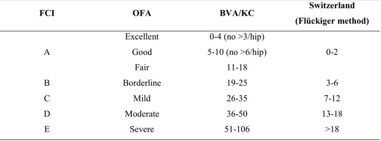
The DJD is evaluated using the SVDV, a universal radiographic view used in dogs older than 1 (Fédération Cynologique Internationale’s [FCI] system) or 2 years (Orthopaedic Foundation for Animals [OFA] system) of age (Corley, 1992; Ginja et al., 2010). This view has been used since the 1960s (Whittington, 1961). The dog is placed in dorsal recumbency on the X-ray table, with hind limbs extended parallel to each other with the stifles internally rotated (Whittington, 1961; Ginja et al., 2010). Many international systems are used to evaluate DJD, including the FCI (Ginja et al., 2010), OFA (Corley, 1992), British Veterinary Association/Kennel Club (BVA/KC) (Gibbs, 1997), and the Flückiger (Flückiger, 1994) method with more influence in continental European countries, USA, UK and Australia, and Switzerland, respectively. As the main guideline of all of these scoring methods is based on the degree of subluxation, joint congruence and remodeling of the femoral head and acetabulum, these scoring systems may be equal (Table 4). However, these direct comparisons between grades and schemes are considered speculative, due to their subjective nature (Soo and Worth, 2015). These scoring systems have some particularities: FCI requires a minimum age of 12 months in medium breeds and the OFA method 24 months; FCI scrutinizers are not certified; FCI, OFA, and BVA/KC are voluntary screening schemes. The SVDV is not strongly evaluative of HJL. It is underestimated. The parallel hip-extended positioning and the internal rotation of stifles twist the hip soft tissues, tightening the tensile elements of the joint capsule and may reduce some degree of luxation (Smith et al., 1990).
FCI OFA BVA/KC Switzerland
(Flückiger method) A
Excellent 0-4 (no >3/hip)
0-2
Good 5-10 (no >6/hip)
Fair 11-18
B Borderline 19-25 3-6
C Mild 26-35 7-12
D Moderate 36-50 13-18
E Severe 51-106 >18
Table 4: Comparison of four canine hip dysplasia scoring systems: Fédération Cynologique Internationale (FCI), Orthopaedic Foundation for Animals (OFA), British Veterinary Association/Kennel Club (BVA/KC) and Flückiger method.
24 3.4.2.3. Other imaging techniques
Ultrasonography in human neonates is the reference technique for the definitive diagnosis of developmental hip dysplasia (Gerscovich, 1997). However, the use of ultrasound in puppies for the confirmation of CHD is not recommended, as the acetabulum cannot be evaluated after 8 weeks of age because femoral head ossification and acetabular chondro-osseous alterations are only evident after this age (Ginja et al., 2009). Increased synovial fluid volumes in hip joints detected by magnetic resonance imaging in 8-week-old puppies were correlated with later HJL and CHD (Ginja et al., 2009). Dynamic ultrasonography was used in puppies at 8–16 weeks of age, to quantify HJL (O´Brien et al., 1997). Hip joint laxity and osseous acetabular structure can be evaluated confidently using computed tomography (Farese et al., 1998).
3.5. Prevention and treatment
Some preventive conservative and surgical treatments have been proposed for young dogs with clinical predisposition for CHD (Manley et al., 2007; Vezzoni et al., 2008). The main conservative management recommendations are based on limiting food consumption and controlled weight-bearing activity to prevent obesity and develop muscular tissues (Kealy et al., 2000; Ginja et al., 2010). Disease-modifying osteoarthritis drugs given by injection are recommended, as they retard breakdown and may promote the synthesis of cartilage matrix and reduce pain and inflammation (Lust et al., 1992). Analgesic or anti-inflammatory medications are effective to manage pain and lameness but should only be used for the short term due to their undesirable side effects. However, the long-term effectiveness of this conservative treatment is questionable, since their ability to prevent the development and progression of osteoarthritis is at best limited (Manley et al., 2007; Vezzoni et al., 2008).
Juvenile pubic symphysiodesis is a surgical treatment used on puppies at 14–20 weeks of age and at risk of developing CHD (Patricelli et al., 2002; Vezzoni et al., 2008), with greater improvements achieved when surgery is performed at 15 weeks of age (Patricelli et al., 2002). Juvenile pubic symphysiodesis is a minimally invasive procedure based on induction of thermal necrosis in chondrocytes of the growth plate of the pubis (Patricelli et al., 2002; Vezzoni et al., 2008). The pubic growth plate undergoes premature closure resulting in an underdeveloped ventral pelvis and normal dorsal development (Manley et al., 2007). This modified pelvic growth results in an increase in acetabular coverage of the femoral head and reduction of subluxation forces (Patricelli et al., 2002). This technique has been recommended
25
in puppies with slight to moderate signs of CHD but not in animals with severe signs of CHD. In the JPS, acetabular ventroversion occurs slowly and in severe cases of CHD the femoral head continues to slide laterally, the dorsal acetabular edge becomes round and femoral head stability is never obtained (Vezzoni et al., 2008).
Triple pelvic osteotomy is a reasonable surgical treatment option for CHD being used in animals between 5 and 12 months old, without radiographic signs of DJD and with minimal or clinical signs of CHD (Manley et al., 2007). However, triple pelvic osteotomy is more effective in preventing the development of DJD when used in dogs younger than 7 months of age (Manley et al., 2007). The pelvis is cut in the pubis, ischium, and ilium, rotated and the ilium fixed with a surgical plate. This surgical procedure results in ventrolateral rotation of the acetabulum and provides immediate increased femoral head stability. However, the hips of dogs with extant osteoarthritic changes or with a high HJL continue to deteriorate and have a less favorable outcome (Manley et al., 2007). When osteoarthritis is already at an advanced stage, treatment should be performed to alleviate pain and maintain joint function (Johnson et al., 1998; Farrell et al., 2007).
Femoral head and neck excision reduces the pain produced by abnormal bone to bone hip joint contact, but it does not effectively maintain the full range of hip motion and limb function. The total hip replacement is the best treatment to preserve long-term limb functionality.
3.6. Genetic and environmental factors in the pathogenesis of canine hip dysplasia
Canine hip dysplasia is a complex polygenic disease due to the small additive effect of many genes (Chase et al., 2004; Zhu et al., 2009). Environmental factors such as sex, age, and body weight can influence the expression and severity of the disease (Kealy et al., 2000). It appears that CHD is not a congenital disease, hips are normal at birth with adequate femoral head and acetabular congruence. The first 60 days of a puppy’s life is thought to be the most critical period in terms of development of the hip joint (Riser, 1975). In this period, the depth of the acetabular cavity and the proximal femoral head and neck conformation are susceptible to modeling according to the stress loading (Wenger and Bomar, 2003).
In a normal congruent hip joint, the normal weight bearing force is transmitted between femoral head and acetabulum across the surface of the articular cartilage. The joint incongruence favors the reduction of contact between the cartilaginous surfaces, the early
26
destruction of chondrocytes by increasing pressure and the cyclic cascade of osteoarthritis. Small synovial joint volume and low intracapsular pressure, high pelvic muscle mass, and a reduced level of the hormones that promote soft tissue relaxation maintain stability and prevent the development of CHD signs (Smith et al., 1990). The HJL is the primary risk factor, well-evaluated in a radiographic study that is associated with CHD development (Smith et al., 2001). Acetabular and proximal femoral head and neck conformation are directly associated with the magnitude of transmitted hip forces (Weigel and Wasserman, 1992). So, genetic factors associated with CHD can be related to hip conformation, cartilage susceptibility to pressure forces, joint soft tissues or even to hormonal factors.
Published heritabilities of CHD traits are variable and commonly range between 0.1 to 0.60 (Breur et al., 2012; Hou et al., 2013). Differences of heritability estimates depend on the trait used, calculation method, selection, the population and sample used for estimation (Silvestre et al., 2007; Hou et al., 2013). For example, heritability reached as high as 0.83 for passive hip laxity in the Estrela Mountain dog breed from Portugal (Ginja et al., 2008). The genetic improvement used in selection of traits with higher heritabilities and similar selection pressures will be bigger per generation (Ginja et al., 2010).
3.7. Selection of breeding stock with low hip scores – progress toward reducing incidences of hip dysplasia
Because there is no ideal medical or surgical treatment, veterinarians in practice are at the forefront, and their main focus is on prevention of CHD through reproductive control (Sánchez-Molano et al., 2014b). Selection of breeding stock has clearly been a priority intervention area of veterinary medicine. Control CHD programs based on radiographic phenotype quality of hips were used in some countries as early as the 1960s (Leighton, 1997; Swenson et al., 1997; Leppänen et al., 2000). These programs were based on radiographic screening of CHD using the SVDV and scores of hip quality based on DJD signs and hip congruence. Selection of breeding stock was based on an individual dog’s hip phenotype and subjective pedigree evaluation performed by the breeder. The results of phenotype-based genetic screening on CHD prevalence and severity are somewhat disparate in different countries and breeds. When the CHD control schemes are voluntary, as the OFA, FCI, and BVA/KC, only the better hip phenotypes are scrutinized and the studies performed using these databases are biased (Ginja et al., 2009; Hou et al., 2013). Using the OFA scoring scheme and the best linear unbiased prediction method for the estimation of breeding values