Niccolò Bonacchi
Dissertation presented to obtain the
Ph.D degree in Biology | Neuroscience
Instituto de Tecnologia Química e Biológica António Xavier | Universidade Nova de Lisboa
Spatial goals and actions in the
orbitofrontal cortex
Subtitle
Subtitle
Niccolò Bonacchi
Dissertation presented to obtain the
Ph.D degree in Biology | Neuroscience
Instituto de Tecnologia Química e Biológica António Xavier | Universidade Nova de Lisboa
Spatial goals and actions in
the orbitofrontal cortex
Subtitle
Subtitle
This work was developed in the context of the International Neuroscience Doctoral Programme (INDP) of the Champalimaud Research Programme, Champalimaud Center for the Unknown, Lisbon, Portugal. The project entitled “Orbitofrontal cortex, actions and locations” was carried out at the Champalimaud Research Programme, Champalimaud Center for the Unknown, Lisbon, Portugal, under the scientific supervision of Zachary Mainen, Ph.D, and under the guidance of the Thesis Committee composed by Christian Machens, Ph.D, and Marta Moita, Ph.D. This work was supported by the fellowship SFRH / BD / 51259 / 2010 from Fundação para a Ciência e Tecnologia, Portugal.
Acknowledgments
The following is an incomplete list of people that contributed directly or indirectly in shaping me and consequently how I see life and how I do science:
My mother, Marzia, for all the unconditional support and for her example of how it is possible to reinvent yourself no matter how old you are if you really want to.
My father, Ronaldo, for showing me the world in a funny and analytical way instilling in me the idea that every thing is interesting and everything has at least one explanation and can be viewed in multiple ways.
My sister, Irene, Horácio, and the new member of the family Zéfinha. My family, Andreia, for the constant support and partnership in this journey of PhD and life, and Olivia, for reframing my life in the best possible way, this is for you.
My first science teacher, Paco Hernandez, one of the best teachers I have encountered so far, for his incredible kindness and enthusiasm for science.
My literature teacher, Helena Azevedo, with whom sadly I cannot share this moment, for your brilliance, patience, attention to detail and almost infinite knowledge of everything.
Zach, aka. the captain, the boss, the senior post-doc, for his incredible generosity and for his openness and curiosity to new and weird ideas, for fostering what I believe is one of the best environments to do science, and for taking a chance on me.
Guillaume, my first scientific ‘mentor’, the approach, enthusiasm and care you shared, makes me (for better or worst) the scientist I am today.
Cindy, my ‘partner in crime’, for the mentoring and shaping of me as a scientist and for the courage to jump into a project with an old clown with weird ideas.
The lab, past and present: Bass, Masa, Eran, Fanny, Eric, the other Nico, Paul, Joana, Rita, Ana, Claudia, Hope, Magor, Gil, Maria, André, Patrícia, Rita, Sara, Mada, Pietro and Dario, boy we are a lot, am I missing someone? It was truly my honor to have the possibility of working with you guys, you have taught me how to do science in the most supportive, insightful, helpful, and fun way possible.
My rapidly diminishing non lab related friends, for sticking around. Moão Jontenegro, Miguel, Basalto, and Ana, for the endless nights talking about life, the universe and everithing. To Cacá for his friendship and late night ramblings. To the FODC and ‘ramboia’ just because.
Special thanks to Mafalda for the help with the goal-directed literature and to Cindy, and Joe, for comments on the manuscript and to Gil Costa for comments and the design of Figure 8 and the cover.
Finally, to the 12 apostles of Neuroscience, the best INDP class ever: Andreia, Libbi, Anna, Sevinç, Raquel, Diogo, David, Pedro, Ali, Thiago, and Simone, for one of the best years of my life!
Resumo
Acredita-se que o córtex orbitofrontal (OFC) esteja envolvido na representação antecipada de objectivos/‘outcomes’ comportamentais que dirigem o comportamento ‘goal-directed’. Entre as propriedades destes ‘outcomes’, representados no OFC, está a sua localização espacial, uma característica fundamental especialmente importante para animais que dependem da sua capacidade de locomoção para foragear. Estudos prévios descreveram correlatos neuronais de escolhas e localização de ‘outcomes’ em ratos enquanto realizavam tarefas espaciais binárias, i.e., em que podiam escolher entre duas alternativas. No entanto, relativamente pouco se sabe sobre as propriedades espaciais destas representações neuronais no OFC.
As tarefas comportamentais usadas anteriormente, apresentam uma extensão espacial da arena comportamental relativamente restrita que não permite a caracterização detalhada das propriedades espaciais destas representações. Para além disso, devido à ausência de um contexto de navegação explícito, factores como a localização da recompensa, a ação que leva à recompensa e a direção em que a recompensa se encontra, estão completamente correlacionados. Consequentemente, os dados obtidos até ao momento não permitem desambiguar os contributos das variadas representações para o funcionamento do OFC, nomeadamente: representações associadas à ação, à direção ou à localização espacial dos ‘oucomes’.
Neste estudo mostramos que o OFC é necessário para manter a performance numa tarefa comportamental, espacialmente estendida, de escolha alternada livre. Este facto é verdadeiro apenas se o sujeito é obrigado a visitar o lado oposto da caixa antes de recolher uma
recompensa, e é falsificado se retirarmos esta contingência ao testarmos uma simples tarefa de inversão espacial. A introdução desta componente espacial, e possivelmente de navegação, parece ser a variável fulcral e explicativa destes resultados.
Para melhor investigar estas propriedades espaciais, desenvolvemos uma tarefa de navegação espacial guiada por odores, usando uma regra allocentrica, onde os estímulos olfactivos são mapeados para localizações espaciais de onde os sujeitos podem recolher uma recompensa. Resolver esta tarefa requer um mapa cognitivo do espaço e uma representação da localização espacial do ‘outcome’. Ao realizarmos uma análise de viés baseada historial de escolhas dos sujeitos, concluímos que esta tarefa comportamental evidencia as localizações espaciais enquanto variáveis de decisão mas não as ações ou trajectórias que os sujeitos executam. Neste estudo, utilizamos registros extracelulares com tetrodos no OFC de ratos para revelar o papel destes neurónios na codificação de localizações espaciais dos ‘outcomes’, das ações, bem como o seu envolvimento na navegação espacial. Encontramos populações neuronais distintas que codificam ações e localizações, e que, dado o design da nossa tarefa, podem ser diferenciadas claramente. Finalmente, propomos o OFC como o local de integração da informação espacial com outras expectativas do ‘outcome’ quando o sujeito age num contexto ‘goal-directed’.
Abstract
The orbitofrontal cortex (OFC) is thought to be involved in the representation of anticipated behavioral outcomes that drive goal-directed behavior. Among the properties of goals or outcomes that may be represented in the OFC is their spatial location, a fundamental feature of goals for animals that rely heavily on locomotion for foraging. Previous studies have described neural correlates of choice and goal location in rats performing spatial two-alternative choice tasks. However, relatively little is known about the spatial properties of these OFC neural representations.
In previous tasks, the constrained spatial extent of the behavioral arena did not allow characterization of the detailed spatial properties of representations. Furthermore, because of the absence of an explicit navigational context, the location of the reward and the choice side were always correlated. Consequently, the data could not disambiguate between representations of the nature of the action, of the direction or of the spatial location of the goal.
Here we show that the OFC is necessary to maintain performance in a spatially extended 2 alternative free choice task only if the subject is required to initiate a trial by visiting the opposite side of the box but not in a simple spatial reversal task. The introduction of this spatial and possibly navigational component seems to be the key variable behind our results.
In order to better investigate these spatial properties we developed an odor guided spatial navigation task where odor stimuli are mapped to outcome locations using an allocentric rule. Solving such task requires a cognitive map of space and a representation of the cued outcome spatial
location. A bias analysis show that rats in this task seem to care about locations more than actions. We use extracellular tetrode recordings in the rat's OFC to reveal its role in coding for outcome locations, actions and spatial navigation. We find that distinct neuronal populations in OFC respond to actions, or locations, and that we are able to clearly differenciate between the two by the nature of the task we developed. We propose the OFC as the site of integration of location information with other outcome expectations in goal-directed behavior.
Abbreviations list
AUC Area under the curve AFC Alternative forced choice AP Anterio-posterior
CCTV Closed circuit television
dPCA Demixed principal component analysis DLS Dorso-lateral striatum
DMS Dorso-medial striatum DV Dorso-ventral
IL Infra limbic cortex
INDP International Neuroscience Doctoral Programme
IR Infra red
LED Light emitting diode ML Medial-lateral OFC Orbitofrontal cortex OSD Odor sampling duration PFC Prefrontal cortex PL Prelimbic cortex
PSTH Peri-stimulus time histogram RL Reinforcement learning
ROC Receiver operator characteristic RTLFSM Real time linux finite state machine SVM Support vector machine
Figure index
Figure 1 - Free choice spatial task 23 Figure 2 - Free choice spatial task raw 29 Figure 3 - Goal accuracy aligned at block switch 30 Figure 4 - Goal accuracy aligned at block switch - no init 31
Figure 5 - Example fit 32
Figure 6 - Rate parameter comparisons 33
Figure 7 - Cannulae placement 34
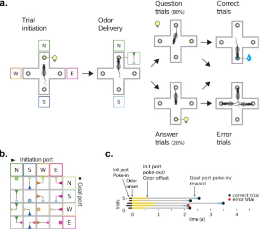
Figure 8 - Odor guided spatial navigation task 43
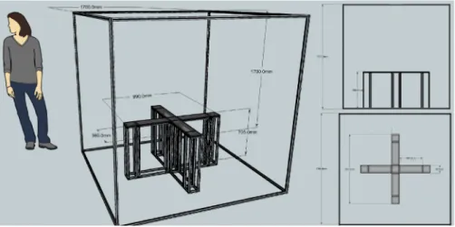
Figure 9 - Testing apparatus 47
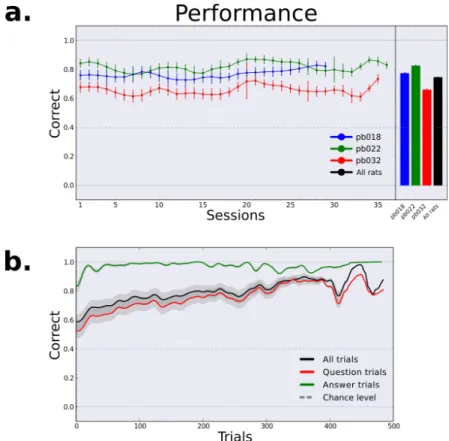
Figure 10 - Performance 49
Figure 11 - Action performance 51
Figure 12 - Error trials 52
Figure 13 - Movement time distributions 53 Figure 14 - Movement time distributions by action 54
Figure 15 - Velocity 55
Figure 16 - Trial history bias 56
Figure 17 - Tetrode placement 67
Figure 18 - Average population responses 70
Figure 19 - ROC @ Goal port in 71
Figure 20 - Significantly modulated cells 72 Figure 21 - Proportion of selective cells 73
Figure 22 - Action selective cells 74
Figure 23 - Proportion of selective cells - no back action trials 75 Figure 24 - Location selective cells 77 Figure 25 - Location selective cells tuning 78
Table of contents
Acknowledgments i
Resumo iii
Abstract v
Abbreviations list vii
Figure index viii
Table of contents ix 1 General Introduction 1 1.1 Chapter summary 2 1.2 Introduction 3 Orbitofrontal cortex 5 OFC Anatomy 5 OFC Function 7
From reversal learning to outcome expectancies 8
OFC and space 12
Final remarks 14
1.3 Bonsai 16
2 Free Choice Spatial Task 17
2.1 Chapter summary 18
2.2 Introduction 18
2.3 Materials and methods 22
Animal Subjects 24 Pre-handling 24 Surgery 24 Recovery 25 Water restriction 25 Training 26 Pharmacological Inactivation 27 Histology 28 Testing apparatus 28 Data analysis 29 2.4 Results 29 2.5 Discussion 34
3 Odor Guided Spatial Navigation Task 39
3.1. Chapter Summary 40
3.2 Introduction 40
3.3 Materials and methods 41
Behavioral Task 41 Animal Subjects 44 Training Protocol 44 Testing apparatus 46 Data analysis 48 3.4 Results 48 3.5 Discussion 57
4 Odor Guided Spatial Navigation Task OFC recordings 63
4.2 Introduction 64
4.3 Materials and methods 66
Surgery 66
Histology 66
Drive, Gold plating and Recording system 67 Event detection & Clustering 68
Data analysis 68 4.4 Results 68 General modulation 69 Feature selectivity 72 4.5 Discussion 79 Odor 79 Reward 81
Action & Location 81
Removal of Back trials 82
Locations 82 Conjunctive coding 83 5 General discussion 87 Final remarks 93 6 References 97 Supplementary figures 111
1 General Introduction
1.1 Chapter summary
General notes on the manuscript:This chapter features the general introduction to the relevant theoretical and conceptual framework that oriented our exploration of behavior and the orbitofrontal cortex (OFC). Chapter 2 revolves around an inactivation experiment in a simple free choice spatial task and its results which led us to further explore and develop a new task. The odor guided spatial navigation task and its behavioral results is the focus of Chapter 3. Chapter 4 will report the results of the neurophysiological recordings we performed in this task. Finally, the thesis will wrap up with a general discussion chapter and the bibliography.
With the exception of the present and last chapters, all other chapters will feature a Chapter summary that, just like this paragraph, will summarize and comment on the chapter. This summary will be followed by an Introduction where we will set up the different themes of each chapter followed by the Methods section that will begin with a description of the behavioral task. The subsequent Results section will exemplify the main findings of the experiment and finally, the Discussion section will summarize the results and bridge to the following chapter. For readability purposes, all bibliography will be presented in a final Reference Chapter. Overall the organization of the thesis revolves around behavior, not only conceptually but also practically. Consequently, Chapter 2, Chapter 3 and Chapter 4 are organized around the 2 behavioral paradigms developed during the thesis work.
1.2 Introduction
Imagine you just arrived at your grandmother’s house. As you walk in, the smell of her famous apple pie greets you, your stomach echoes a gurgle and your mouth salivates slightly. You say hello to everyone, embrace your grandparents and start talking, you haven’t seen each other for a while and you really want to spend some time with them and catch up. As you are talking however, in the back of your mind, this persistent image of apple pie grows to the point you’re trying to find a polite way of asking for some pie. Reading your mind (as they do), your grandma asks if you want some pie. “Of course! …”, you say smiling, and almost as to attone for the fact that you haven’t been listening for the last 30 seconds, you utter: “...don’t bother getting up I’ll fix myself a plate! Do you want some?”. Sure says grandpa, who's sitting on the couch. “None for me.” answers grandma while she gets up anyway to set the table for dinner. As you enthusiastically stand up with the image of your grandma’s apple pie etched in your brain, your stomach growls again, and you go... but where? You know there is pie in the house, you smelled it, you have some prior of where pies ‘live’ in general and some prior about where your grandma sets her pies to cool down. Maybe unsurprisingly, you decide to check the toilet seat in the bathroom, find the pie and fix a piece for you and grandpa.
The reader might, up to a point, have a similar story (details about relatives notwithstanding), and I’d also venture the guess that the last sentence was found to be somewhat strange. The only strange thing about the last sentence was the substitution of the location of the apple pie from ‘kitchen counter’ to ‘toilet seat in the bathroom’. This substitution is received as surprising only because an expectation about the location of the apple pie exists. Objects in the world have properties that usually
correspond to our sensory experience of them, they have a color, a shape, a size, a texture, a resistance, a temperature, a weight, an odor, and a taste. What objects also have, is a context. Context, unfortunately, is quite a generic word used by people with multiple backgrounds. From the arts and social sciences, to biology and physics, the definition of context can refer to very different things.
Journalism, that is arguably tasked with the accurate report of events, has used the 5 ‘W’ rule to write a news story, meaning: who, what, when, where and why. This is thought to be the best way to get an unbiased and accurate depiction of some event. A scientific experiment, is also a report of an event, and experimentalists, also worried with biases and concerned with accuracy, usually think of their experiments in similar terms: subjects (who), objects (what) and context (when, where and why). Temporal/rithmic information, spatial/locational information, as well as motivational context are critical criteria that should be considered when developing tasks and interpreting behavioral results.
As the reader might have gathered already, this thesis will focus on locations and although locations are not exactly a primary sensory experience, we hope our little story has demonstrated that they are an integral part of the description of an object when this object becomes a behavioral goal. So, while for the identity of an object its physical location might be superfluous, it becomes critical if this object needs to be acted upon in some way. During the decision and implementation of a goal-directed behavior, the spatial information about an object, as well as other ‘secondary properties’ like timing, are probably integrated with the sensory information that defines this object, somewhere in the brain (effectively ‘contextualizing’ the object). We propose, based on our own
results as well as previous studies, the orbitofrontal cortex (OFC) as the region that serves this function.
Our hope is that this thesis will contribute to the understanding of goal-directed behavior, spatial processing and the function of OFC in a spatial context and help pave the way for the emerging functional theory of OFC in the brain.
We will start by briefly introducing the OFC at the anatomical and functional levels introducing the relevant concepts, previous works and ideas about OFC that gave rise to this project.
Orbitofrontal cortex
The orbitofrontal cortex (OFC) in primates and orbital cortex in rodents refers to the ventral surface of the frontal lobe, it is called this way because of its close proximity to the eyes. It receives projections from a considerable number of other brain areas including visual, olfactory, somatosensory and visceral/gustatory cortices. Besides its many other functions that we will introduce in this section, OFC is interestingly considered the secondary gustatory cortex and around 8% of neurons respond to different gustatory stimuli and are sensitive to devaluation protocols (Thorpe et al., 1983; Nakano et al., 1984; Rolls et al., 1989, 1990).
OFC Anatomy
The OFC is the target of many different areas both directly and indirectly through the medial dorsal nucleus of the thalamus (Carmichael and Price, 1995a, 1995b). These projections that include but are not limited to: striatal, somatosensory, olfactory, and viscera inputs, carry sensory
and reward related information that can be integrated in the OFC. OFC also possesses medial-prefrontal and limbic reciprocal connections two major areas in decision-making, implicating it in related functions (Ongür and Price, 2000; Carmichael and Price, 1995a). While medial OFC shares reciprocal connection with the ventro-medial prefrontal cortex, central and lateral sections of the OFC receive reciprocal projections mainly from visceral afferents (Carmichael and Price, 1996; Ongur and Price, 2000). OFC’s connectivity pattern is largely consistent between species from rodents to primates (Krettek and Price, 1977a, 1977b, 1978; Ferry et al., 2000; Ongür and Price, 2000; Kondo et al., 2003, 2005; Price, 2007; Kondo and Witter, 2014). In contrast to primates that have granular and agranular prefrontal cortices (PFC), PFC in rats, and consequently OFC, is exclusively agranular (Ongür and Price, 2000). This fact poses as a limitation to the use of morphology to support comparisons of brain areas in different species. Homology between species can be thus asserted at the connective and functional level. Both of these criteria are currently under debate, in fact some go as far as questioning if rodents have a prefrontal cortex all together (Preuss, 1995; Uylings et al., 2003).
Nonetheless, connection similarities, of both inputs and outputs, have been reported especially pertaining to the caudal agranular OFC in primates and rodents (Croxson et al., 2005; Price, 2007). Furthermore, thalamic, amygdalar complex (especially baso-lateral amygdala), anterior hippocampus, hypothalamus and nucleus accumbens reciprocal projections show remarkable similarities (Deacon et al., 1983; Groenewegen, 1988; Carmichael and Price, 1995a, 1996; Haber et al., 1995; Cavada et al., 2000; Ongür and Price, 2000; Ramus et al., 2007; Mailly et al., 2013). Similar impairments are also observed in lesions studies to the amygdala and OFC (Jones and Mishkin, 1972; Gaffan and
Murray, 1990; Schoenbaum et al., 1999, 2000, 2002, 2003b; Schoenbaum and Setlow, 2001; Fellows and Farah, 2003; Pears et al., 2003; Wallis and Miller, 2003; Mariano et al., 2009). In fact, the strong reciprocal connectivity between baso-lateral amygdala and OFC has been hypothesized as contributing to the emotional and motivational aspects of learning (Davis, 1992; Holland and Gallagher, 1999; Schoenbaum et al., 2000; Baxter and Murray, 2002).
OFC Function
It is known that OFC lesions or inactivations during contingency reversals (reversal learning), strongly affect performance (Teitelbaum, 1964; Jones and Mishkin, 1972; Schoenbaum et al., 2002, 2003a; Bohn et al., 2003; Izquierdo et al., 2004). At the same time however, learning new stimulus-action associations is thought to be independent of OFC as the acquisition of new associations is not affected by these lesions (Schoenbaum et al., 2002; Chudasama and Robbins, 2003). This means that although OFC is not important for the initial stimulus-action associations per se, it becomes necessary if these previously learned associations need to be updated.
The Iowa gambling task (Bechara et al., 1994) has been used in humans to assess impairments in evaluating risk and future rewards. Subjects are asked to pick cards from a number of decks that can yield gains and losses. Losses are distributed in different amounts and probability in such a manner that, over time, some decks will be ‘good’ decks and some will be ‘bad’ decks yielding more losses than gains. Humans with OFC lesions performing this task choose decks with higher losses over time and demonstrate, what the authors called: “impairments in future consequences”. However, a later study (Fellows and Farah, 2005)
demonstrated that a slight modification in the experimental design was sufficient to remove the impairment previously observed. While the original task only presented rewards for the first 10 trials for each deck, this modification involved shuffling randomly the gains and losses since the beginning. Having only rewarded trials at the beginning was supposed to help subjects assess the statistics of the gains quickly. However, this practice resulted in the subjects learning the gains and then having to ‘reverse’ or update that learning once the losses started to appear. Moreover, in a series of studies using an analogue of the shuffled version of the Iowa gambling task for rodents (Zeeb et al., 2009; Zeeb and Winstanley, 2011, 2013), the authors also report that inactivating OFC causes no impairment in selecting the best option overall. These results suggest that the hypothesized lack of ability to evaluate future losses resulting from OFC lesions can be better explained as a deficit in the ability to update previously learned associations as assessed by reversal learning paradigms.
From reversal learning to outcome expectancies
Behaviorally, the inability to reverse or update previously learned associations, could be explained if OFC is required for inhibiting a learned response. Presumably, in order to learn something new, mapped to the same behavioral output, subjects need the ability to, first and foremost, inhibit the previously learned response. Indeed, OFC is necessary for animals to be sensitive to devaluation protocols (Critchley and Rolls, 1996; Gallagher et al., 1999; Izquierdo et al., 2004; Pickens et al., 2005; Plassmann et al., 2007; Roesch et al., 2009). However, multiple studies have reported OFC as not necessary for response inhibition (Schoenbaum et al., 2002, 2003a; Pickens et al., 2003).
Several results however, have suggested that the association between reversal learning and OFC was not the full story. Some of these results suggested a bigger picture for OFC beyond reversal learning. OFC has been shown to be embedded in a hierarchical network that is responsible for outcome identity (Keiflin et al., 2013) and outcome location reversals (Young and Shapiro, 2009) but not strategy switches (which are attributed to prelimbic/infralimbic cortex – PL/IL). Meaning that OFC would not be necessary to learn reversals, but sufficient to overrule other brain areas that had learned them.
The actual involvement of OFC in reversal learning altogether has also been questioned, at least in primates by Rudebeck and colleagues (Rudebeck and Murray, 2011; Rudebeck et al., 2013). These authors suggest that, in primates, the reversal effects previously observed were due to the removal of fibers of passage. In fact, instead of using the usual aspiration method, in their study they performed lesions to OFC using excitotoxic methods, which target specifically cell bodies, and fail to observe the reversal effects previously described. Temporal lobe and limbic system damage seem to reproduce reversal learning impairments in primates (Murray et al., 1998; Izquierdo et al., 2005; Chudasama et al., 2009) and, although one can also find the same projections from temporal and limbic areas to mainly the ventral and medial orbital areas, in rats (Carmichael and Price, 1995a, 1995b; Schmahmann et al., 2007; Kondo and Witter, 2014; Timbie and Barbas, 2014), the same observation made by Rudebeck and colleagues has, to our knowledge, yet to be reported.
Furthermore, if OFC neurons were to be responsible for the reversal impairments observed, one would predict that neurons that are sensitive to a particular reward would, upon reversal, either stop firing or reverse
their tuning to now represent the new reward. However, studies that compared OFC neurons with amygdala neurons in both rats and primates, reveal a much higher change in preference for amygdala neurons, whilst OFC neurons tend to maintain their preferred responses (Thorpe et al., 1983; Schoenbaum et al., 1999; Paton et al., 2006; Stalnaker et al., 2006).
Several more studies, show OFC to be important for more than reversal effects and together they contribute to paint a picture of a broader functional scope of OFC. Specifically, a considerable number of reports have surfaced implicating OFC in the representation of outcome properties and the representation of cues associated with specific outcomes. These properties include diverse features, amongst which: identity and taste (McDannald et al., 2011; Jones et al., 2012; Keiflin et al., 2013), size and economic value (Tremblay and Schultz, 1999; Schultz, 2000; Hikosaka and Watanabe, 2004; Padoa-Schioppa and Assad, 2006; Jones et al., 2012), uncertainty (Kepecs et al., 2008; Kepecs and Mainen, 2012; Lak et al., 2014; Zariwala et al., 2013), regret (Steiner and Redish, 2012, 2014), and spatial location (Corwin et al., 1994; Feierstein et al., 2006; Roesch et al., 2006). Reward prediction errors (Sutton and Barto, 1998) have also been shown to require OFC for proper computation (Takahashi et al., 2011) and the additivity and transitivity (or inferred values) properties in an economic value framework seem to be important factors that explain the modulation of firing rates of OFC neurons (Jones et al., 2012; Takahashi et al., 2013). Attempting to integrate these ever growing and incredibly varied results, several functional hypotheses of OFC have arisen. One such hypothesis postulates its role in facilitating behavioral and associative flexibility of downstream areas by encoding “outcome expectancies” (Schoenbaum
and Eichenbaum, 1995; Schoenbaum et al., 1998, 1999, 2003a, 2007). OFC’s involvement in reversal learning loss of fuction experiments would thus be a consequence of the inability to represent outcome expectancies or properties. This suggests that a more generic function for OFC should be considered as, possibly, an integrating information hub for outcomes, cues and context (Wallis, 2006, 2007; Mainen and Kepecs, 2009; Schoenbaum and Esber, 2010).
More recently, a more general role of OFC in decision-making and learning has been proposed. This proposal implicates OFC in the representation of a cognitive map (Tolman, 1949) of ‘task space’ (Wilson et al., 2014), and OFC would be responsible for learning and representing hidden states. The authors of this study used a series of model-free and model-based reinforcement learning (RL) models to revisit some of the classical results known from the loss of function OFC literature. Their hypothesis was that OFC would represent hidden states. Hidden states or, non-stimulus-bound states are posited in opposition to states that can be differentiated by some sensory stimulus that would act like a cue that informs the animal of the current state. In this view, inactivating OFC would result in an impoverished state space over which the RL agent had to learn. As it turns out, this simple manipulation was able to recapitulate a great number of classical OFC inactivation results. This result is particularly interesting because it links RL, specifically model-based RL (Sutton, 2012) to goal-directed behavior through a particular brain structure. A goal-directed action is defined in opposition to a habitual action and is an action performed on the basis of the consequences the action will cause rather than in response to a stimulus (Adams and Dickinson, 1981; Colwill and Rescorla, 1985, 1986). Goal directedness can be assessed experimentally by outcome devaluation
and contingency degradation (Dickinson, 1985; Dickinson and Balleine, 1994; Balleine and Dickinson, 1998). When planning a goal-directed action, the goal, or outcome desired, is not present and needs to be imagined in order to accurately implement a decision. Furthermore, because this outcome is a desired future state that is not immediately present it is effectively a hidden state. Outcomes, or goals, can be represented in the brain either as a categorical variable, where a population of neurons represent this category specifically, or as a vector of sensations, where the categorical property could be considered only if one knows the precise combination of sensations the animal is sensitive to. In this context, representing goals, consequences, outcome properties or hidden states is arguably equivalent.
Tolman’s cognitive map is thus reinterpreted from an actual spatial map to a more abstract state map, that might or might not have a particular relationship with physical space. Interestingly, however, the brain area that is essential for spatial navigation, the hippocampus (O’Keefe and Conway, 1978; Wilson and McNaughton, 1993) is also known to affect memory formation (Scoville and Milner, 1957; Squire, 2009), suggesting an intimate relationship between spatial variables, memory and goal-directed behavior.
OFC and space
This thesis will expand on previous results (Corwin et al., 1994; Feierstein et al., 2006; Roesch et al., 2006; Young and Shapiro, 2009) that implicate OFC in the coding of spatial, or spatial-like features in the context of navigation. A more detailed description of these studies can be found in Chapter 2 and Chapter 3 in the introduction and discussion sections.
So why would space be important? Kant, in the Critique of Pure Reason, described time as an a priori notion that, together with other a priori notions such as space, allows us to comprehend sense experience. It would follow that spatial variables, not only must be represented somewhere in the brain but should also be used as a fundamental cognitive anchor for our not-only-sensory experience. The most obvious case where spatial variables would be involved in decisions is in the case of spatial navigation.
Spatial processing is important for animals as they move about in the world, moreso for rodents that rely on foraging for survival. Rats, for example, live in intricate underground burrow systems (Pisano and Storer, 1948; Calhoun, 1963), and rely on exploration of their surroundings for food (Barnett, 2007). A delicate exploration-exploitation balance is important as rats will lower their probability of predation the less they explore but increase the probability of running out of resources if no exploration attempt is made (Charnov, 1976). Knowing where the food is and how to get there becomes paramount to properly allocate the correct amount of time and resources to exploiting one particular patch of resources, or exploring the environment to find another. Furthermore, the location, direction or just general area exploration efforts should focus on, should be informed by a cognitive spatial map of the animal’s surroundings. Lastly, in case of danger, the relative location of the animal’s home is fundamental in order to rapidly plan an escape route. Navigation can be accomplished using different types of cognitive strategies, the 2 extreme cases of which are called egocentric and allocentric navigation. The egocentric reference frame is centered on the subject and defines positions and orientations as a sequence of actions
relative to a single localizing cue, usually visual, that resets the initiation. Allocentric reference frames are centered on an area map and are built using a configuration of different cues where the subject is one of these cues (Dolins and Mitchell, 2010; Lihoreau, 2010). Different brain areas have been involved with one or the other type of navigation and because these are conceptual, extreme cognitive strategies, perhaps unsurprisingly, the neuronal substrates that enable them, have been found to have complex interactions and a somewhat mixed strategy (Iaria et al., 2003; Ekstrom et al., 2014). Nonetheless, several studies have found striatum, caudate nucleus and putamen to be important for egocentric navigation (Maguire et al., 1998; Rubio et al., 2012; Chersi and Burgess, 2015) whereas hippocampus and para-hippocampal regions have shown involvement in allocentric navigation (Hartley et al., 2003; Rubio et al., 2012; Chersi and Burgess, 2015).
Final remarks
Several pieces of evidence seem to implicate OFC in spatial processing, among them we find: OFC’s reciprocal projections to hippocampus (Carmichael and Price, 1995a); hippocampal involvement in allocentric navigation (Dolins and Mitchell, 2010; Lihoreau, 2010); OFC’s involvement in goal-directed behavior; the presence of spatial like features in OFC (Feierstein et al., 2006; Roesch et al., 2006); and the fact that OFC is required for allocentric navigation (Corwin et al., 1994). Furthermore, if the hippocampus is responsible for providing ‘contextual’ information to the rest of the brain (Moser et al., 2008) and, at the beginning of the chapter, we defined context as the where, the when, and the why, looking at location correlates in OFC would support the hypothesis that the hippocampus ‘contextualizes’ prospective sensory objects, at least in terms of location. In any case, whether OFC is
involved in spatial navigation, or the hippocampus is involved in providing contextual information to the representation of expected outcomes, the involvement of OFC in the representation of outcome expectancies would still hold as long as we consider locations as just another outcome expectancy (i.e. a property of a sought outcome). Examining OFC’s spatial properties becomes important especially considering the limitations of previous studies. While some studies that look at spatial properties of OFC (Corwin et al., 1994; Young & Shapiro, 2009) have an explicit navigational context, they are framed in terms of reversal learning and not of location representation in the context of outcome expectancies. Contrary to this, studies that have focused on outcome expectancies and spatial features (Feierstein et al., 2006; Roesch et al., 2006), used small behavioral boxes, with no navigational contextor demands, and don’t separate locations, direction, or actions. If OFC is involved in integrating spatial information with other outcomes expectations, effectively representing location as one of the outcome expectancies referred to above, then this representation, while relevant for learning, should be persistent even after learning.
Our proposal is thus, to investigate the representation of outcome locations in the OFC in overtrained animals performing a task that has a precise navigational context.
1.3 Bonsai
While thinking about the implementation of a navigational task, we rapidly decided that such an experiment would require a fast and customizable tracking system. While tools to this purpose are commercially available, their implementation was highly optimized for particular physical setups and didn’t allow low level control of parameters. Rapid and flexible prototyping of experimental designs is paramount to any exploratory endeavour at the basis of the development of a new behavioral paradigm. Considering this, in collaboration with Gonçalo Lopes, another PhD student, we started to develop our own video tracking system which rapidly evolved into a full fledged generic framework that processes data streams: Bonsai (Lopes, Bonacchi, et al., 2015). Bonsai has been published in Frontiers of Neuroinformatics and has been adopted by several labs around the world for, among other things, the integration of behavioral protocols, electrophysiological recordings and real-time video processing.
Bonsai: an event-based framework for processing and controlling data streams
The design of modern scientific experiments requires the control and monitoring of many parallel data streams. However, the serial execution of programming instructions in a computer makes it a challenge to develop software that can deal with the asynchronous, parallel nature of scientific data. Here we present Bonsai, a modular, high-performance, open-source visual programming framework for the acquisition and online processing of data streams. We describe Bonsai's core principles and architecture and demonstrate how it allows for flexible and rapid prototyping of integrated experimental designs in neuroscience. We specifically highlight different possible applications which require the combination of many different hardware and software components, including behavior video tracking, electrophysiology and closed-loop control of stimulation parameters.
2 Free Choice Spatial Task
Unpublished data
Author contributions: Bonacchi N. and Mainen Z.F. designed the studies. Bonacchi N. built the apparatus, ran the experiments, analyzed the data and wrote the manuscript.
2.1 Chapter summary
This chapter reports the rationale, implementation and results of the free choice spatial task inactivation experiment. This was our first attempt of introducing spatial locations as relevant decision variables for animals performing a decision-making task. We will start by introducing the historical and conceptual rationale behind the development of this task, present the results, and discuss the implications for the rest of the thesis.
2.2 Introduction
Behavioral tasks used to study OFC function never focused on spatial components, with few notable exceptions (Corwin et al., 1994; Feierstein et al., 2006; Roesch et al., 2006; Young and Shapiro, 2009). Even these exceptions were arguably not designed specifically to examine spatial representations in the context of navigation and location. For example, behavioral tasks in these studies generally did not explicitly parse out action versus direction.
At the time I joined the Mainen Lab, the task that was used was no exception. The two-alternative choice odor discrimination task (Uchida and Mainen, 2003; Kepecs et al., 2008) was designed in a relatively small behavior box and the task entailed the animals to remain mostly stationary when interacting with the apparatus. This task is a particular case of a 2 AFC (Alternative Forced Choice) task that uses as guiding stimuli a mixture of 2 odors where the relative concentration of the individual odors is used as a way of changing the difficulty of the choice on a trial by trial basis. This task is one particular example of a category
of tasks one might call sensory decision making tasks under uncertainty. These type of tasks are designed to look at sensory processing and usually add a source of uncertainty to the stimulus in order to manipulate its difficulty parametrically. This emphasis on the stimulus as the relevant decision variable as well as the trial by trial difficulty manipulations, allows the experimenter to build classical psychometric functions by measuring behavioral output variables like accuracy.
Although this task, as previously mentioned, is not optimized to study spatial features, a 2006 paper (Feierstein et al., 2006) used a pure odor variation of this behavioral paradigm, and was one of the first to describe spatial-like variables in OFC. The authors found OFC cells, appropriately called goal cells, that significantly changed their firing rate for particular goal locations. These cells fired both in the presence or absence of rewards, and to some extent independently from the action just performed. Finally, these cells fired for the same goal location even independently of stimulus identity when multiple stimuli were associated with the same reward location or direction. In other words, these cells seem to care about the goal location/direction but not: the presence of reward, the action performed or the stimulus that led the animal there. Furthermore, by looking at the choice moment and at the trial reinitiation moment, the authors were able to describe a set of cells that were selective for particular left or right actions. Nonetheless, we know hippocampal place cells and grid cells in the entorhinal cortex, demonstrate an increase in size and spacing of the associated place fields as one navigates dorso-ventrally (Sargolini et al., 2006; Brun et al., 2008; Stensola et al., 2012). If a place field like response in OFC is to be found – one could speculate that there could be an increase in place field and most probably a conjunctive aspect of place and other goal expectancies that are characteristic of OFC already. Given the reduced size of the behavior box and the proximity between pokes it
could very well be the case that the ‘action’ cells found by Feierstein and colleagues (2006) were not correlated with a left or right action per se but might have been representing locations of a wider place field. There is the possibility that the location/direction selective cells reported and the action cells were one and the same population sensitive to different size place fields.
Another study (Lak et al., 2014) using the same task, performed inactivations and found that the absence of a functioning OFC affected the time animals are willing to wait for a reward, both depending on trial difficulty and expected outcome. Most importantly for our purpose, rats could perform the task with no impairment in accuracy regardless of stimulus difficulty. This means that to “solve” the task, or more accurately, for implementing the initial decision of where to go given a particular stimulus, OFC was not being used. So, while OFC cells were found to be causally involved in the decision to stay or wait for a reward depending on the trial difficulty, they didn’t seem to be involved in the initial decision of where to go.
From these two studies we can conclude that OFC is not necessary to solve the two-alternative choice odor discrimination task. At the same, however, and in an apparent contradictory fashion, OFC seems to represent some spatial variables i.e., the location or direction of the goal. On these basis, we set out to explore a behavioral task, with a higher spatial component, where animals have to implement decisions that necessarily require OFC activity, i.e., we tried to find a task where OFC would be ‘used’ and therefore required to solve the task. If successful, and by inactivating OFC we find a behavioral effect, this alone would falsify the claim made by Feierstein and colleagues that OFC is
monitoring task variables but not involved in the decision per se. In any case, given all the above, some characterization of the spatial properties of OFC neurons seemed to be an interesting direction.
Considering that the goal cells Feierstein et al. (2006) reported could be OFC cells sensitive to outcome locations, we decided to introduce an obvious spatial component to the outcome. The easiest way of making space a relevant feature, was actually inspired from the discovery of grid cells (Fyhn et al., 2004; Hafting et al., 2005) where a simple increase in size of the recording arena was the key change from previous work that allowed for such discovery. This increase in space would also, possibly help, in teasing apart action selective cells reported previously, from location selective cells that cover more than one port.
Finally, considering the reversal learning literature, we decided that a change in contingencies would probably be helpful in engaging OFC especially if the reversal was in the spatial dimension.
With these things in mind we modified the two-alternative choice odor discrimination task in a number of significant ways:
1. We increased the size of the box to 1 m2
2. We located the initiation port on the opposing wall of the reward ports
3. We removed stimuli
4. We made the reward change places (spatial reversal)
With these modifications we hoped that goal location would become a more relevant feature. Firstly, by making the box’s footprint bigger and separating the pokes further apart; and secondly, by making the animal move from initiation port to reward port on every trial. Lastly, by removing stimuli, we hoped to make the animals focus on ‘where’ the
reward would be rather than on ‘what’ odor was delivered. Removing stimuli also had the added benefit of not needing special stimulus training and thus hopefully reduce training times. Finally, the introduction of a spatial reversal component of the reward would hopefully engage OFC and also contribute to highlight the reward location property.
2.3 Materials and methods
All experiments and procedures were approved by the Champalimaud Foundation Bioethics Committee and the Portuguese National Authority for Animal Health, Direcção-Geral de Alimentação Veterinária (DGAV). After having optimized a training protocol, we designed the testing phase to ascertain necessity by pharmacologically inactivating the OFC in 2 conditions: in the presence of an initiation port, and in its absence. The order of events was: cannula implantation surgery; water restriction; testing with no initiation port; and testing with initiation port.
Behavioral Task
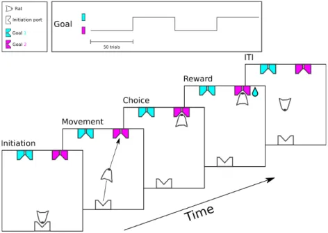
The task was initiated by poking in the lit initiation port located on one side of the box; rats could subsequently retrieve a drop of water from either the left or right reward ports located on the other side of the box as shown in Figure 1. No stimulus was delivered and water rewards switched location every fifty trials starting from a random side. A poke in the currently rewarded port was scored as correct and contrarily a poke in a non rewarded port was scored as an error (Figure 2 top panel).
Figure 1 - Free choice spatial task Task timeline and structure
Rats were exposed to two different task conditions: with or without initiation port. When the initiation port was not present, the task structure remained exactly the same but rats did not need to initiate a trial from the (now absent) initiation port. In fact, after a uniformly distributed random inter trial interval of 2 to 4 seconds a new trial was automatically initiated allowing them to stay at the rewarded port and just collect the rewards. In either condition, the best that any animal could do is one mistake per block-switch plus or minus one mistake if they happen to start from the wrong port. This is because there was no way to predict when the block would switch unless, of course, rats could count all the trials. From our data we concluded that rats don’t seem to be able to count to 50. As soon as they make the first mistake however, they should be able to know the location of the reward given only 2 reward ports were available.
Animal Subjects
A total of 15 Long-Evans male rats were used for the experiment. Data from all rats was used to optimize training protocol. 6 rats were submitted to the surgical implantation of guide cannulas, 4 of these rats were used for investigating OFC inactivation in the Free Choice Spatial Task. During both training and testing rats had ad libitum food and motivation was obtained by water restriction. Body weight was kept higher than 85%, other health indicators were also monitored daily for the duration of the experiment.
Pre-handling
In order to reduce the stress on the animal during surgery and during subsequent behavioral tasks, each animal is handled for 3-5 days before surgery. During this familiarization procedure the animals are placed for ~20 minutes in the behavioral box in which they will later undergo behavioral training and testing, in addition each animal is handled by the experimenter for ~10 minutes.
Surgery
All surgical procedures for cannulae implantation were carried out under aseptic conditions. Anesthesia was initiated and maintained with isoflurane inhalation at ~2% (1.5-3% ) in O2, at a flow rate of 0.5 lpm. Isofluorane adjustments were made according to paw withdrawal reflex. After craniotomy, guide cannulae (24-gauge Plastics One, Roanoke, VA) were stereotaxically implanted in each hemisphere and targeted using a rat brain atlas (Paxinos and Watson, 2006), 2 mm above OFC (AP: +3.72, ML: +/-2.5, DV: +4.2 from skull surface). Stainless steel stylets
were inserted into the guide cannulae to ensure patency (protruding 0.5 mm below the tip of the guide cannulae).
Recovery
Postoperative analgesia was administered, ketoprofen (5 mg/kg, IP) or Buprenorphine (0.05-0.1 mg/kg, SQ) and lidocaine was applied topically to the surgical site. To prevent infection, an antibiotic (0.3% gentamicin sulfate) is applied (once daily for 2-3 days) to the surgical site. To assist in rehydration, a prewarmed isotonic Lactated Ringer's solution may be given (15 ml/kg, SC). During the postoperative recovery (2-4 hrs), the animal is placed in an absorbent blanket on a microwavable heating pad. Body temperature and breathing rate are monitored during this period. The animal is then returned to its home cage and allowed to recover for at least 5 days. DietGel® Boost and Recovery purified high calorie dietary supplement from ClearH2O® is administered for 2 days and
water consumption is closely monitored, activity and appearance are used to assess postoperative recovery and as a warning sign for postoperative pain. Conditions such as non-healing of skin margins, wound infection, seizures or abnormal behavior (e.g. hyperactivity, stereotypy) were considered parameters indicating early endpoint.
Water restriction
For behavioral training and testing, the animal was placed on a water restriction schedule. Water restriction is always ceased at least 2 days before surgery and 5 days post surgery. During water restriction, food is continuously available and hydration is monitored by the CR animal facility staff, that checked water consumption and skin elasticity. Animals received water (>10 ml) during the behavioral session and 15-30 min of free water access at a variable time after the behavioral session.
Drinking time was adjusted to maintain 85-90% of free-drinking weight. During the weekend, animals are given free water. If the weight after the weekend exceeds the ‘free drinking weight’, the standard is adjusted to the weight of the day of beginning of the week. For immature animals, this is calculated by comparison to a cage of age-matched non water-restricted controls.
Training
The training protocol had two phases corresponding to the two testing conditions explained above. After at least one week of recovery from surgery, rats were placed on a water restriction schedule and behavioral sessions started. Rats were initially exposed to the behavior box for a short period of time ~15 minutes with no pokes lights or sounds in the box in order to recall their pre-handling experience. After all rats have gone through this recall the actual training started. A port was selected pseudo-randomly to be the first rewarded port and from there on every 50 trials the reward would switch to the other goal port. Each drop of water rewarded was preceded with an 80ms 3 KHz tone in order to cement a strong association between the tone and the reward. Poking in the non rewarded port was initially ignored. After animals had reached training criterion (>200 trials per session or 3-4 block switches, block size = 50 trials) pharmacological inactivation protocol of the first condition could begin. After this testing phase an initiation port was introduced in the opposing wall. Poking this port would yield the previously associated tone that would now work as a bridging stimulus. Rats were trained to poke in the initiation port before going to either reward port. After the correct port was found error trials were introduced, meaning an 80ms white noise burst was played if the animal chose the wrong port and no water reward. Animals remained in this new
configuration until they reached the same criterion with an acceptable performance (>80% correct). Once this criterion was obtained, the pharmacological inactivation protocol in this second condition started.
Pharmacological Inactivation
Animal subject were tested in two different conditions, both in the presence of only the goal ports and in the presence of an initiating port located on the opposite wall. The goal was to get one session a day interleaving inactivated sessions with vehicle sessions for 6 days yielding 3 vehicle and 3 muscimol sessions per subject, per condition. Inactivation and control sessions were counterbalanced. Temporary inactivation was achieved via localized injections of γ-aminobutyric acid (GABAA) receptor agonist muscimol (Sigma Alderich) under light anesthesia induced by 1-2% isoflurane (for about 6 min during which hind leg reflex never disappeared over the course of infusion). On each testing day the stylets were replaced with 33-gauge (Plastics One) injector cannulae protruding 2.0 mm below the tip of guide cannulae. One minute after proper bilateral placement of the injectors, muscimol (0.4µl of 0.125 µg/µl solution or 0.05 µg of muscimol) or sterile saline (0.9%; 0.4 µl) was injected over a 4 minute period at the rate of 0.1 µl/min on each side. Fluid was infused via 0.38 mm diameter polyethylene tubing (Intramedic, New York, NY) attached to the injector on one end and to two 2 ml Hamilton syringe (Hamilton, Reno, NV) on the other end. The syringes were driven with a syringe pump (Harvard Apparatus, MA). Injections were confirmed by monitoring the movement of mineral oil fluid in the tubing via a small saline bubble. After infusions were complete, the injector cannulae were left in place for 4 minutes and then replaced with stylets. Behavioral testing began about 45 minutes after infusion. (Martin and Ghez, 1999) showed that the maximal extent
of muscimol spread, using this procedure, was 1.5 to 2 mm within 10-20 minutes of injection.
Histology
Upon completion of behavioral tests, rats were injected with 0.4µl of evans blue solution to mark both the location as well as to give an indication of the spread of the muscimol injection. After 24 hours all animals were subsequently deeply anesthetized and then transcardially perfused with PBS and a saline 4% paraformaldehyde solution. Brains were removed, postfixed, and sectioned in 50 μm coronal slices using a fixed-tissue vibratome (VT1000S, Leica Instruments, Germany). Standard Cresyl Violet staining (Nissl staining) immunohistochemistry was performed in order to better visualize brain areas for cannula placement estimations.
Testing apparatus
The testing apparatus consisted of a custom built box with a footprint of ~1 m2 built with 20 mm aluminum rails and M4 screws with pre and post
assembly nuts from MISUMI Group Inc. 6mm thick white, high density polyethylene (HDPE) modules were used as ‘tiles’ to construct and apply the box’s surface. Sensors and actuators from IslandMotion™ were assembled using HDPE single modules of 120x120x6mm, which ensured the possibility of fastly and flexibly adapt the behavioral box to most possible configurations.
A Point Gray camera, Flea3 1.3 MP Color USB3 Vision (Sony IMX035) was used to monitor and track subject’s behavior. The Bonsai framework (Lopes et al., 2015) was used to interface with the camera. A real time linux finite state machine (RTLFSM) and Bcontrol (behavioral control system) were used to program the task.
Data analysis
All data were analyzed using custom scripts developed with the Python programming language and relevant libraries (Python Software Foundation. Python Language Reference, version 2.7 and 3.5. Available at http://www.python.org).
2.4 Results
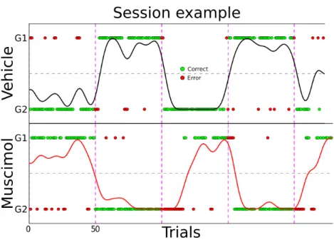
We found that the inactivation of OFC during this task yielded an impairment in the recovery of performance after a block switch as compared with vehicle sessions. This effect was visible at the single session level where rats tended to make more errors after a block switch in inactivated sessions as shown in Figure 2.
Figure 2 - Free choice spatial task raw
Raw data example session for one muscimol (bottom panel) and one vehicle (top panel) session. Red and green dots represent error and correct single
trials; black and red curves are the smoothed local averages using a Gaussian kernel.
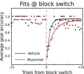
The average performance aligned at a block switch (Figure 3 left panel) for the comparison of the first vehicle session with the first muscimol session of an example rat also shows this difference as a slower recovery of performance after a block switch. This effect, although smaller, was still present in the average across sessions (Figure 3 right panel).
Figure 3 - Goal accuracy aligned at block switch
Average goal accuracy aligned at block switch for example session (left) and average session (right); black and red curves represent vehicle and muscimol sessions respectively; dashed lines are standard error of the mean.
This effect was only present in the initiation port condition. Comparing Figure 3 its equivalent in the condition where the initiation port was not present (Figure 4) we find no effect of OFC inactivation if rats are allowed to stay at the rewarded port and switch whenever the water stops coming.
Figure 4 - Goal accuracy aligned at block switch - no initiation port Average goal accuracy aligned at block switch for example session (left) and average session (right); black and red curves represent vehicle and muscimol sessions respectively; dashed lines are standard error.
To quantify this delay in recovery of performance we performed exponential fits (example in Figure 5) for all rats and all sessions using:
f ( x)=−e
−bx+
c
Where b was the free parameter and c was fixed to be 90% of the mean performance pre block switch. These fits yielded a consistent difference in rate between muscimol and vehicle conditions. Inactivated sessions almost always had a lower rate than vehicle sessions (Figure 6 right panel).
Figure 5 - Example fit
Example of exponential fits of accuracy data; muscimol sessions in red and vehicle sessions in red
This effect is greatest if one compares the first inactivated session with the first vehicle one (colored lines in Figure 6 left panel) and diminishes with following comparisons. The only exception was in the case of one particular subject (cyan line in Figure 6 left panel) which upon histological verification was found to have had an error in targeting mostly in D/V positioning of one of the cannulas (Figure 7 left hemisphere). Figure 6 right panel shows all the fitted rate values for all muscimol sessions plotted against the vehicle sessions. Most rats fall beneath the unity line indicating a lower fitted rate for muscimol sessions than for vehicle ones.
Figure 6 - Rate parameter comparisons
Left panel: Comparison of vehicle and muscimol values for the rate of the fitted exponential. Vehicle sessions in black, muscimol sessions in red. Colored lines underline the first session comparison for every subject. Right panel: Muscimol sessions fitted rate parameter as a function of Vehicle sessions, Colors represent individual subjects. Error bars are standard deviation; Markers with error bars are the average parameter value per subject.
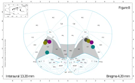
Figure 7 shows the placement of the tip of the cannulas after histological examination. All anterio-posterior measurements were estimated from the ubiquitous Paxinos and Watson’s The Rat Brain in Stereotaxic Coordinates, which unfortunately describes the average Wistar-Kyoto rat brain and not the Long-Evans strain. As a result of the slight differences between these two species although the aimed A/P (Anterior / Posterior) target was 3.72 mm after histological examination of the subject’s brains we found that we consistently hit A/P 4.2 mm. Because this atlas shows the average brain, it is understandable that the further away from the center (interaural zero) one targets, the bigger the error. These coordinates were kept throughout the study.
Figure 7 - Cannulae placement
Diagram of cannulae placement after histological examination, OFC target areas in gray; different colors represent different subjects.
2.5 Discussion
Two main conclusions can be drawn from this set of experiments:
We can conclude that inactivating OFC in these task conditions causes a decrease in performance, leading us to believe that OFC is necessary to implement the choice of where to go. The fact that this impairment is aligned to the block switches can be interpreted as evidence of an impairment in selecting between two different and opposing actions based on reward history and not a result of general apathy, confusion or other motor effects caused by the inactivation. This by itself would point to the reversal of reward locations as being a significant behavioral factor. However, although a reversal component is present and probably a factor, a priori we would expect to be outside of what is classically considered reversal learning as the reward’s spatial reversals have been pre-trained and rats have extensive exposure to the task’s statistics. The learning experiment we did not do, would have been to compare how
long animals take to switch location with or without OFC function. In this case however, the number of animals tested would have to be much larger in order to compare between rats and moreover, if this hypothetical experiment would have worked we wouldn’t have learned anything new and if it failed we would have had no way of knowing why. Secondly, comparing the inactivations in the two different conditions (with and without initiation port) we conclude that one of the relevant behavioral changes seems to be the movement from initiation port to reward port. The difference between the left panels of Figure 3 and Figure 4 is striking and seem to imply some change in the nature of the task. Just by making the animals move through space, by making them ‘go’ to the reward port ~1m away, animals seem to be entering a different state, maybe engaging the navigational system that cares about locations and trajectories and specifically goal locations.
Corwin et al. (1994) in fact, reported that electrolytic lesions to VLO (ventrolateral orbital cortex) impact learning allocentric but not egocentric navigation tasks. In this 23 year old study, animals were tested in two different tasks: the cheeseboard task and the adjacent arm maze task (Kesner et al., 1989); these tasks accentuate the importance of allocentric spatial localization and egocentric spatial lateralization respectively. Latencies to reach reward were significantly higher in VLO lesioned animals when compared with sham controls only in the cheesboard task and not in the adjacent arm maze task, leading the authors to conclude that OFC is necessary for allocentric navigation. One possible explanation of OFC’s involvement in allocentric navigation could be related to its involvement in the representation of spatial locations. In fact, to plan an allocentric action it is necessary to represent