Paula Cristina Paulo Videira da Silva
DIFFERENTIATION, DEVELOPMENT AND GROWTH OF THE
BLACKSPOT SEABREAM (
Pagellus bogaraveo) MUSCLE
Dissertação de candidatura ao grau de Doutor
em Ciências Biomédicas submetida ao Instituto
de Ciências Biomédicas Abel Salazar da
Universidade do Porto.
Orientador – Prof. Doutor Eduardo Jorge Sousa
da Rocha
Categoria – Professor Catedrático
Afiliação – Instituto de Ciências Biomédicas Abel
Salazar da Universidade do Porto
Co-orientador – Profª Doutora Luísa Maria
Pinheiro Valente
Categoria – Professor Associado
Afiliação – Instituto de Ciências Biomédicas Abel
Salazar da Universidade do Porto
Paula Cristina Paulo Videira da Silva
DIFFERENTIATION, DEVELOPMENT AND GROWTH OF THE
BLACKSPOT SEABREAM (
Pagellus bogaraveo) MUSCLE
Dissertation for applying to a Doctor degree in
Biomedical Sciences submitted to the Institute of
Biomedical Sciences Abel Salazar
Supervisor – Prof. Doctor Eduardo Jorge Sousa
da Rocha
Category – Full Professor
Affiliation – Institute of Biomedical Sciences Abel
Salazar, University of Porto
Co-supervisor – Profª Doctor Luísa Maria
Pinheiro Valente
Category – Associated Professor
Affiliation – Institute of Biomedical Sciences Abel
Salazar, University of Porto
Esta Tese é dedicada aos meus adorados filhos
This Thesis is dedicated to my dearest children
No cumprimento do disposto no Decreto-Lei nº 216/92 de 13 de Outubro, declara-se que a autora desta Tese participou na concepção e na execução do trabalho experimental que esteve na origem dos resultados apresentados, bem como na sua interpretação e na redacção dos respectivos manuscritos.
Nesta tese inclui-se ainda quatro artigos científicos publicados em revistas internacionais resultantes de uma parte dos resultados obtidos no trabalho experimental, referenciado como:
Silva, P., Valente, L. M. P., Galante, M. H., Andrade, C. A. P. Monteiro, R. A. F. & Rocha,
E. (2009). Dietary protein content influences both growth and size distribution of anterior and posterior muscle fibres in juveniles of blackspot seabream (Pagellus
bogaraveo, Brunnich). Journal of Muscle Research and Cell Motility. Em Publicação. Silva, P., Valente, L. M. P., Olmedo, M., Galante, M. H., Monteiro, R. A. F. & Rocha, E.
(2009). Hyperplastic and hypertrophic growth of lateral muscle in blackspot seabream Pagellus bogaraveo from hatching to juvenile. Journal of Fish Biology 74, 37-53.
Silva, P., Rowlerson, A. M., Valente, L. M. P., Olmedo, M., Monteiro, R. A. F. & Rocha, E.
(2008). Muscle differentiation in blackspot seabream (Pagellus bogaraveo, Brunnich): histochemical and immunohistochemical study of the fibre types. Tissue
and Cell 40, 447-458.
Silva, P., Andrade, C. A. P., Timóteo, V. M. F. A., Rocha, E. & Valente, L. M. P. (2006).
Dietary protein, growth, nutrient utilization and body composition of juvenile blackspot seabream, Pagellus bogaraveo (Brunnich). Aquaculture Research 37, 1007-1014.
In compliance with what is stated in Decret-Law nº 216/92 of October 13th, it is hereby declared that the author of this thesis participated in the creation and execution of the experimental work leading to the results shown, as well as in their interpretation and the writing of respective manuscripts.
This thesis also includes three scientific papers published in international journals originating from part of the results obtained in the experimental work referenced to as:
Silva, P., Valente, L. M. P., Galante, M. H., Andrade, C. A. P. Monteiro, R. A. F. & Rocha,
E. (2009). Dietary protein content influences both growth and size distribution of anterior and posterior muscle fibres in juveniles of blackspot seabream (Pagellus
bogaraveo, Brunnich). Journal of Muscle Research and Cell Motility. In Press.
Silva, P., Valente, L. M. P., Olmedo, M., Galante, M. H., Monteiro, R. A. F. & Rocha, E.
(2009). Hyperplastic and hypertrophic growth of lateral muscle in blackspot seabream Pagellus bogaraveo from hatching to juvenile. Journal of Fish Biology 74, 37-53.
Silva, P., Rowlerson, A. M., Valente, L. M. P., Olmedo, M., Monteiro, R. A. F. & Rocha, E.
(2008). Muscle differentiation in blackspot seabream (Pagellus bogaraveo, Brunnich): histochemical and immunohistochemical study of the fibre types. Tissue
and Cell 40, 447-458.
Silva, P., Andrade, C. A. P., Timóteo, V. M. F. A., Rocha, E. & Valente, L. M. P. (2006).
Dietary protein, growth, nutrient utilization and body composition of juvenile blackspot seabream, Pagellus bogaraveo (Brunnich). Aquaculture Research 37, 1007-1014.
Agradecimentos _______________________________________________________ 3 Acknowledgements ____________________________________________________ 7 Abstract _____________________________________________________________ 13 Resumo ______________________________________________________________ 17 Résumé _____________________________________________________________ 21 General Introduction ___________________________________________________ 25 The blackspot seabream, Pagellus bogaraveo (Brunnich) ________________ 27 Muscle morphology and fibre types in teleost fish ______________________ 30
Slow-red muscle fibres ___________________________________________ 31 Fast-white muscle fibres __________________________________________ 33 Intermediate-pink muscle fibres ____________________________________ 34
Methods used to study muscle growth _______________________________ 34 Fish muscle growth _______________________________________________ 36
Hypertrophy ___________________________________________________ 36 Hyperplasia ____________________________________________________ 37
Effect of intrinsic factors on muscle growth ___________________________ 40
Genetic effects _________________________________________________ 40 Hormonal control ________________________________________________ 41
Effect of extrinsic factors on muscle growth ___________________________ 41
Diet __________________________________________________________ 41 Temperature ___________________________________________________ 42 Exercise ______________________________________________________ 44
References ______________________________________________________ 44
Chapter II - Expression of the myosin light chains 1, 2 and 3 in the muscle of
blackspot seabream (Pagellus bogaraveo, Brunnich) during development ______ 93
Chapter III – Ultrastructural observations and morphometric data on the
muscle from blackspot seabream (Pagellus bogaraveo, Brunnich) _____________ 109
Chapter IV - Hyperplastic and hypertrophic growth of lateral muscle in the
blackspot seabream Pagellus bogaraveo (Brunnich) from hatching to juvenile ____ 125
Chapter V - Influence of temperature on muscle fibre hyperplasia and
hypertrophy in larvae of blackspot seabream (Pagellus bogaraveo, Brunnich) ___ 151
Chapter VI - Dietary protein content influences both growth and size
distribution of anterior and posterior muscle fibres in juveniles of blackspot
seabream Pagellus bogaraveo (Brunnich) _________________________________ 175
Chapter VII - Dietary protein, growth, nutrient utilization and body composition of juvenile blackspot seabream Pagellus bogaraveo (Brunnich) _______________ 203
Agradecimentos
Estes cinco anos de doutoramento, os últimos três em paralelo com actividade docente, foram apenas mais uma etapa de um percurso que teve o seu começo no mesmo ano em que o Mundo assistiu ao início de mais um milénio. Na altura, ainda a viver na cidade que me viu nascer (Vila Real) e a trabalhar na Universidade de Trás-os-Montes e Alto Douro (UTAD), a minha Alma Mater, eu andava indecisa sobre as acções a tomar, pois acreditava que tinha encontrado na Ciência a minha motivação, mas não sabia o que fazer para consegui-la levar a cabo. Foi a Professora Doutora Luísa Valente e o Professor Doutor Emídio Gomes que me indicaram qual o caminho que devia seguir e que me ensinaram que para o fazer com êxito temos de ser perseverantes e, sobretudo, ter confiança em nós próprios. Pelo incentivo, ajuda e amizade eu expresso a ambos o meu muito obrigada. Um agradecimento especial para a Professora Doutora Luísa Valente que, para além do incentivo, me acompanhou nesta caminhada sempre de uma forma muito presente, preocupada e amiga, primeiro como orientadora de Mestrado e depois como co-orientadora desta Tese.
A propósito de uma colaboração num projecto, foi também a Professora Doutora Luísa Valente que me apresentou o Professor Doutor Eduardo Rocha. Desde cedo fiquei encantada com a sua capacidade intelectual e de trabalho e, como tal, quando decidi fazer o meu doutoramento fora da UTAD para explorar novas realidades científicas, não hesitei em convidá-lo para ser meu orientador. E o primeiro agradecimento que publicamente lhe quero fazer é o facto de tão, prontamente, me ter dito que sim. Foram cinco anos de um excelente convívio profissional e pessoal onde diariamente renovei a minha muito agradável surpresa inicial. E mais importante do que a sua sabedoria é a benevolência com que transmite os seus vastos conhecimentos. Muito obrigada por tudo e faço votos para que possamos continuar a trabalhar juntos.
Para fazer o meu primeiro ensaio experimental, tive que atravessar parte do Oceano Atlântico e ir para o Centro de Maricultura da Calheta, na Madeira. E se do ponto de vista profissional tudo correu dentro do esperado, isso deveu-se à preciosa ajuda do Doutor Carlos Andrade e do Doutor Timóteo Viriato, a qual quero muito sinceramente agradecer. No entanto, não foi fácil estar doze semanas separada dos que amo, principalmente dos meus filhos, como disse Fernando Pessoa sob o heterónimo de Barão de Teive: “Descobri, pela falta, como se descobre a valia de tudo, que a afeição me era necessária; que como o ar, se respira e não se sente”. Durante todo o tempo em que permaneci na Madeira não a consegui ver como um jardim, mas senti o perfume das suas flores, tinham o cheiro da amizade que fiz com a Piedade e o Álvaro que tanto me ajudaram a suavizar as saudades que eu sentia de casa.
De regresso ao Porto, iniciei trabalhos no Instituto de Ciências Biomédicas de Abel Salazar (ICBAS). Não quero deixar passar este momento sem manifestar a minha homenagem e gratidão a esta instituição pela sua filosofia científica e pedagógica da procura por manter o espírito da interdisciplinaridade e que por isso tão bem sabe receber quem de fora a procura. E se o ICBAS tem sido o meu lar, todos que trabalham no Departamento de Microscopia, principalmente no Laboratório de Histologia e Embriologia, têm sido a minha outra família. O chefe desta família, o Senhor Professor Doutor Rogério Monteiro, é um sábio pelo seu espírito inteligente, de carácter único e grandioso, firme, infalível, subtil, ubíquo, lúcido, amante do bem e amável para com os outros. Neste momento tão importante da minha vida queria apenas dizer que lhe serei eternamente grata por tudo o que fez por mim e que é um enorme privilégio poder conviver com o Sr. Professor. Desta grande família permitam-me ainda que destaque outra pessoa, que bem pode ser considerada como a matriarca da mesma. Falo, obviamente, da Sra. D. Helena Galante que como qualquer boa “mãe” a todos sabe ajudar e amar. Não sei como recompensá-la por toda a ajuda prestada no laboratório. Quero apenas que saiba que sou uma mulher mais feliz porque gosto de si. A todos os outros colaboradores do Laboratório de Histologia e Embriologia deixo aqui esta pequena e singela mensagem: ”Neste dia especial, quero a Deus agradecer, esta família sem a qual eu não conseguia viver”.
Na referida filosofia da interdisciplinaridade seguida pelo ICBAS são elementos chave os docentes que dela fazem parte, alguns dos quais merecem da minha parte um agradecimento especial. Ao Professor Alexandre Lobo da Cunha, pela amabilidade que teve em aceitar pertencer à minha comissão de acompanhamento e por todas as ocasiões em que tive oportunidade de partilhar da sua, sempre, bem-humorada companhia. Ao Professor Doutor Luís Baldaia estou grata pela sua preciosa ajuda na tradução do resumo desta Tese para Francês. Gostaria ainda de agradecer ao Professor Doutor Rui Henrique (Director do Serviço de Anatomia Patológica, do Instituto Português de Oncologia, IPO, no Porto) pela disponibilização do crióstato necessário em uma das etapas deste projecto e principalmente pela sua amizade.
Este é capaz de ser um dos doutoramentos realizados no ICBAS com maior número de viagens e quiçá quilometragem, pois para além da já referida viagem à Madeira e das viagens realizadas entre Vila Real e o Porto (cinco dias por semana durante 3 anos), muitas foram as vezes que me desloquei a Vigo para efectuar colheitas. Quero agradecer a todos os funcionários do Centro Ocenográfico de Vigo que sempre com tanta alegria e carinho me receberam, em especial à Doutora Mercedes Olmedo e à Doutora Blanca Álvarez-Blázquez, sem a ajuda das quais não seria possível a realização das referidas colheitas. Viajei ainda até ao Algarve onde fui muito bem recebida pela Professora
Doutora Deborah Power e pela Doutora Nádia Silva que me ajudaram a dar os primeiros passos pela área da Histologia Molecular (através do uso da hibridização in situ) e cujos ensinamentos foram de tal maneira cativantes que me estimularam a querer explorar melhor esta área. Mas como em Ciência devemos esforçar-nos por trabalhar sempre com os melhores, ainda fiz mais uma viagem, desta feita a Londres para trabalhar no King’s College com a Professora Doutora Anthea Rowlerson, uma referência mundial em estudos de músculo de peixe, a quem muito tenho que agradecer pois foi a pessoa que mais me ensinou sobre a musculatura de peixe e sobre as suas técnicas de estudo qualitativo. Foi uma honra ter tido os seus ensinamentos, baseados numa já longa e internacionalmente reconhecida carreira científica dedicada ao músculo, e um prazer poder ter desfrutado da sua companhia.
Esta etapa do meu percurso não teria sido possível sem financiamento, por isso gostaria também de agradecer à Fundação para a Ciência e Tecnologia (FCT) a atribuição da bolsa de doutoramento (SFRH-BD-14068-2003) e o financiamento através do projecto POCTI/CVT/39239/2001) e ainda do financiamento plurianual dos Professores Doutores Rogério Monteiro e Eduardo Rocha, enquanto investigadores do Centro de Investigação Marinha e Ambiental – Laboratório Associado, via Laboratório de Estudos Celulares e Moleculares.
Uma palavra ainda de agradecimento e amizade à Maria da Graça pela forma sempre divertida como me ajudou a melhorar o meu Inglês.
E se é ao abrigo umas das outras que as pessoas vivem, eu tenho sido abrigada pelo carinho de muitas das quais tenho que destacar as amigas Dolores Resende, Paula Lemos, Alice Andrade e Helena Andrade.
Um agradecimento especial a toda a minha família em particular à minha irmã Anabela, companheira desta e de outras viagens. Citando alguém que todos conhecemos (Albert Einstein): “Há só duas maneiras de estar na vida. Uma é pensando que nada é um milagre. Outra é pensando que tudo é um milagre”. Quero que saibas que tu és sem dúvida um dos milagres da minha vida. Aos meus pais Alberto e Maria Rosa quero agradecer por me terem educado de acordo com princípios elevados; por me terem ajudado na busca do meu “verdadeiro eu” de uma forma profunda e vital; por me terem advertido que devo correr riscos, porque o maior risco da vida é nada arriscar e que a cada desafio devo sempre olhar para o alto da montanha e não deixar que nada mais baixo do que a montanha me distraia. Muito obrigada por estarem sempre do meu lado e por carregarem comigo ao colo nos momentos mais difíceis da minha vida.
E porque os últimos são sempre os primeiros, a minha última palavra de agradecimento é para com o meu marido Alfredo José e filhos Ana Rita e Manuel João. Não vos quero pedir desculpa pelos momentos em que estive ausente, mal-humorada, cansada e sem
paciência, porque as desculpas não se pedem, evitam-se, e eu de facto não as consegui evitar, quero apenas vos agradecer por existirem na minha vida. Acreditem, este só é um dos momentos mais felizes para mim porque o posso partilhar convosco.
Acknowledgements
The past five years devoted to my Doctoral Thesis, in parallel with regular teaching duties from the second year on, were but a further stage on a long road, which started exactly at the time the World entered its new millennium. At that particular moment in time, still living in Vila Real, my birthplace, and working at the University of Trás-os-Montes and Alto Douro (UTAD), my Alma Mater, I was feeling somewhat confuse about which decisions should I take since I truly believed to have found my working interest in Science, although, I did not know, then, how to pursue such a goal. Professor Luísa Valente and Professor Emídio Gomes, with their wisdom and discernment, showed me the right path and taught me that to succeed in life one has to be persevering and self-assured. For their incentive, help and friendship, I express my sincere gratitude. A special thank to Professor Luísa Valente who, besides her continuous encouragement, was an ever present and friendly role model, first as Master degree supervisor and later as Doctoral degree co-supervisor. It was the also Professor Luísa Valente who introduced me to Professor Eduardo Rocha whose amazing intellectual and working abilities captivated me immediately. As a result, when I decided to make for my Doctoral Thesis outside the UTAD, in order to explore new scientific realities, I did not hesitate for a single moment to invite him to be my supervisor. And my first public expression of gratitude is mainly due to the fact that he accepted that invitation so promptly. We have had five years of an excellent professional and personal relationship during which my initial very pleasant surprise was daily renewed. More important than his outstanding knowledge is, no doubt, the benevolence he shares it with others. I am very grateful for everything and sincerely hope we may continue to work together.
In order to carry out my very first experiment, I needed to partially cross the Atlantic Ocean and visit the Calheta Mariculture Centre, in the Madeira Island. If professionally all went according to the expectations, this was no doubt due to the help and collaboration received from Dr. Carlos Andrade and Dr. Timóteo Viriato to whom I am deeply indebted. It was not easy, though, to be far from those I loved, mostly from my children, for 12 long weeks. And, quoting our poet Fernando Pessoa, under the heteronym of Barão de Teive: "I discovered, due to absence, how every single thing is truly valued, that affection was absolutely essential to me; such as the air we breathe but do not feel or touch". During my whole stay in Madeira I could not perceive the island as a garden but I definitely enjoyed the perfume of the blooming flowers. It was the scent of my friendship with Piedade and Alvaro who helped mitigate my ever present "saudades".
Back to Oporto, I started to work at the Institute of Biomedical Sciences of Abel Salazar (ICBAS). I most definitely wish to express my gratitude and respect to this institution for its
scientific and pedagogic philosophy regarding its continuous efforts aimed at keeping the interdisciplinary spirit alive and thus being able to welcome so warmly those who address it. If the ICBAS has been my home for the past few years, all those who work in the Department of Microscopy, mostly in the Laboratory of Histology and Embryology, have been my other family. The chief of this family - Professor Rogério Monteiro - is a true wise scholar, due to his intelligence, unique and grandiose character, steady, infallible, subtle, ubiquitous, lucid, a true lover of the good and kind to all. In this particular moment, so very important in my life, I would like to express him my immense gratitude for everything he has done and to say how privileged and honoured I feel to know him. Another person of extreme relevance is, no doubt, the person who may well be considered the family’s matriarch. Obviously, I mean Ms Helena Galante who, as any good mother, knows how to love, care and help everybody. I can never repay you for all your help in the lab. I want you to know, at least, that loving and respecting you made me a happier woman. To all the remaining collaborators of the Laboratory of Histology and Embryology I would like to leave this short and heartfelt message: “On this special day, I wish to thank God, for I would never live without this sweet, caring group.”
Key elements to the above mentioned interdisciplinary philosophy followed by the ICBAS are the members of its teaching staff, and some deserve a very special thank from me. Thus, Professor Alexandre Lobo da Cunha for having so graciously accepted to be part of my Doctoral supervising commission and for his always pleasant and good-humoured company. Professor Luís Baldaia my gratitude for his priceless help in the translation of the Thesis abstract into French. Professor Rui Henrique (Director of the Pathologic Anatomy Service of the Portuguese Oncology Institute, IPO, in Porto) for making the indispensable cryostat available and, most of all, for his friendship.
My Doctoral Thesis may be the one of those with more journeys and perhaps the highest kilometre-consuming that ever took place at ICBAS. Besides my already mentioned flight to Madeira and the regular trips between Vila Real and Oporto (5 days a week during a 3 year period), many were the times I drove to Vigo/Spain for sample gathering. I am under very special obligation to all the staff at Vigo Oceanographic Centre for its always warm and friendly welcome. Special thanks to Dr. Mercedes Olmedo and Dr. Blanca Álvarez-Blázquez who made the gathering of such samples possible. I travelled to Algarve too, and I was always most welcomed by Professor Deborah Power and Dr. Nádia Silva, who initiated me in the area of Molecular Histology (using in situ hybridization methodology) and whose attractive teachings fuelled me into exploring this area further. It is a well-known fact that in Science one should always dream reaching the top and work with the best. So, I packed my suitcases again and went off to London, going now to work at King's College with Professor Anthea Rowlerson, a world reference in fish muscle, to whom I am
extremely grateful as she was the person who taught me the most about the subject, offering not only her immense experience but even her personal protocols. Again, it was an honour and a pleasure to have benefited from both her company and knowledge, based on a long and internationally recognized scientific career dedicated to muscle. This specific stage of my chosen course would not be possible without financing. I would like, therefore, to express my gratitude to the Science and Technology Foundation (FCT) for the scholarship (SFRH-BD-14068-2003) and respective grant (POCTI/CVT/39239/ 2001) and also to FCT pluriannual projects attributed to the Professors Rogério Monteiro and Eduardo Rocha, as researchers of Centre for Marine and Environmental Research – Laboratory Associated, via the Laboratory of Cellular and Molecular Studies.
A word of thanks and friendship to Maria da Graça who helped me to improve my English language, in an ever amusing and unconventional way.
And if people shelter and care for each other during their lives, I have been lucky enough to be sheltered by the loving care of so many. I want to name my dear friends Dolores Resende, Paula Lemos, Alice Andrade and Helena Andrade.
A very special thanks to my family in general and, in particular, to my sister Anabela, my fellow companion of this and other adventures. Quoting someone we all know (Albert Einstein): "There are only two ways to live one’s life: Thinking that nothing is a miracle or believing that everything is a miracle." I want you to know that you are, no doubt, one of my life’s amazing miracles. I also wish to thank my parents Alberto e Maria Rosa, for having brought me up according to their high moral values; for having helped me in the continuous search for my real self in a deep and vital way, and for advising me that risks are to be taken since the highest risk in life is to never risk anything. I learned from you that each challenge should make me look to the mountain top, not letting that smaller or meaner things distract me from my chosen path. Thank you very much for having been there whenever I needed and for loving and helping in the most difficult times of my life. Last but not least, my very final thank is for my husband Alfredo José and my children Ana Rita and Manuel João. I do not apologize for those moments when I was absent, ill-humoured, tired or irritable as apologies should not be made. They should instead be avoided; in fact, I was unable to avoid them. I just want to thank you all for being part of my life. Please, believe me when I say that this is one of the happiest moments in my life, it is because I may share it with you.
Abstract
This Thesis covers the muscle differentiation and development in the blackspot seabream (Pagellus bogaraveo, Brunnich), a fish under consideration as a potential candidate for Mediterranean aquaculture. Firstly, we used a panel of myosin isoform-specific antibodies, as well as histochemical staining for myosin ATPase and for SDH enzyme activity (a marker for mitochondrial content and oxidative metabolism), to identify developmental transitions in myosin expression in the muscle of blackspot seabream, from hatching to juveniles and adult. Secondly, we used in situ hybridization to characterise the spatial and temporal expression of embryonic myosin light chain 1 (MLC1), 2 (MLC2) and 3 (MLC3) gene expression during the fish early life. The combination of these two approaches contributed to understanding the differentiation of blackspot seabream muscle during development. Overall, these studies showed that changes in fibre phenotype reflected developmental events in the lateral muscle of blackspot seabream, and although many features resembled events also observed in other fish species, there were some particular characteristics too. Data supported that, at hatching, several layers of deep fast-white fibres were covered by a superficial fibre monolayer. At this age, the superficial monolayer was already differentiated from the deep muscle in what regards its myosin composition and MLC expression. At 5 days (transition from endogenous to exogenous feeding), slow-red fibres appeaslow-red near the lateral line nerve as shown by immunohistochemistry using anti-myosin sera, although these fibres did not contain any of the three MLC transcripts studied. At 40 days (weaning), an additional fibre type was identified: a typical ‘pink’ or also called intermediate type. These fibres were identified by their intermediate SDH activity and alkaline-stabile mATPase activity. We also observed a higher degree of similarity between the intermediate-pink and the fast fibre MLC isoforms, suggesting that these two types of fibres have the same origin. Also at 40 days of age, slow-red and fast-white muscle layers transitions occurred, from larval myosin isoforms to the ones typical of the adult muscle. Thus, the three main fibre types observed in the later juvenile and adult blackspot seabream stages appeared by the end of larval life, and were typically located within the myotome as observed in other species during the stratified hyperplastic growth phase. At 70 days (juveniles), small fibres with a distinct ATPase profile and MLC expression appeared throughout the fast-white muscle, marking the onset of “mosaic” hyperplasia. The persistence of early MLC isoforms in growing blackspot seabream suggested that de novo myogenesis process that occurs in growing muscle could be similar to that observed during embryonic development. Unexpectedly, in juveniles and adults, the slow-red muscle layer could be resolved into two distinct types. Therefore, we conducted an electron microscopy study in order to clarify whether the two distinct
slow-red fibres types could have different fine structural features. It was concluded that the basic ultrastructural aspects of the blackspot seabream skeletal muscle did not seem to vary in comparison to those described for other fish species. It was also proved that, despite the different phenotype, all slow-red fibres seemed to have the same fine structure. Overall, the slow-red muscle was characterised by high amounts of mitochondria (21%) and lipid droplets (4%). In contrast, just 1% of fast-white fibre volume was occupied by mitochondria and no lipid droplets were found in fast-white fibres, suggesting that this seabream uses the slow-red muscle specifically and only for sustained or slow swimming.
Although changes in muscle fibre phenotype (especially in contractile protein expression) can be correlated to hyperplastic mechanisms of growth, this does not give a measure of muscle growth. Therefore, the lateral muscle growth of blackspot seabream was measured using a morphometric approach in order to determine the relative contributions of hyperplasia and hypertrophy to muscle growth, from hatching until the juvenile stage. Our morphometric data of both fibre types showed that growth throughout the various stages resulted from both hypertrophy and hyperplasia of muscle fibres. The fibre number and the fibre area of both fibre types were correlated, linearly and positively, with the body length. A pause in both hypertrophy and hyperplasia occurred in the first five days after hatching. Thereafter and until the end of the larval period (23 days) larvae muscle growth occurred simultaneously by hypertrophy and by marked hyperplasia, mainly of the fast-white fibres. We believe that the noted increase in the number of fibres did correspond, in fact, to the stratified hyperplastic growth observed in other fishes. That increase in the fibre number is in accordance with the embryonic character of the fast-white fibres shown by the immunohistochemical and in situ hybridization studies. Between 70 and 100 days, a distinct hyperplastic process started in the fast-white muscle, resulting in a threefold increase in the total number of fibres over that period. This was reflected in a wide range of fibre diameters displaying the mosaic appearance, as demonstrated in transverse body sections. This hyperplastic mosaic growth perfectly agreed with the result obtained with blackspot seabream, in the histochemical and in situ hybridization studies in which, at this age, new small fibres were noted in the fast-white muscle; different from the large fibres in their ATPase reactivity and MLC expression. The growth of the slow-red muscle in the blackspot seabream juveniles seemed to be due to cellular hypertrophy rather than to hyperplasia, as shown by a progressive increase in mean fibre area and in the values of the proportion of hypertrophy contribution to slow-red muscle area. Overall, hyperplasia provided the main relative contribution to the increase of fast-white muscle cross-sectional area, whereas the slow-red muscle area mainly benefited from hypertrophy. By 180 days, neither hyperplastic nor hypertrophic growth had ceased.
After studying the differentiation and development of the skeletal muscle of the blackspot seabream, we tried to promote the understanding of how external factors could influence growth mechanisms in this fish at several development stages. Temperature is probably the single most important factor affecting muscle growth during the embryonic and larval stages. Therefore, we investigated the influence of two water temperatures (14 and 18 ºC, used in incubating early embryos and sustaining the vitelline phase) on the blackspot seabream axial muscle cellularity, at two important development moments: hatching and mouth opening. It was concluded that the temperature of 18 ºC accelerated the development rate of the fish and influenced the larval muscle growth dynamics, via differential effects on the production of new fast-white muscle fibres (hyperplasia) at the post-opercular level. Facing this interesting result, and as a higher fibre number in young fish was previously related to improved growth potential in other fish species, we stressed the importance of further investigation to find out whether this early temperature effects can be maintained in posterior developmental stages or influence future growth outcomes.
The level of dietary protein has a particularly important effect on fish growth, especially in juveniles. For that reason, we performed two parallel studies: one aimed at determining the effect of graded dietary protein level on growth, nutrient utilisation and body composition of juvenile blackspot seabream, and other at evaluating the effect of diet protein level on the muscle cellularity and growth. From the results of both studies, it was concluded that a protein content slightly higher than 40% seems to be the most adequate for the blackspot seabream, simultaneously maximizing growth performance, nutrient use and further growth potential. Moreover, a higher total fast-white fibre number in the posterior part of the fish was found in fish fed those diets.
All these histological studies were carried out in two different body locations, namely at post-opercular and post-anal levels. It was concluded that the fibre phenotype profiles and ultrastructural features were the same for all myotomes studied, regardless of their position along the cranio-caudal axis. It was proved, however, that during ontogeny rostro-caudal differences exist in fibre cellularity, with a consistently higher number of smaller sized slow-red fibres being found at caudal level when compared to the post-opercular level. It was also proved that the axial musculature of blackspot seabream embryo/larvae reacted differently to temperature influence according to the body location. All these conclusions strongly support the need to look at different body locations when evaluating muscle cellularity in fish, namely in growth and aquaculture related studies.
To the best of our knowledge, the studies described in this Thesis were the first ones performed in the skeletal muscle of blackspot seabream. When compared with studies in other species, we can say we even used more complete approaches in a broad context, such as the cranio-caudal analysis, or else promoting unbiased fast-measuring strategies
(such as the 2D-nucleator) in the study of muscle fibres. If by one hand the Thesis adds data about differentiation, development and growth of Pagellus bogaraveo muscle, by other hand new questions of prospective relevance were raised, concerning both fundamental questions and farming. Further work is worth and needed for solving at least some of the questions.
Resumo
Esta Tese descreve a diferenciação e o desenvolvimento do músculo do goraz (Pagellus
bogaraveo, Brunnich), uma espécie que está em consideração como potencial candidata
à aquacultura mediterrânica. Numa primeira fase, e com o objectivo de identificar mudanças na expressão da miosina do músculo do goraz ao longo do seu desenvolvimento, desde a eclosão até ao adulto, utilizamos um painel de anticorpos específicos para as isoformas da miosina, bem como colorações histoquímicas para detecção da actividade enzimática da ATPase da miosina e da SDH (um marcador para o conteúdo em mitocôndrias e para o metabolismo oxidativo). Numa segunda fase, utilizamos hibridização in situ para caracterizar a expressão espacial e temporal do gene para a miosina de cadeia leve embrionária 1 (MLC1), 2 (MLC2) e 3 (MLC3) durante a fase inicial da vida do peixe. A combinação destas abordagens contribuiu para compreender a diferenciação do músculo do goraz durante o seu desenvolvimento. De uma forma geral, estes estudos mostraram que alterações no fenótipo da fibra muscular reflectem as modificações que ocorrem no crescimento do músculo do goraz e, apesar de muitas das características serem semelhantes às encontradas em outras espécies, foram descobertas, também, algumas diferenças. Os dados mostraram que, na eclosão, várias camadas de músculo rápido (branco) estavam revestidas por uma monocamada superficial de fibras. Nesta idade, tal monocamada era já diferente da camada muscular profunda relativamente à sua composição em miosina e expressão das MLCs. Aos 5 dias de idade (transição da alimentação endógena para a exógena), fibras musculares lentas (vermelhas) apareceram junto do nervo da linha lateral, conforme o demonstrado imunohistoquimicamente com o uso de soro antimiosina. No entanto, estas novas fibras não continham nenhum dos 3 transcritos de MLC estudados. Aos 40 dias (“desmame”), um tipo de fibra adicional foi identificado: o típico “rosa”, também chamado de tipo intermédio. Estas fibras foram identificadas por possuírem uma actividade da SDH intermédia e uma actividade ATPásica estável para pH’s alcalinos. Foi também observado um elevado grau de similitude entre as MLC das fibras musculares intermédias e as rápidas, sugerindo que estes dois tipos de fibras têm a mesma origem. Também aos 40 dias ocorreram modificações nas camadas de fibras lentas (vermelhas) e de rápidas (brancas), observando-se uma transição das isoformas larvares para as isoformas típicas do adulto. Ou seja, no goraz, os três tipos de fibras principais apareceram no final da vida larvar, ocupando posições no miótomo consideradas típicas da fase de crescimento hiperplástico estratificado. Aos 70 dias (juvenis), pequenas fibras, com perfil histoquímico bem marcado para a ATPase, apareceram no músculo (branco) rápido, marcando o início do crescimento hiperplástico em mosaico. A persistência das
isoformas de MLC embrionárias no goraz em crescimento sugere que este processo de miogénese de novo, que ocorre no músculo em crescimento, pode ser idêntico ao que ocorre durante o desenvolvimento embrionário. Inesperadamente, nos juvenis e nos adultos, a camada muscular lenta vermelha pôde ser subdividida em dois tipos de fibras. Nessa sequência, realizámos um estudo de microscopia electrónica com o objectivo de verificar se esses tipos de fibras lentas possuíam características ultra-estruturais diferentes. Foi observado que as características ultra-estruturais básicas do músculo-esquelético do goraz não parecem diferir das de outras espécies de peixes. Demonstrou-se, também, que apesar das diferenças fenotípicas, todas as fibras musculares lentas (vermelhas) apresentavam a mesma ultra-estrutura. De uma forma geral, o músculo lento (vermelho) distinguia-se por possuir fibras com uma elevada quantidade de mitocôndrias (21%) e de gotículas lipídicas (4%). Pelo contrário, apenas 1% do volume das fibras rápidas (brancas) era ocupado por mitocôndrias, não tendo sido quantificadas gotículas lipídicas nestas fibras. Os dados quantitativos fizeram-nos sugerir que o goraz utiliza específica e exclusivamente o músculo lento (vermelho) para uma natação lenta e sustentada.
Apesar da alteração no fenótipo das fibras musculares (especialmente na expressão das proteínas contrácteis) poder ser correlacionada com os mecanismos de hiperplasia, a mesma não nos dá uma medida do recrutamento de novas fibras. Assim, o crescimento do músculo lateral do goraz foi medido utilizando uma abordagem morfométrica, de forma a determinar as contribuições relativas de hiperplasia e hipertrofia celular. Os resultados morfométricos dos dois tipos principais de fibras mostraram que o crescimento ao longo das várias fases resultou quer de crescimento hipertrófico quer hiperplástico das fibras musculares. O número e a área dos dois tipos de fibras correlacionaram-se, linear e positivamente, com o comprimento corporal. Nos primeiros cinco dias após a eclosão ocorreu uma pausa na hipertrofia e na hiperplasia. A partir daí, e até ao final da fase larvar (23 dias), o crescimento muscular ocorreu simultaneamente por hipertrofia e por uma marcada hiperplasia, principalmente das fibras musculares rápidas. Acreditamos que este aumento significativo no número de fibras corresponde, de facto, ao crescimento hiperplástico estratificado observado em outras espécies de peixes. Aquele aumento do número de fibras está de pleno acordo com o carácter embrionário das fibras musculares rápidas observado nos estudos de imunohistoquímica e hibridização in situ. Entre os 70 e os 100 dias, um crescimento hiperplástico distinto no músculo rápido (branco), resultou num aumento de 3 vezes do número de fibras nesse período. Este resultado reflectiu-se no aparecimento de fibras de diferentes diâmetros, dando ao músculo rápido (branco) uma aparência em mosaico, indubitavelmente observado em cortes transversais do corpo. Este crescimento hiperplástico confirmou, assim, o resultado obtido nos estudos histoquímicos e de hibridização in situ, nos quais, nesta idade, foram identificadas novas
fibras, diferindo das maiores na reactividade da ATPase e expressão das MLC. O crescimento do músculo lento (vermelho) dos juvenis do goraz parece estar mais relacionado com a hipertrofia celular do que com a hiperplasia, como foi demonstrado pelo progressivo aumento da média da área da fibra e na contribuição da hiperplasia e da hipertrofia para a área total de músculo lento (vermelho). De uma forma geral, a hiperplasia foi o mecanismo que mais contribuiu para o crescimento da área de músculo rápido (branco), enquanto a área de músculo vermelho beneficiou principalmente da hipertrofia. Aos 180 dias, nem o crescimento hiperplástico nem o hipertrófico tinham cessado.
Depois de estudar a diferenciação e desenvolvimento do músculo-esquelético do goraz, tentámos contribuir para a compreensão da influência de alguns factores externos nos mecanismos de crescimento nesta espécie, em diferentes fases do seu desenvolvimento. A temperatura é, provavelmente, o mais importante dos factores a influenciar o crescimento muscular durante as fases embrionária e larvar. Assim, nós investigamos a influência de duas temperaturas da água (14 e 18 ºC, usadas na incubação e mantidas até ao final da fase vitelina) na celularidade do músculo axial do goraz. Este efeito foi avaliado em dois momentos importantes do seu desenvolvimento: eclosão e abertura da boca. Concluímos que a temperatura de 18 ºC acelerou a velocidade de desenvolvimento do goraz e a dinâmica de crescimento muscular, via efeitos diferenciais na produção de novas fibras musculares rápidas brancas (hiperplasia), na zona pós-opercular. De acordo com este interessante resultado e com o facto de ter sido previamente descrito em outras espécies que um maior número de fibras musculares nos peixes melhora o seu potencial de crescimento, nós realçamos neste trabalho a importância de continuar esta investigação para descobrir se as diferentes respostas iniciais à temperatura são mantidas em fases posteriores, ou mesmo se influenciam as fases seguintes de crescimento muscular. O nível de proteína na dieta tem um efeito particularmente importante no crescimento do peixe, especialmente em juvenis. Por esta razão, realizámos dois estudos paralelos: um com o objectivo de determinar o efeito do nível de proteína no crescimento, no aproveitamento de nutrientes e na composição corporal dos juvenis de goraz; e outro para determinar o efeito do nível de proteína no crescimento e celularidade muscular. Pelos resultados obtidos nos dois estudos concluímos que um nível proteico ligeiramente superior a 40% parece ser o mais adequado para maximizar, simultaneamente, a “performance” de crescimento, aproveitamento de nutrientes e potencial de crescimento futuro, uma vez que foi encontrado na parte posterior destes peixes um maior número de fibras musculares rápidas brancas.
Todos os nossos estudos histológicos foram realizados em duas localizações corporais diferentes, nomeadamente na zona pós-opercular e na pós-anal. Concluiu-se que perfil fenotípico e as características estruturais das fibras eram as mesmas em qualquer um
dos miótomos estudados, independentemente da sua posição ao longo do eixo crânio-caudal. No entanto, foi provado que existem diferenças na celularidade ao longo do corpo e durante a ontogenia, verificando-se consistentemente um maior número de pequenas fibras musculares lentas (vermelhas) na zona caudal, quando comparada com a zona pós-opercular. Ficou também provado que a musculatura axial do embrião/larva reage diferentemente à influência da temperatura de acordo com a sua localização. Todas estas conclusões sustentam fortemente a necessidade de se analisar diferentes localizações corporais quando se está a avaliar a celularidade muscular nos peixes, nomeadamente em estudos de crescimento e de aquacultura.
Tanto quando sabemos, os estudos descritos nesta Tese foram os primeiros a ser realizados em músculo-esquelético do goraz, e também a utilizar abordagens espaciais mais completas num amplo contexto de estudo, tais como a análise crânio-caudal, e, ainda, os primeiros a promover estratégias de medição rápidas e não-enviesadas (como o “2D-nucleator”) no estudo das fibras musculares. Com este estudo, passaram a existir novos dados sobre a diferenciação, desenvolvimento e crescimento do músculo do
Pagellus bogaraveo, levantando-se novas questões relevantes para o futuro, quer no
âmbito de assuntos fundamentais quer da criação intensiva do goraz. Parece justificar-se pois encetar esforços para tentar responder a pelo menos algumas destas questões.
Résumé
Cette Thèse décrit la différenciation et le développement du muscle chez le pageot rose (Pagellus bogaraveo, Brunnich), espèce présentant un potentiel dans l'aquaculture méditerranéenne. Dans un premier stade, et avec l'objectif d'identifier des changements dans l'expression de la myosine du muscle du pageot rose au long de son développement, depuis l'éclosion jusqu'à l'adulte, nous utilisons un panneau d'anticorps spécifiques pour les isoformes de la myosine, ainsi que des colorations histochimiques pour la détection de l'activité enzymatique de l'ATPase de la myosine et de SDH (un marqueur pour le contenu dans des mitochondries et pour le métabolisme oxydative). Dans un secondes phases, nous utilisons hybridation in situ pour caractériser l'expression spatiale et temporale du gène pour la myosine embryonnaire à chaîne légère 1 (MLC1), 2 (MLC2) et 3 (MLC3) pendant la phase initiale de la vie du poisson. La combinaison de ces abordages a contribué pour comprendre la différenciation du muscle du pageot rose pendant son développement. D'une forme générale, ces études ont montré que des modifications dans le phénotype de la fibre musculaire reflètent les modifications qui se produisent dans la croissance du muscle du pageot rose et, malgré beaucoup de ces caractéristiques être semblables aux trouvées dans d'autres espèces, quelques différences ont été observées. Les données ont montré que, pendant l'éclosion, plusieurs couches de muscle rapide (blanc) étaient enduites par une monocouche superficielle de fibres. À cet âge, telle monocouche était déjà différente de la couche musculaire profonde à l'égard de son contenu en myosine et expression des MLCs. Aux 5 jours d'âge (transition de l'alimentation endogène pour l'exogène), des fibres musculaires lentes (rouges) sont apparues près du nerf de la ligne latérale, comme démontré par immunohistochimie avec l'utilisation de sérum antimyosine. Néanmoins, ces nouvelles fibres ne contenaient aucune des 3 transcriptions de MLC étudiées. Aux 40 jours (« sevrer «), un type de fibre supplémentaire a été identifié : la typique « rose », aussi appellé de type intermédiaire. Ces fibres ont été caractérisées comme possédant une activité de SDH intermédiaire et une activité ATPase stable pour pH' s alcalin. Nous avons aussi observé un élevé degré de similitude entre les MLC des fibres musculaires intermédiaires et les rapides, en suggérant que ceux-ci deux types de fibres aient le même origine. Aussi aux 40 jours se sont produites des modifications dans les couches de fibres lentes (rouges) et rapides (blanches), en s'observant une transition des isoformes larvaires pour les isoformes typiques de l'adulte. C'est-à-dire, chez le pageot rose, les trois types de fibres principales sont apparus à la fin de la vie larvaire, en occupant des positions dans la myotome considérées typiques de la phase de croissance hyperplastique stratifié. Aux 70 jours (juvéniles), de petites fibres, au profil histochimique
bien marqué pour l'ATPase, sont apparues dans le muscle (blanc) rapide, en marquant le début de la croissance hyperplastique en mosaïque. La persistance des isoformes de MLC embryonnaires chez le pageot rose en croissance suggère que ce processus de myogenèse de novo, qui se produit dans le muscle en croissance, puisse être identique auquel se produise pendant le développement embryonnaire. Inopinément, chez nos juvéniles et nos adultes, la couche musculaire lent rouge a pu être subdivisée dans deux types de fibres. En conséquence, nous avons réalisé une étude de microscopie électronique avec l'objectif de vérifier si ces types de fibres lentes possédaient des caractéristiques ultrastructurelles différentes. Il a été observé que les caractéristiques ultrastructurelles basiques du muscle squelettique du pageot rose ne semblent pas différer de celles d'autres espèces de poissons. Il s'est démontré, aussi, que malgré des différences phénotypiques, toutes les fibres musculaires lentes (rouges) présentaient la même ultrastructure. D'une façon générale, le muscle lent (rouge) se distinguait par posséder des fibres avec une quantité élevée de mitochondries (21%) et des gouttelettes lipidiques (4%). Par contre, seulement 1% du volume des fibres rapides (blanches) était occupé par des mitochondries, les gouttelettes lipidiques dans ces fibres n'ayant pas été quantifiées. Les données quantitatives suggèrent que le pageot rose utilise spécifique et exclusivement le muscle lent (rouge) pour une natation lente et soutenue. Malgré la modification du phénotype des fibres musculaires (spécialement dans l'expression des protéines contractiles) pouvoir être corrélé avec les mécanismes hyperplasiques, cela ne nous donne pas une mesure du recrutement de nouvelles fibres. Ainsi, la croissance du muscle latéral du pageot rose a été mesurée en utilisant un abordage morphométrique, de manière à déterminer les contributions relatives de l’hyperplasie et de l’hypertrophie cellulaire. Les résultats morphométriques des deux types principaux de fibres ont montré que la croissance, au cours des plusieurs phases, a résulté soit de la croissance hypertrophique soit hyperplasique des fibres musculaires. Le nombre et le secteur des deux types de fibres se sont corrélés, linéaire et positivement, avec la longueur corporelle. Pendant les cinq premiers jours après l'éclosion s'est produite une pause dans la hypertrophie et dans la hyperplasie. À partir de là, et jusqu'à la fin de la phase larvaire (23 jours), la croissance musculaire s'est produite simultanément par hypertrophie et par une hyperplasie marquée, principalement des fibres musculaires rapides. Nous croyons que cette augmentation significative dans le nombre de fibres correspond, de fait, à la croissance hyperplastique stratifiée observée dans d'autres espèces de poissons. Cette augmentation du nombre de fibres est d’accord au caractère embryonnaire des fibres musculaires rapides observé par nos études d'immunohistochimie et d’hybridation in situ. Entre les 70 et 100 jours, une croissance hyperplastique distincte dans le muscle rapide (blanc), a résulté dans une augmentation de 3 fois du nombre de fibres dans cette
période. Ce résultat s'est reflété dans l'aspect de fibres de différents diamètres, en donnant au muscle rapide (blanc) une apparence dans mosaïque, indubitablement observé dans des coupes transversales du corps. Cette croissance hyperplastique a confirmé, ainsi, le résultat obtenu dans nos études histochimiques et d'hybridation in situ, qui montre que, a cet âge, sont identifiées de nouvelles fibres, en différant des plus grands dans la réactivité de l'ATPase et expression de MLC. La croissance du muscle lent (rouge) des juvéniles du pageot rose semble être plus en rapport avec l’hypertrophie cellulaire qu'avec l’hyperplasie, comme il a été démontré par la progressive augmentation de la moyenne de la section de la fibre et dans la contribution de l’hyperplasie et de l’hypertrophie pour la section total de muscle lent (rouge). D'une forme générale, l’hyperplasie a été le mécanisme qui plus a contribué à la croissance de la section du muscle rapide (blanc), pendant que la section du muscle rouge a bénéficié principalement de l’hypertrophie. Aux 180 jours, ni la croissance hyperplastique ni l’hypertrophique avaient cessé.
Après étudier la différenciation et le développement du muscle squelettique chez le pageot rose, nous avons essayé de contribuer à la compréhension de l'influence de quelques facteurs externes dans les mécanismes de croissance chez cette espèce, pendant les différentes phases de leur développement. La température est, probablement, la plus importante des facteurs capables d'influencer la croissance musculaire pendant les phases embryonnaire et larvaire. Ainsi, nous avons étudié l'influence de deux températures de l'eau (14 et 18ºC, utilisées dans l'incubation et maintenues jusqu'à la fin de la phase vitelline) dans la cellularité du muscle axial du pageot rose. Cet effet a été évalué pendant deux moments importants de son développement : l’éclosion et l’ouverture de la bouche. Nous concluons que la température de 18 ºC a accéléré la vitesse de développement du pageot rose et la dynamique de croissance musculaire, par des effets différentiels dans la production de nouvelles fibres musculaires rapides blancs (hyperplasie), dans la zone post-operculaire. Conformément à ce intéressant résultat et avec le fait d'avoir été préalablement décrits chez d'autres espèces que un plus grand nombre de fibres musculaires chez les poissons améliore son potentiel de croissance, nous montrons dans ce travail l'importance de continuer cette recherche pour découvrir si les différentes réponses initiales à la température sont maintenues dans des phases postérieures, ou même si elles influencent les phases suivantes de croissance musculaire.
Le niveau de protéine dans le régime alimentaire a un effet particulièrement important dans la croissance du poisson, spécialement chez les juvéniles. Pour cette raison, nous avons réalisé deux études parallèles : un avec l'objectif de déterminer l'effet du niveau de protéine dans la croissance, dans l'exploitation d'éléments nutritifs et dans la composition
corporelle des juvéniles du pageot rose ; et l’autre pour déterminer l'effet du niveau de protéine dans la croissance et cellularité musculaire. Par les résultats obtenus dans les deux études nous concluons qu'un niveau protéique légèrement supérieur à 40% semble être ajusté pour maximiser, simultanément, la «performance» de croissance, d'exploitation d'éléments nutritifs et le potentiel de croissance future, vu qu'il a été trouvé dans la partie postérieure de ces poissons un plus grand nombre de fibres musculaires rapides blancs. Toutes nos études histologiques ont été réalisées dans deux localisations corporelles différentes, notamment dans la zone post-operculaire et dans la post-anal. Il s'est conclu que profil phénotypique et les caractéristiques structurelles des fibres étaient les mêmes dans quelconque une des myotomes étudiées, indépendamment de sa position au long de l'axe crânio-caudal. Néanmoins, il a été prouvé que existent des différences dans la cellularité au long du corps et pendant l'ontogénie, en se vérifiant d’une façon cohérente un plus grand nombre de petites fibres musculaires lentes (rouges) dans la zone caudale, quand comparée avec la zone post-operculaire. Il a été aussi prouvé que la musculature axiale de l'embryon/larve réagit différentement à l'influence de la température en fonction de sa localisation. Toutes ces conclusions soutiennent fortement la nécessité d'analyser différentes localisations corporelles quand nous voulons évaluer la cellularité musculaire chez les poissons, notamment dans des études de croissance et d'aquaculture. Tant ce qui nous savons, les études décrites dans cette Thèse ont été les premiers à être réalisés dans le muscle squelettique chez le pageot rose, et aussi à utiliser des abordages spatiaux plus complets dans un suffisant contexte d'étude, tels que l'analyse crânio-caudal, et, encore, les premiers à promouvoir des stratégies de mesure rapides et non biaisées (comme la « 2D-nucleator ») dans l'étude des fibres musculaires. Avec cette étude existent de nouvelles données sur la différenciation, développement et croissance du muscle du Pagellus bogaraveo, en se soulevant nouvelles questions importantes pour l'avenir, soit dans le contexte de sujets fondamentaux soit de la création intensive du pageot rose. A notre avis, il se justifie de commencer dés maintenant les efforts pour essayer de répondre, au moins, à quelque de ces questions.
Preface
In a broad sense, the aim of this Thesis was to increase the cytological and histological knowedge of the muscle fibre types from the axial musculature of the blackspot seabream (Pagellus bogaraveo, Brunnich), and to use such information for: i) studying hypertrophic and hyperplastic mechanisms of muscle enlargement; and ii) assessing the influence of selected external factors on the growth of this fish. In the first part of this Chapter, the focus will primarily be on the biology of blackspot seabream. The rest of the Chapter aims summarizing what is known about the mechanisms of muscle patterning and growth in fish. First, the characteristics of the various fish muscle fibre types and their development are discussed. After that, a summary is given of methods used to measure muscle fibre hypertrophy (size increase) and hyperplasia (number increase), during post-embryonic growth. Then, the main features of hypertrophic and hyperplastic growth are covered. Finally, various biotic and abiotic factors that can affect muscle growth are considered, especially in the context of aquaculture. The Chapter concerns mainly fish that are important aquaculture species, especially those raised by intensive methods.
The blackspot seabream Pagellus bogaraveo (Brunnich)
Marine fish farming has undergone a significant growth in recent years. In Portugal, marine aquaculture is currently concentrated on the production of gilthead seabream (Sparus aurata, Linnaeus) (aquaculture production in 2006: 1,519 tons (7,502,000 €), European seabass (Dicentrarchus labrax, Linnaeus) (aquaculture production in 2006: 1,530 tons (8,153,000 €) and turbot (Psetta maxima, Linnaeus) (aquaculture production in 2006: 214 tons (1,499,000 €) (INE, 2007). The marine teleost blackspot seabream has a high market price (captured fish, unloaded in Portugal docks in 2006: 1,114 tons; average price of unloaded fish: 10.35 €/kg) (INE, 2007) and it is considered a candidate species for intensive aquaculture in Mediterranean. Yet, studies on blackspot seabream in captivity are extremely scarce and deal mainly with reproduction and disease control, larvae and juveniles culture techniques (Olmedo et al., 1998; Peleteiro et al., 2000; Micale et al., 2002; Mladineo, 2003) and more recently with nutritional requirements (Palmegiano et al., 2007; Figueiredo-Silva et al., 2009; Valente et al., 2009).
The blackspot seabream (Fig. 1) has the following taxonomy classification: • Super-class - Pisces • Class – Actinopterygii • Super-order – Teleostei • Order - Perciformes • Super-family – Percoidea • Family – Sparidae
• Species – Pagellus bogaraveo
Figure 1: External anatomy of Pagellus bogaraveo (Brunnich). Adapted from original picture hosted at: (a) http://filaman.ifm-geomar.de/Photos/ThumbnailsSummary.php?ID=890;
(b) www.fishing.pl/ryby/ryby_swiata/morlesz_bogar.
The fish body is long, fusiform and bilaterally symmetric, characteristics which are found in other percoid fish. With a large head, a small mouth, a snout shorter than the eye diameter (Muus & Nielsen, 1999), the species presents a grey back, which changes gradually to silver plated white in the abdomen. This fish shows a distinctive black spot above the pectoral fin base. Its dorsal fin has 12-13 spines, although its anal fin is supported by 3 spines only.
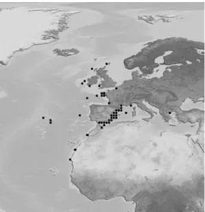
The blackspot seabream is a benthopelagic fish commonly found in the Mediterranean (Fig. 2). It can also be found in the Atlantic from Norway (65ºN) to Cape Blanco, Madeira and the Canaries, exceptionally farther south, in depths from the inshore waters above various bottoms (rocks, sand and mud), down to 400 m in the Mediterranean and 700 m in the Atlantic (Bauchot & Hureau, 1990).
Figure 2: Geographical distribution of Pagellus bogaraveo (Brunnich). The image is hosted at http://fishbase.ifm-geomar.de.
The species exhibits protandric hermaphroditism, with males becoming females when they reach a length of 20-30 cm. In an adult population, most of the individuals are females (Krug, 1990). In the natural environment, the blackspot seabream reaches sexual maturity at age of 5 years, at a mean weight and length of 650 g and 35 cm, respectively (Sánchez, 1983). While in captivity, the first spawning occurs at 4 years of age when a mean weight and length of 800 g and 30 cm, respectively, is attained (Fernández-Pato et
al., 1990). The reproducing stage in the natural environment varies in terms of latitude and
longitude. Generally, spawning occurs at the end of winter between January and April (Bauchot & Hureau, 1990), reaching peak spawning activity between February and May (Sánchez, 1983; Krug, 1990). In captivity, spawning coincides more or less with the above periods, as it has been observed in the north-west of the Iberian Peninsula (Olmedo et al., 1998). Fecundity estimates ranges from 73,000 to 1,500,000 oocytes for female fish with lengths swinging between 29 and 41 cm (Krug, 1990). The eggs float and fingerlings swim in big groups near the shoreline. When they grow, they dive into deeper zones (Morales, 1986). In captivity (Instituto Español de Oceanografia, Cabo Estay, Vigo, Spain), embryonic life (from fertilization to hatching) lasts 54 hours only. Larvae are maintained in the incubators at the natural temperature with running water until the day prior to mouth opening (at 115 h after hatching) when they are transferred to the larva culture tanks at 18 ± 1 ºC. To our knowledge, the blackspot seabream has been mainly fed on diets specifically developed for other marine fish, namely gilthead seabream, and few available publications exist on the specific nutritional requirements of blackspot seabream (Figueiredo-Silva et al., 2009; Valente et al., 2009). Generally, the feeding plan consists in rotifers (Brachionus plicatilis, Mueller) (5 - 35 days), Artemia AF380 (30 - 35 days) and
metanauplius (35 – 45 days) followed by innert microdiets (45 - 60 days) (Krug, 1990; Peleteiro et al., 1997; 2000; Olmedo et al., 1998). Ribeiro et al. (2008) analysed the activity of digestive enzymes at different development stages of blackspot seabream raised with this feeding plan and observed that the pattern of digestive enzymes activity was related to organogenesis and the type of the food used.
The first growth experiments performed with blackspot seabream juveniles relied on fish caught in the natural environment. Fish exhibit lower growth rates (Peleteiro et al., 1994; Olmedo et al., 2000) when compared to other farmed Sparidae, such as gilthead seabream (Aksnes et al., 1997; Santinha et al., 1999; Izquierdo et al., 2003), and high lipid deposition in muscle, liver and particularly around viscera (Linares et al., 2001). These results clearly point the need of optimizing culture conditions and improving specific diets for supporting the consolidation of blackspot seabream farming.
Muscle morphology and fibre types in teleost fish
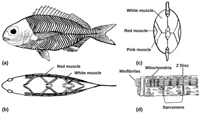
The swimming muscle of fish comprises about approximately 40 – 60% of the total body mass (Bone, 1978). The muscle tissue is organized in a series of myotomes on either side of the body. These units are separated by sheets of connective tissue, the myosepta. The myosepta are attachment sites for the muscle fibres and play a role in force transmission. The myosepta form a series of overlapping cones which are stacked along axes parallel to the midline, giving rise to the characteristic W-shape appearance of myotomes in longitudinal section (Van Leeuwen, 1999) (Fig. 3a). Each myotome contains: 1) a superficial wedge-shaped region lying directly beneath the lateral line, where the muscle fibres run parallel to the body axis; 2) and a deeper part where the muscle fibres are arranged in a helical fashion, forming angles of up to 40º (Sänger & Stoiber, 2001) (Fig. 3b). The functional significance of this arrangement is that it is thought to allow similar degrees of sarcomere shortening at different body flexures (Rome et al., 1988). A horizontal septum divides the axial musculature into a dorsal (epaxial) and a ventral (hypaxial) part (Bone, 1978).
The muscle fibre is made up of many myofibrils. A cylindrical myofibril consists of thousands of microscopic units called sarcomeres – each being about 2 µm long – that are connected end-to-end (Fig. 3d). The components of sarcomere include two types of myofilaments, made of proteins called actin and myosin. When a nerve impulse reaches a skeletal muscle fibre, it causes a change in electric potential across the plasma membrane. Skeletal muscle fibres can quickly convert this elecytric signal into despolarization through a rise of cytosolic Ca2+ from the sarcoplasmic reticulum, and then initiates the contraction. Myosin is a motor protein that interacts with actin filaments and
couples the hydrolysis of ATP. In skeletal muscle, contraction is regulated by four proteins associated with the actin filament: tropomyosin and troponins C, I and T. The cytolosic Ca2+ concentration influences the position of these proteins on the thin filaments, which in turn controls myosin-actin interaction (Purves et al., 2001). Myosin is a major component of the contractile apparatus, consisting of two heavy chains and two pairs of light chains. Each heavy chain consists of a structural α-helical rod and a globular head. The globular head region is known to contain the light chain and actin binding sites as well as the ATPase activity (Sellers, 2000).
In contrast to mammalian muscle, which is characterised by a mosaic pattern of fibre types, the axial muscle of teleosts shows anatomically distinct zones of fibre types. The muscle fibres are separated in two layers: subdermal slow-red muscle, with oxidative metabolism and slow contraction; and deep fast-white muscle, with glycolytic metabolism and fast contraction (Fig. 3c). In several fish, intermediate-pink muscle located in the transitional zone between slow-red and fast-white muscle can be distinguished (Fig 3c), with fibres characterized as fast contracting with intermediate resistance to fatigue and intermediate speed of shortening (Johnston et al., 1975; Bone, 1978).
Slow-red muscle fibres
Slow-red muscle fibres are located in a superficial strip that forms a thicker layer at the major horizontal septum. In general, the superficial slow-red fibres run parallel to the longer axis of the body. Slow-red fibres are relatively small (ca. 25 - 45 μm) and usually comprise around 10 - 30% of the myotomal muscle, according to the locomotion mode (Sänger & Stoiber, 2001). Active pelagic families, like the Scombridae, have a higher proportion of slow fibres compared to the myotomes of benthic predators and deep-sea fish, which are almost entirely composed of fast muscle (Johnston, 1981).
In most muscle cross sections, the proportion of slow-red fibres increases from rostral to caudal regions (Bone, 1978; Sänger & Stoiber, 2001), but this depends upon the lifestyle of fish species. For example in the scup (Stenotomus chrysops, Linnaeus), the total area occupied by the slow-red muscle in body cross sections was larger in the mid and posterior body positions (Zang et al., 1996). On the contrary, another study on variations in the total slow-red muscle cross-sectional area found it more anteriorly developed in thunniform swimmers (Ellerby et al., 2000). Slow-red fibres in all fish are profusely innervated (Bone, 1978).
Sustained swimming speeds are supported by the contraction of the slow-red muscle fibres using aerobic pathways and mostly carbohydrates and/or lipids as fuels (Sänger & Stoiber, 2001). The continuous use of the superficial slow-red muscle requires a good
supply of oxygen-carrying blood. This is provided by extensive vascularization and a high concentration of myoglobin in the muscle cells, which endows its red colour (Bone, 1978). Other major general ultrastructural features of this muscle type are a high amount of subsarcolemmal and inter-myofibrillar mitochondria of the lamellar type and lipid droplets. The relative numbers of these intracellular components vary dependent upon species and environmental influences. The myofibrils are roughly polygonal in shape, except the most peripheral ones, which exhibit a radial alignment (Sänger & Stoiber, 2001).
Figure 3: Myotomal muscle in fish. Myotomes are shown as they appear in the lateral (a) and dorsal view (b) of fish. A longitudinal band of slow-red muscle runs down the midline of each side of the fish (b). The fast-white muscle is visible above (epaxial) and below (hypaxial) the slow-red muscle band. When viewed transversely (c), the lateral bands of slow-red muscle are “V” shaped, while the bulk of the myotome is fast-white muscle. In some fish species, an intermediate-pink muscle is observed between these layers (c). The muscle fibres are composed of smaller cylindrical units called myofibrils (d) which are made of many small structures called sarcomeres. Figures based on: (a) Bone, 1978; (b) Rome et al., 1988; (c) Johnston et al., 1975; (d) Johnston, 2001.
The slow-red muscle contains relatively high concentrations of respiratory and citric acid cycle enzymes, required for energy generation for slow sustained aerobic swimming (the most commonly measured of these being succinic dehydrogenase (SDH) (Johnston & Lucking, 1978; Higgins, 1990). Histochemical analysis of slow-red muscle also shows an alkaline-labile mATP activity (Veggetti et al., 1993; Assis et al., 2004; Aguiar et al., 2005).
(a) (b) (d) (c) Red muscle White muscle Red muscle White muscle Pink muscle Miofibrilas Mitochondria Z Disc Sarcomere