ESCOLA DE CIÊNCIAS E TECNOLOGIA
DEPARTAMENTO DE QUÍMICAIsolation and molecular characterization of the
fatty acid synthase gene from an obese pig breed
André Filipe Barreto Albuquerque
Supervisors:
Professor José Manuel Mota Ruivo Martins
Doctor Marta Sofia S. V. C. Ferreira Laranjo
Master course in Biochemistry
Dissertation
“Success consists of going from failure to failure without loss of enthusiasm.”
Winston Churchill
“You must be the change you wish to see in the world.”
I
TABLE OF CONTENTS
TABLE OF CONTENTS I
LIST OF ILLUSTRATIONS III
LIST OF TABLES V ABBREVIATION LIST VI ACKNOWLEDGEMENTS XIII RESUMO XIV ABSTRACT XV I. INTRODUCTION 1
II. LITERATURE REVIEW 3
1. THE ALENTEJANO PIG - HISTORICAL AND LOCAL BACKGROUND 3
1.1. THE SWINE 3
1.2. PRODUCTION SYSTEM -THE INFLUENCE OF MONTADO AND MONTANHEIRA 4
2. SWINE-BASED RESEARCH 6
2.1. SWINE AS HUMAN BIOMEDICAL MODEL 6
2.2. SWINE BIOMEDICAL MODELS FOR OBESITY AND TYPE-2DIABETES STUDIES –FATNESS GENETICS 8
2.3. POTENTIAL OF THE ALBREED 11
3. LIPID METABOLISM 11
3.1. INTRODUCTION TO LIPIDS –THE FATTY ACIDS 12
3.2. FATTY ACID CATABOLISM 14
3.3. DIETARY LIPID ABSORPTION 21
3.4. DE NOVO SYNTHESIS 23
3.5. BIOSYNTHESIS OF LONG-CHAIN SFAS,MUFAS AND PUFAS 30
4. FATTY ACID SYNTHASE 32
4.1. STRUCTURE AND ACTIVITY OF THE MAMMALIAN FASCOMPLEX 32
II
III. MATERIAL AND METHODS 36
1. SLAUGHTER CONDITIONS AND TISSUE COLLECTION 36
2. RNA EXTRACTION 36
3. COMPLEMENTARY DNA SYNTHESIS 36
4. CDNA AMPLIFICATION BY PCR 37
4.1. SEQUENCING STRATEGY 37
4.2. PRIMER DESIGN 38
4.3. PCRPROTOCOL OPTIMIZATION 39
5. ELECTROPHORESIS AND PURIFICATION OF PCR PRODUCTS 40
6. CLONING OF PCR PRODUCTS 41
6.1. CLONING VECTORS 42
6.2. PLASMID-INSERT LIGATION 44
6.3. TRANSFORMATION CONDITIONS 44
6.4. PLASMID DNA EXTRACTION 44
7. RESTRICTION ASSAY 44
8. DNA SEQUENCING AND ANALYSIS 45
9. RAPID AMPLIFICATION OF CDNA ENDS 45
9.1. 3’RACE APPROACH 46
IV. RESULTS 48
1. FRAGMENT AMPLIFICATION DETECTION 48
2. DETECTION OF THE PRODUCT AFTER RESTRICTION ASSAY 49
3. MOLECULAR CHARACTERIZATION OF THE SEQUENCED RESULTS 49
V. DISCUSSION 55
VI. CONCLUSIONS AND FUTURE PERSPECTIVES 58
REFERENCES 59
APPENDIX I – COMPLETE LIST OF TESTED PRIMERS 65
III
LIST OF ILLUSTRATIONS
ILLUSTRATION 1. INSULIN RESISTANCE INDUCED BY OBESITY MIGHT INVOLVE A COMPLEX NETWORK OF ENDOCRINE, INFLAMMATORY AND NEURONAL PATHWAYS. ... 9 ILLUSTRATION 2. THE STRUCTURE OF PALMITIC ACID OR PALMITATE (C16:0), ONE OF THE MOST
COMMON FAS FOUND IN ANIMALS. CARBON COUNT STARTS FROM THE CARBOXYLIC GROUP (1), THE FIRST CARBON AFTER THE CARBOXYL CARBON IS CALLED Α (2) WHILE THE NEXT IS KNOWN AS Β (3) AND THE FOURTH AS Γ (4). ... 12 ILLUSTRATION 3. DISTINCT TYPES OF FATTY ACID–CONTAINING COMPOUNDS. A. THE STRUCTURE OF
PHOSPHATIDYLCHOLINE (GLYCEROLPHOSPHATE-BASED LIPID), A PHOSPHOLIPID BASED ON A GLYCEROL BACKBONE ALONG WITH A PHOSPHATE GROUP, A CHOLINE GROUP AND TWO STEARIC ACIDS (C18:0); B. THE STRUCTURE OF A TRIACYLGLYCEROL COMPRISING A GLYCEROL AND THREE DISTINCT FA, FROM TOP TO BOTTOM: PALMITIC ACID (C16:0), OLEIC ACID (C18:1, Δ9) AND
ALPHA-LINOLENIC ACID (C18:3, Δ9, 12, 15); C. THE STRUCTURE OF THE ANIMAL WAX CERYL MYRISTATE, AN
ESTER OF CERYL ALCOHOL AND MYRISTIC ACID (C14:0). ... 13 ILLUSTRATION 4. EXAMPLE OF FATTY ACIDS CLASSIFICATION USING THE DELTA NOMENCLATURE, FROM
TOP TO BOTTOM: STEARATE (C18:0), TRANS-OLEATE (C18:1, Δ9) AND CIS-OLEATE (C18:1, Δ9). ... 14
ILLUSTRATION 5. HYDROLYSIS OF A TRIGLICERIDE INTO GLYCEROL AND FAS BY ADIPOCYTE LIPASES. .... 15 ILLUSTRATION 6. FORMATION OF ACYL ADENYLATE AND PYROPHOSPHATE IN THE FIRST REACTION OF
THE FA ACTIVATION PROCESS. ... 16 ILLUSTRATION 7. THE SECOND ACTIVATION STEP COMPRISES THE FORMATION OF THE THIOESTER ACYL
COA. ... 16 ILLUSTRATION 8. TRANSFER OF THE ACTIVATED ACYL INTO THE MITOCHONDRIAL MATRIX INVOLVES A
PREVIOUS REACTION, ON THE OUTER MITOCHONDRIAL MEMBRANE, WITH CARNITINE FORMING ACYL CARNITINE. ... 17 ILLUSTRATION 9. SUMMARY OF THE ACYL GROUP TRANSPORTATION FROM CYTOSOL TO THE
MITOCHONDRIAL MATRIX, A PROCESS MEDIATED VIA ACYL CARNITINE TRANSLOCASE. ... 17 ILLUSTRATION 10. Β-OXIDATION INVOLVES 4 MAIN STEPS: OXIDATION, HYDRATION, OXIDATION AND
THIOLYSIS. ... 18 ILLUSTRATION 11. AFTER Β-OXIDATION, THE MAIN RESULTANT PRODUCT, ACETYL COA, ENTERS THE
CITRIC ACID CYCLE FOR E- PRODUCTION THAT CAN BE TRANSPORTED VIA NADH AND FADH 2
CARRIERS TO ENTER THE RESPIRATORY CHAIN FOR ATP SYNTHESIS. ... 19 ILLUSTRATION 12. THE FULL OXIDATION OF A MONOUNSATURATED FATTY ACID (MUFA) INVOLVES THE
ACTION OF AN ISOMERASE TO CHANGE THE DOUBLE BOND LOCATION AND CONFIGURATION. ... 20 ILLUSTRATION 13. THE FULL OXIDATION OF A POLYUNSATURATED FATTY ACID INVOLVES THE ACTION
OF AN ISOMERASE AND A REDUCTASE. ... 21 ILLUSTRATION 14. ABSORPTION OF DIETARY LIPIDS IN VERTEBRATES. ... 22 ILLUSTRATION 15. THE METABOLIC KEY ROLE PLAYED BY THE CIC PROVIDING A LINKAGE BETWEEN
GLYCOLYSIS AND LIPOGENESIS. HMGCOA, 3-HYDROXY-3-METHYLGLUTARYL-COA; PYC, PYRUVATE CARRIER. (A) PYRUVATE DEHYDROGENASE, (B) CITRATE SYNTHASE, (C) ATP-CITRATE LYASE, (D) ACETYL-COA CARBOXYLASE, (E) FATTY ACID SYNTHASE, (F) 3-HYDROXY-3-METHYLGLUTARYL-COA REDUCTASE, (G) CYTOSOLIC MALATE DEHYDROGENASE, (H) MITOCHONDRIAL MALATE
DEHYDROGENASE, (I) MALIC ENZYME. ... 24 ILLUSTRATION 16. THE FIRST STEP OF DE NOVO SYNTHESIS COMPRISES THE CARBOXYLATION OF ACETYL
IV
ILLUSTRATION 17. THE ACETYL COA CARBOXYLASE IS A THREE SUBUNIT POLYPEPTIDE THAT MEDIATES THE CARBOXYLATION OF ACETYL COA TO MALONYL COA. ... 25 ILLUSTRATION 18. REGULATION OF ACETYL COA CARBOXYLASE BY
PHOSPHORYLATION/DEPHOSPHORILATION. ... 26 ILLUSTRATION 19. CONTROL ON THE FA BIOSYNTHESIS BY HORMONAL TRIGGERS AND OTHER
SUBSTRATE FACTORS. ... 26 ILLUSTRATION 20. THE BASIC STEPS OF ONE CYCLE IN FA SYNTHESIS. ... 28 ILLUSTRATION 21. BIOSYNTHESIS OF MUFAS AND PUFAS DERIVE FROM PALMITATE SYNTHESIS WITH
ACCESSORY ENZYME SYSTEMS. FAS SHADED PINK CANNOT BE PRODUCED BY MAMMALS AND HAVE TO BE INGESTED IN THE DIET. ... 30 ILLUSTRATION 22. THE DESATURATION OF A FA INVOLVES AN ELECTRON-TRANSPORT CHAIN. ... 31 ILLUSTRATION 23. FINAL CYCLIC REACTION IN PALMITATE SYNTHESIS MEDIATED BY FAS COMPLEX. ... 33 ILLUSTRATION 24. FRONT VIEW AND LINEAR ORGANIZATION SCHEMES OF THE MAMMALIAN FAS WITH
RESPECTIVE CATALYTIC SITES COLORED. ... 33 ILLUSTRATION 25. THE BASIC STEPS IN POLYMERASE CHAIN REACTION; DNA DENATURATION, PRIMER
ANNEALING AND EXTENSION. ... 37 ILLUSTRATION 26. PARTIAL SEQUENCING STRATEGY FOR THE FATTY ACID SYNTHASE GENE... 38 ILLUSTRATION 27. THE BASIC STEPS OF GENE CLONING. ... 41 ILLUSTRATION 28. PGEM®-T EASY VECTOR MAP AND POINTS OF INTEREST, NAMELY, AMPR AND LACZ
GENE LOCATIONS. ... 42 ILLUSTRATION 29. PNZY28 MAP AND POINTS OF INTEREST, NAMELY, AMP AND LACZ GENE LOCATIONS.
... 43 ILLUSTRATION 30. OPERATION OF RESTRICTION ENZYMES – ECORI. ... 45 ILLUSTRATION 31. SUMMARY OF THE FASN 3’ END AMPLIFICATION BY RACE. ... 46 ILLUSTRATION 32. EXAMPLES OF AMPLICONS GENERATED IN PCR ASSAYS USING DISTINCT PRIMERS
OBSERVED BY 1% AGAROSE GEL ELECTROPHORESIS. ... 48 ILLUSTRATION 33. EXAMPLE OF AN INSERT RELEASE GEL AFTER THE RESTRICTION ASSAY. ... 49 ILLUSTRATION 34. NUCLEOTIDE SEQUENCE ALIGNMENT OF TOTAL SEQUENCED PRODUCT WITH SWINE
FASN SEQUENCE NM_001099930.1. ... 53 ILLUSTRATION 35. PROTEIN SEQUENCE ALIGNMENT OF TOTAL SEQUENCED PRODUCT WITH SWINE
V
LIST OF TABLES
TABLE 1. MAJOR ADVANTAGES OF A SWINE BIOMEDICAL MODEL. ... 7 TABLE 2. COMPLETE LIST OF PRIMERS USED IN FASN SEQUENCING ALONG WITH RESPECTIVE SEQUENCE,
GENE LOCATION REGARDING TO SWINE FASN RECORD NM_001099930.1 ON GENBANK,
ANNEALING TEMPERATURE AND AMPLIFIED PRODUCT SIZE. ... 38 TABLE 3. LIST OF PRIMERS USED FOR FASN 3’ END SEQUENCING. ... 47 TABLE 4. CHARACTERIZATION OF THE IDENTIFIED POLYMORPHISMS. NOMENCLATURE OF MUTATIONS
VI
ABBREVIATION LIST
7TM – Seven transmembrane receptors ACC – Acetyl CoA carboxylase
ACP – Acyl carrier protein AFW – Abdominal fat weight AL – Alentejano
AMP – Adenosine monophosphate AMPK – AMP-activated protein kinase ATP – Adenosine triphosphate
BFT – Backfat thickness BMI – Body mass index bp – Base pairs
BSA – Bovine serum albumin BW – Body weight
C – Carbon
ºC – Celsius degree
ca. – circa
cAMP – Cyclic adenosine monophosphate cds – Coding sequence
cDNA – Complementary deoxyribonucleic acid CiC – Citrate carrier
CO2 – Carbon dioxide
CoA – Coenzyme A
CVD – Cardiovascular disease DH – Dehydratase
VII
DMSO – Dimethyl sulfoxide
dNTPs – Deoxynucleotide Triphosphates DNA – Deoxyribonucleic acid
EC – Enzyme classification number
E. coli – Escherichia coli
EDTA – Ethylenediaminetetraacetic acid
e.g. – exempli gratia
EGCG – Epigallocatechine-3-gallate ER – Enoyl reductase
FA – Fatty acid
FAD – Flavin adenine dinucleotide
FADH2 – Reduced form of flavin adenine dinucleotide
FAS – Fatty acid synthase
FASN – Fatty acid synthase gene FFA – Free fatty acid
FW – Forward g – Grams g – Gravitational acceleration h – Hour H2O – Water HDL – High-density lipoprotein kD – KiloDalton KS – Ketoacyl synthase KR – Ketoacyl reductase IKKβ – IκB kinase-β IL – Interleukin
VIII
IPTG – Isopropyl-[beta]-D-thiogalactopyranoside IRS – Insulin receptor substrate
IUBMB – International Union of Biochemistry and Molecular Biology IUPAC – International Union of Pure and Applied Chemistry
JNK – Jun kinase Kb – Kilobase kDa – Kilodalton kJ – Kilojoule
LDL – Low-density lipoprotein LEPR – Leptin receptor gene LPS – Lipopolysaccharide M – Molar
MAT – Malonyl-acetyl transferase
MCP – Monocyte chemoattractant protein min – Minute
mg – Milligrams MJ – Megajoule mL – Milliliters mM – Millimolar
M-MLV – Moloney murineleukemia virus mRNA – Messenger RNA
MUFA – Monounsaturated fatty acid
NAD+ – Oxidized form of nicotinamide adenine dinucleotide NADH – Reduced form of nicotinamide adenine dinucleotide
NADPH - Reduced form of nicotinamide adenine dinucleotide phosphate NCBI – National Center for Biotechnology Information
IX
nsSNP – Non-synonymous single nucleotide polymorphism O2 – Molecular oxygen
P – Pig
PCR – Polymerase chain reaction PKC – Protein Kinase C
PUFA – Polyunsaturated fatty acid QTL – Quantitative trait locus rpm – Revolutions per minute RNA – Ribonucleic acid RNase – Ribonuclease RT – Reverse transcription
RT-qPCR – Reverse transcription followed by quantitative polymerase chain reaction RV – Reverse
s – Second
SCD – Stearoyl CoA desaturase SFA – Saturated fatty acid
SNP – Single nucleotide polymorphism
SOCS – Suppressor of cytokine signaling proteins
SREBP1-c – Sterol regulatory element binding protein 1-c SUB – Subcutaneous adipose tissue
TBE – Tris-Borate-EDTA TE – Thioesterase
TLR – Toll-like receptor TNF – Tumor necrosis factor
Tris – Tris (hydroxymethyl) aminomethane U – Unit
X UV – Ultraviolet V – Volt w/v – weight/volume X-Gal – 5-Bromo-4-chloro-3-indolyl-β-D-galactopyranoside µL – Microliters Nucleotide bases A Adenine T Thymine G Guanine C Cytosine
Amino acid nomenclature follows International Union of Pure and Applied Chemistry (IUPAC) and International Union of Biochemistry and Molecular Biology (IUBMB) recommendations (1984)
A Alanine C Cysteine D Aspartate E Glutamate F Phenylalanine G Glycine H Histidine I Isoleucine K Lysine L Leucine
XI M Methionine N Asparagine P Proline Q Glutamine R Arginine S Serine T Threonine V Valine W Tryptophan Y Tyrosine
XII
This work is funded by FEDER Funds through the Operational Programme for Competitiveness Factors - COMPETE and National Funds through FCT - Foundation for Science and Technology under the Strategic Projects C/AGR/UI0115/2011 and PEst-OE/AGR/UI0115/2014.
XIII
ACKNOWLEDGEMENTS
First of all, I would like to thank ICAAM (Instituto de Ciências Agrárias e Ambientais Mediterrânicas) of the Universidade de Évora for providing the necessary conditions to make this work possible, particularly to professor Maria Ivone Clara for letting me stay at the Laboratório de Virologia Vegetal, where this project was carried out.
Thanks are due to Associação Nacional de Criadores de Porco Alentejano (ANCPA) for allowing the collection of samples from AL carcasses that enabled this work possible.
I would like to thank my supervisors, Professor José Manuel Martins and Doctor Marta Laranjo, for all their help, support, guidance, patience and availability.
I would also like to thank to Maria Mário and Luizeta Palma, technicians of Laboratório de Virologia Vegetal, for all support and assistance throughout this project.
Special thanks to Professor Rosário Félix and Doctor Carla Varanda for the warm welcoming in the lab, in which we spent so many days of scientific discussions, joy and helpful counseling, without them, this work would have never been possible. I could not forget to mention, all my friends and colleagues, particularly Helena Gaspar, Mônica Portella, Fernando Eliziário, Susana Santos, Mohamed Salem Zellama, Danielle Martinha, Mara Silva, Rodrigo Silva, Ana Paço, Miguel Landum, Margarida Santana, Clarisse Brígido, Ana Alexandre and to everyone else involved, my sincere and honest regards.
Finally, this work is dedicated to my family, particularly my brother and my mother, who, despite all, endured through the worst times alongside me during this critical stage of my life.
XIV
RESUMO
Isolamento e caraterização molecular do gene fatty acid synthase num porco obeso
O porco Alentejano é uma raça autóctone do sul de Portugal geneticamente idêntica ao porco Ibérico. Esta raça obesa é caraterizada por um crescimento lento e uma capacidade adipogénica precoce e elevada. A formação do tecido adiposo em suínos é obtida em 80% via síntese de novo, apesar de também poder ocorrer síntese independente de ácidos gordos intramusculares. O porco Alentejano apresenta a característica genótipo
“poupador” (thrifty genotype), responsável pelo elevado potencial e maior capacidade de
deposição de tecido adiposo que outras raças comerciais magras.
O objetivo deste estudo foi determinar a sequência genética do enzima lipogénico
fatty acid synthase (FAS) do tecido adiposo subcutâneo de um porco obeso de raça
Alentejana. De acordo com a pesquisa feita, esta é a primeira publicação de uma sequência parcial deste gene em porcos Alentejanos. Através da clonagem de produtos de PCR, foram detetados três polimorfismos: c.6361C>T, c.6632C>A e c.6693C>T., sendo que os últimos dois conduzem a alterações no resíduo traduzido.
Palavras chave: Suíno; raça Alentejana; fatty acid synthase; ácidos gordos; lipogénese; deposição; adipócito; síntese de novo; gordura intramuscular; sequenciação.
XV
ABSTRACT
The Alentejano (AL) pig is an autochthonous breed genetically similar to the Iberian pig. This obese breed is characterized by slow growth rates and precociously high lipogenic activity. De novo synthesis is responsible for about 80% of all fatty acid (FA) synthesis in swine adipose tissue, although independent intramuscular FA synthesis can also occur. The AL breed features the thrifty genotype, responsible for a higher potential and ability for fat deposition than other industrial lean breeds.
The aim of this study is to determine the gene sequence of the porcine fatty acid synthase (FAS) lipogenic enzyme of an obese AL pig in subcutaneous adipose tissue. To our knowledge this is the first publication of a partial sequence of this gene in AL pigs. Through cloning of polymerase chain reaction (PCR) assayed products three polymorphisms were detected: c.6361C>T, c.6632C>A and c.6693C>T, the latter two change the translated residue.
Key Words: Swine; Alentejano breed; lipogenesis; deposition; adipocyte; de novo synthesis; intramuscular fat; Fatty acid synthase; sequencing
1
I.
INTRODUCTION
Sus scrofa domesticus, commonly known as domestic pig, is a eutherian mammal
that has shared a natural bond with humans since the beginning of its domestication all across Eurasia around 10000 years ago up to modern breeding practices and biomedical models (Groenen et al., 2012).
Pigs are commonly raised for meat, which is the most important animal protein food source, and feed a majority of the global population. As an important meat source in a world with increasing and demanding food needs, particularly due to developing countries and exponential human population growth, this particular livestock species has increased its global population by 13 percent in the last two decades (FAO, 2013).
As a component of meat, fat, particularly saturated fat, is generally considered as deleterious to the health of the consumers, and is associated with higher chances on developing heart disease. Therefore animal feed strategies and processing technologies are nowadays increasingly used to alter meat composition to be more consistent with human dietary guidelines. Nevertheless, these strategies and techniques need to be based on the knowledge of the genetic potential and physiological characteristics of the animals to be efficient.
The perception of the adipose tissue being solely an energy storage organ has been extended to include an endocrine organ that plays a pivotal role in relation to energy homeostasis and metabolism. Apart from secreting free fatty acids, adipose tissue produces and releases numerous proteins and substances with autocrine, paracrine and endocrine functions.
The Alentejano (AL) pig, Sus mediterraneus, is an autochthonous breed, reared in the southern area of Portugal. This breed has been scarcely selected through centuries and due to its trend to fat deposition, is smaller in size and presents poorer meat yields than modern commercial breeds. This precocious and high trend of fat storage has been found not only in AL and Iberian pigs, but also in other animal species and even in humans, being named as “thrifty” genotype. In fact, the thrifty genotype is an adaptive mechanism to the environment allowing accommodation to cycles of feasting and famine (Neel, 1962) One intrinsic mechanism of the thrifty genotype has been recently associated in humans with a syndrome known as leptin resistance. Leptin resistance has been identified with disruptions of signal transduction processes at the level of leptin receptors with effects on food behavior and obesity (Lubis et al., 2008). Thus, leptin resistant individuals are obese genotypes with elevated leptin levels but unable to suppress feeding when food is in excess (Myers et al., 2008). In the Iberian pig a leptin receptor gene polymorphism has also been detected, with effects on food intake, body weight and fat deposition (Munoz et al., 2009; Ovilo et al., 2005).
2
Porcine genome sequencing it’s of most importance because it provides a valuable feature for this livestock species progress, while enables continuous studying for multiple disease-causing variations extending the swine potential as a biomedical model. (Groenen et al., 2012)
This study’s main goal is to sequence a lipogenic enzyme gene, fatty acid synthase (FAS), of an obese AL pig. Genotyping was performed in the most important tissue involved in the lipid metabolism, subcutaneous adipose tissue.
3
II.
LITERATURE REVIEW
1. The Alentejano pig - Historical and Local Background
1.1. The Swine
The autochthonous pig breed Alentejano (AL) or Black pig is a traditionally raised breed that produces high quality meat products thanks to its particular production system and very particular genotypic features (Freitas et al., 2007; Lopez-Bote, 1998).
AL breed is reared in the southern region of Portugal and descends from Sus scrofa
mediterraneus lineages, just as the genetically similar Iberian pig.Crosses between these two Sus mediterraneus descendants are common throughout the years. The Iberian pig term is accepted and used among many authors, to refer the several breeds existing in the Iberian Peninsula region (Martins et al., 2012; Nunes, 1993).
The Iberian Peninsula constitutes a strong historical and geographical unit that throughout history contributed to the formation of many local animal breeds, including the AL/Iberian pig. Nevertheless, there still is considerable genetic differentiation and relatively high genetic diversity found in Iberian breeds, most of them considered genetic resources (Canon et al., 2011; Matos, 2000; Nunes, 1993).
This Iberian swine branch presents, generally, several features: dark skin and hair color (if present); pointed snout; short and strong limbs. These morphological traits reflect the adaptation of the swine to the local conditions, particularly in terms of resistance to sunstroke and high temperatures (Fabuel et al., 2004; Nunes, 1993). Reproductive and zootechnical parameters are presented in several works (e.g. Freitas et al. (2006)).
Swine production can be divided into three distinct types depending on the final product: meat (e.g. Duroc, German Landrace, Pietrain); bacon (e.g. Danish Landrace, Tamworth) and fat (e.g. AL/Iberian, Gascon, Casertana). Meat-type pig breeds are characterized by high daily weight gains and are slaughtered with a low body fat content and constitute the main source of pork in developed countries, while fat-types have a lower BW gain and produce carcasses rich in fatness (Freitas et al., 2007; Switonski et al., 2010).
The AL is of smaller size and fatter, besides holding higher intramuscular fat (IMF) content than other Iberian breeds. This obese breed is characterized by slow growth rates
4
and precociously high lipogenic activity. Traditionally, these pigs are fed until they reach a body weight (BW) between 150 and 170 kg and 18-24 months of age, when they are ready for slaughter (Lopez-Bote, 1998; Martins et al., 2012; Nunes, 1993).
Unlike modern lean-type breeds, the AL is a local raised unselected breed with considerable genetic differentiation and poor productive traits. Nevertheless, until the second half of the 20th century, the AL pig breed was, economically, the most important
Portuguese pig breed. Their meat, rich in IMF, is mainly used for the manufacture of high-quality dry-cured products. More recently, an increased interest on these products and in fresh meat from Al pigs has been observed among consumers, sustained by their intrinsic quality and healthiness (Freitas et al., 2007; Freitas et al., 2006; Gonzalez-Anover et al., 2010; Teixeira and Rodrigues, 2013) (see below).
Swine fatty acid (FA) synthesis is mainly through de novo synthesis (around 80% or more on adipogenic breeds) in adipose tissue with the possibility of independent intramuscular FA synthesis (Hood and Allen, 1973; Martins et al., 2012). Besides a higher content of fat, carcasses of AL and Iberian pigs generally present a higher content of oleic acid and (Estévez et al., 2006; Martins et al., 2012), consequently, a lower content of saturated fatty acids (SFA) commonly associated with the development of cardiovascular diseases (CVDs) (Estévez et al., 2006).
The production of AL and Iberian pigs is deeply rooted to the Mediterranean ecosystem. The contribution of this swine breed to the preservation and sustainability of its ecosystem is not common among the swine production industry (Lopez-Bote, 1998; Matos, 2000; Pugliese and Sirtori, 2012). In fact, local pig breeds and their production systems have been able to respond to the high criteria and expectations of modern society in regard to environment, animal welfare, and food quality and healthiness (Pugliese et al., 2013).
1.2. Production System - The Influence of Montado and Montanheira
Alentejo, the southern region of Portugal where AL pig is reared, is full of extensive plains of poor, substantially degraded soils where livestock production represents an important local economic activity. The agro-silvo-pastoral system characteristic of this region is based on the montado. (Freitas et al., 2006).
The feed availability offered by the Portuguese montado is seasonal and also varies from year to year (Lopez-Bote, 1998; Nunes, 1993).
Traditionally, a diverse diet is supplied to pigs in extensive free-range conditions in long production cycles. Fattening periods during late October until February take full advantage of the montado landscape opposing the periods of scarcity where feed supplementation based on cereals/commercial diets might be required. The fattening
5
montanheira period explores local natural resources such as native pastures and Quercus rotundifolia and Quercus suber fruits (acorns), and is perfectly adapted to the AL
extensive production. Acorns are low in protein, rich in the monounsaturated fatty acid (MUFA) oleate (C18:1) and pastures on polyunsaturated fatty acids (PUFA) linoleic (C18:2) and linolenic (C18:3) acids, besides fiber, protein and antioxidants such as alpha-tocopherol. Native pasture consumption also contributes for the detoxification due to the tannin content of acorns, while complementing the acorns low-protein content. In the traditional production systems, pigs are finished in the montanheira, where they are submitted to a quick and intense fattening period (Gonzalez-Anover et al., 2010; Matos, 2000; Nunes, 1993).
Modern explorations maintained the most dominant and interesting traits of the traditional production system, particularly, fattening pigs in montanheira associated with the exploration of reproductive sows. Nevertheless, there isn’t a uniform production system since mating season, feed management, weight and age of slaughter varies among farms (Freitas et al., 2006).
More recently, an evolution in this production system has been verified, with the objective of a more intensified production. The feeding of pigs for example,is based on commercial MUFA-enriched diets having alpha-tocopherol supplementation up to 200 mg/kg in order to simulate the effects of the montanheira in the traditional free range feeding system (Daza et al., 2005). The growing interest on the semi-extensive camping system for swine management (requiring fewer workers) is another example (Matos, 2000).
Nowadays, pigs used to produce hams and dry-cured products are slaughtered at 150-170 kg BW and 14 to 24 months of age and are finished in montanheira (high quality products) or semi-extensive production systems (lower quality products). When sausages are the final product, pigs are slaughtered at 120-140 kg, and 14-20 months of age. Finally, for fresh meat, pigs are slaughtered at 90-100 kg and 8-14 months of age. These two final products are generally obtained from pigs reared in the semi-extensive and intensive production systems (Freitas et al., 2006).
Pork is one of the most produced and consumed meat-types in the world, despite the negative assumptions broadly made. The widespread opinion about the unhealthy effects of this meat is mainly related to its SFA and cholesterol content (FAO Food and Agricultural Organization of the United Nations., 2013a; Teixeira and Rodrigues, 2013; Wood et al., 2008).
Commercial lean-type pig breeds, developed to reduce pork total lipid content, have better productive traits when compared to AL and other unselected breeds. They are raised on intensive systems, using advanced technology which translates into an improvement in terms of carcass meat yield and manufacturing of low cost products. Nevertheless, animal products raised traditionally foster now more interest among consumers. This current interest might be associated with the characteristics of the extensive production systems that guarantee quality to the final product, increasing
6
consumer interest in organic and natural pig production. The high IMF content in AL/Iberian pork products is responsible for much appreciated organoleptic traits such as tenderness, juiciness and flavor (Gonzalez-Anover et al., 2010; Pugliese et al., 2013; Switonski et al., 2010).
AL and Iberian products represent an important part of traditional pork production and have significant commercial value. Furthermore, the distinctive FA profile of its meat, rich in unsaturated fatty acids (UFAs) and lower SFA content, contributes to an improved consumer approval of these products.
2. Swine-based research
2.1. Swine as Human Biomedical Model
The use of animal models for research of human diseases is a general practice that provides important information for development of prevention strategies and appropriate treatments. Better models rely on anatomic and physiological similarities to enable reliable extrapolations to humans.
Rodent subjects are widely adopted for human health studies. Nevertheless, rodent models occasionally fail to fully mimic human conditions, so an increasing need for animals closer to humans to further study diseases such as cystic fibrosis, diabetes or obesity, among others, is required (Cirera et al., 2014; Spurlock and Gabler, 2008; Walters et al., 2012).
In the last three decades, the adoption of pigs as human biomedical research model has broadly increased, particularly for general surgical models of most organs and systems, for cardiovascular research and in digestive system models, and more recently in transplantation and xenographic research through genetic engineering (Hamamdzic and Wilensky, 2013; Walters et al., 2012; Whyte and Prather, 2011).
Completion of the swine genome is currently simplifying and broadcasting the use of swine models in several research areas. For instance, it is now possible to search for single nucleotide polymorphisms (SNPs) of specific human disease genes and compare them to the ones existing in pigs. Through modern genomic modification techniques, swine gene functions can be altered, aiding the development of novel disease models based on transgenes on areas as diverse as cardiovascular disease, xenotransplantation and neurodegenerative diseases (Walters et al., 2012; Whyte and Prather, 2011).
7
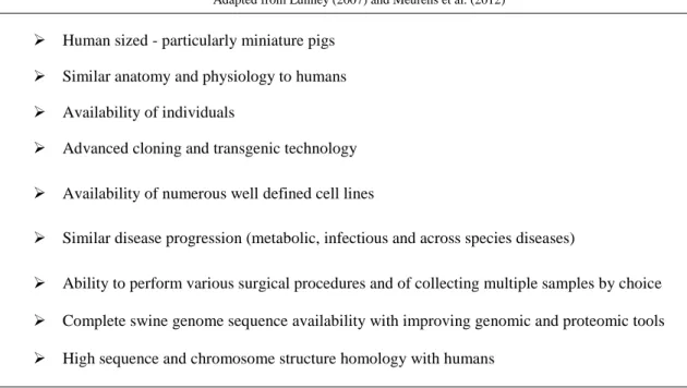
The vast conserved homology saved by both, swine and human, genomes is one of the main features that support the extensive use of swine as biomedical models. Both species are omnivorous and their organs share similar functional features. Furthermore, resemblances in size, anatomy and physiology make the swine a suitable model for research in gene and cell therapy, xenograft and allograft procedures and other fields of regenerative medicine. Similar disease progression has allowed the possibility of a broad range of distinct biomedical swine research models. All-season breeding and large litter sizes provides great availability of individuals that hold short generation intervals (12 months) and gestation lengths (114 days), plus an early sexual maturity (5-8 months) and short lifespan (10-20 years). Handling pigs from the same litter, or cloned or transgenic pigs, eases genetic mapping. Choosing the right breed and age allows the execution of various surgical and non-surgical procedures generally used in human medicine, including catheterization, heart surgery, valve manipulation, endoscopy and broncho-alveolar lavages. In opposition, some of these procedures are found hard to practice in other animal models including rodents (Lunney, 2007; Meurens et al., 2012; Rothschild and Ruvinsky, 2011).
Swine also allow the possibility to perform time studies, image internal vessels and organs, and collect repeated peripheral samples and, at kill, detailed tissue samples. Access to well defined cell lines from different tissues eases gene expression and drug susceptibility testing, for example. Moreover, swine full genomic code is already available and its high sequence and chromosome structure homology with humans will provide a further improvement to its genomic and proteomic tools. Swine models are inexpensive and ethically more acceptable than primates and other animals since they are used for human feeding as well. Table 1 summarizes the general advantages of swine biomedical models (Lunney, 2007; Meurens et al., 2012).
Table 1. Major advantages of a swine biomedical model.
Human sized - particularly miniature pigs Similar anatomy and physiology to humans Availability of individuals
Advanced cloning and transgenic technology Availability of numerous well defined cell lines
Similar disease progression (metabolic, infectious and across species diseases)
Ability to perform various surgical procedures and of collecting multiple samples by choice Complete swine genome sequence availability with improving genomic and proteomic tools High sequence and chromosome structure homology with humans
8
2.2. Swine Biomedical Models for Obesity and type-2 Diabetes Studies – Fatness Genetics
Obesity is a chronic disease characterized by excessive body fat in tissues due to over accumulation of fat stores at the adipocyte level. In Humans, obesity is effectively diagnosed when body mass index (BMI) is superior to 30 kg m-2. Obesity is a major health risk factor for a number of chronic diseases, including diabetes, cardiovascular diseases, and cancer. Among adults, worldwide prevalence of obesity is over 10% and prevalence of obesity and overweight ascend up to more than 30%. In Europe and Northern America these results reach 60 and 70% of the adult population, respectively. Despite these alarming results, trends are expected to climb even more in coming years (FAO Food and Agricultural Organization of the United Nations., 2013b; Spurlock and Gabler, 2008).
Biochemical mechanisms responsible for obesity haven’t yet been fully identified and while rodent models have been extensively experimented for obesity research, their translational utility is less effective than pigs. This fact seems to be related with some differences at metabolic and physiological levels between rodents and humans. For example, several adipokines, such as adipsin or resistin, cytokines secreted by the adipose tissue, appear to have different activities (Arner, 2005; Spurlock and Gabler, 2008). Furthermore, swine, as humans, are devoided of brown adipose tissue postnatally. This resemblance is very important since brown adipose tissue can induce heat production and therefore regulate energy balance (Harms and Seale, 2013; Spurlock and Gabler, 2008).
Several studies suggest that obesity is highly correlated with macrophage accumulation in adipose tissue. Activated macrophages secrete various cytokines and chemokines involved in systemic inflammation such as tumor necrosis factor (TNF)-α, interleukin (IL)-1, IL-6 and monocyte chemoattractant protein (MCP)-1, the later contributing to infiltration of adipose tissue with immunocytes (Harford et al., 2011; Spurlock and Gabler, 2008; Weisberg et al., 2003; Xu et al., 2003).
As obesity, type-2 diabetes is a major cardiovascular risk factor, namely for developing coronary heart disease (Haffner et al., 1998).
Chronic inflammation induced by obesity can develop insulin resistance. It is widely known that high plasma FA concentrations are closely related to obesity because expansion of the adipose tissue entails FA release from the adipocyte depots. Furthermore, obesity-associated changes induce alterations in the secretion patterns of adipokines that modulate insulin signaling (see illustration 1, path B). Recent in vitro studies have demonstrated that SFAs stimulate tissue inflammation the same way as bacterial lipopolysaccharides (LPS), through activation of toll-like receptors (TLR)-4 (illustration 1, path G) which indirectly promote cell inflammation with further production of pro-inflammatory TNF-α and IL-6 through the activation of protein kinases such as Protein Kinase C (PKC, EC 2.7.11.13), IκB kinase-β (IKKβ, EC 2.7.11.10), Jun kinase (JNK, EC 2.7.11.24) and the inhibitor of nuclear factor-κB (NF-κB, EC 2.7.11.10) (illustration 1,
9
path E). These kinases are responsible for the serine phosphorylation of insulin receptor substrates (IRS) that inhibits insulin signaling (illustration 1, path D). Moreover, the adipokine induced family proteins SOCS-3 conflict with IRS-1 and IRS-2 by phosphorylation or proteosomal degradation (illustration 1, path F). Main consequences to insulin sensitivity are an increased glucose synthesis in the hepatocyte, while on the myocyte the glucose uptake is decreased (Bjorntorp et al., 1969; Chait and Kim, 2010; Qatanani and Lazar, 2007; Spurlock and Gabler, 2008).
Another adipokine closely related to obesity and inflammation, in pigs as in humans, is adiponectin. Adiponectin is a protein secreted exclusively by adipose tissue that modulates lipid metabolism by inducing AMP-activated protein kinase, AMPK (EC 2.7.11.31), activity which deactivates acetyl CoA carboxylase, ACC (EC 6.4.1.2) and, consequently, suppresses carbohydrate transformation into lipid reserves by inhibiting de
novo fatty acid synthesis. A small number of studies showed that lower concentrations of
circulating adiponectin are found in obese and among insulin resistant subjects, suggesting anti-inflammatory effects. On the other hand, leptin is an adipocyte secreted hormone that regulates physiological functions in energy shortage periods and, when absent, leads to major metabolic disturbances due to increased insatiability. Adiponectin and leptin are found to be reciprocally regulated (Brennan and Mantzoros, 2006; Qatanani and Lazar, 2007; Spurlock and Gabler, 2008; Wu et al., 2003).
Anti-inflammatory effects by adiponectin have been reported in both swine and human adipose tissue. Production of pro-inflammatory cytokines as IL-6 and TNF by activated macrophages is suppressed by adiponectin. On the other hand, IL-6 and TNF
Illustration 1. Insulin resistance induced by obesity might involve a complex network of endocrine, inflammatory and neuronal pathways.
10
regulate adiponectin expression by inhibiting its mRNA expression (Spurlock and Gabler, 2008).
Further studies, using more swine subject models, might uncover more or confirm about the crucial role that these and other factors might have in inflammation, obesity and obesity-related diseases. These studies will include the search for gene mutations, polymorphisms and allele comparison in relevant genes, taking advantage of the recent availability of the full swine genome sequence and development of techniques such as SNP microarrays (Bruun et al., 2003; Spurlock and Gabler, 2008; Switonski et al., 2010; Wulster-Radcliffe et al., 2004; Yokota et al., 2000).
Evaluation of genetic predisposition through comparative genomic studies on obesity candidate genes and search for fatness quantitative trait loci (QTLs) are current fields of obesity research in expansion. All these obese swine traits have high polygenic background. To date, more than 100 loci have been identified and associated with obesity-related traits by genome-wide association studies (GWAS) as well as several candidate genes. Nevertheless, this approach is revealing to be rather limited since the individual expression value of current identified candidate genes is quite low when explaining fatness phenotypic traits. This is thought to be related with the high polygenic background involved in the determination of fatness traits, as well as the possibility of epistatic effects between potential genes (Cirera et al., 2014; Schook et al., 2005; Switonski et al., 2010). Modern pig breeding has also found interest in studying swine genetic background in order to improve livestock production. Most important and studied swine fatness traits are backfat thickness (BFT), abdominal fat weight (AFW), IMF content and FA composition. BFT and AFW are related to fattening efficiency while IMF and FA composition affect quality meat traits and display nutritional value. IMF levels vary between breeds and muscle type. In Longissimus dorsi, for example, one of the most valuable muscles, the average IMF content in selected breeds is about 1.7%, while in AL/Iberian pigs this is much higher and may attain 6%. Furthermore, lean-type pig breeds are selected towards lower levels of BFT and growth of leaner tissues (Daza et al., 2006; Switonski et al., 2010).
Limitations occur when translating conclusions about porcine candidate genes to humans and vice-versa in fatness deposition. While studies of human obesity focus on BMI, traits studied in swine are concerned with different aspects of fatness which span candidate genes that may not prove to be as relevant for human obesity research. Nevertheless, particular animal models, such as the obese Ossabaw pig, are becoming increasingly valuable assets to understand the development of human obesity, obesity-induced insulin resistance and ultimately the metabolic syndrome and cardiovascular diseases. (Switonski et al., 2010)
11
2.3. Potential of the AL Breed
Several recent research studies about human obesity and development of metabolic syndrome have been done with Ossabaw pigs as models. For example, this breed can easily and efficiently simulate human atherosclerosis. This swine breed is exclusively found on Ossabaw Island (Georgia, USA) and features a thrifty genotype which drivens it to feast in excess, easily become obese and develop all pathologies clustering metabolic syndrome such as obesity with severe visceral adipose expansion, primary insulin resistance, glucose intolerance, dyslipidemia and hypertension. Presence of three out of five of these symptoms is required to characterize metabolic syndrome in humans. Ossabaw swine can become overly obese even when fed with diets with no added fat but rich in carbohydrates. Recent studies suggest that these pigs are descendent from an Iberian branch which may explain their morphological and metabolic similarities (Brisbin and Sturek, 2009; Faris et al., 2012; Hamamdzic and Wilensky, 2013; Spurlock and Gabler, 2008; Toedebusch et al., 2014).
Understanding of the pathophysiology of metabolic syndrome has been delayed by the lack of efficient human-like models. However, Ossabaw breed happens to be one of the best models for studying metabolic syndrome in humans due to the easiness in developing all its associated pathologies at once. As the Ossabaw strain, AL pigs hold high propensity for obesity, due to an adaptive thrifty genotype, including a great capacity to accumulate intramuscular and epidermal fat (Hamamdzic and Wilensky, 2013).
Iberian pigs hold a polymorphism on the leptin receptor gene (LEPR) causing a condition commonly known as leptin resistance, characterized by failure in feeding suppression due to high concentrations of leptin found in obese individuals. This genetic feature results in implications at food intake, BW and fat deposition patterns, promoting obesity development (Munoz et al., 2009; Ovilo et al., 2005).
Recently, a study conducted by Torres-Rovira et al. (2012) concluded that within three months, Iberian sows with access to an ad libitum diet enriched with saturated fat, developed all five criteria associated to metabolic syndrome. This suggests that AL/Iberian pigs could be a valuable human model for further comprehension on mechanisms involved in obesity and its associated comorbities.
3. Lipid Metabolism
This chapter will be focused in introducing mammalian lipids as important energy stores in adipose tissue, and their successive transformations, depending on the current metabolic needs.
12
3.1. Introduction to Lipids – The Fatty Acids
Lipids are a class of biomolecules that display a variety of distinct biological and structural functions. Lipids integrate cell membranes, carry fat soluble vitamins, and play vital roles as enzyme cofactors, hormones, and electron carriers, among others. Nevertheless, their main function lies in providing energy (Berg et al., 2012; Hussain et al., 2013; Lehninger et al., 2008).
Animals store energy mainly in the form of fat so that proteins don’t have to be used, allowing them more important roles such as building and repairing tissues. In addition, storing calories as fat is more efficient than with the same mass of carbohydrates in the form of glycogen. Energy stores offer survival for long periods of food deprivation and its associated efficiency is thought to offer important advantages to animals during evolution (Lehninger et al., 2008; Semenkovich, 1997).
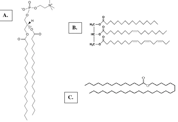
Animals store energy in the form of FAs, the simplest class of lipids. They contain a methyl group, a long hydrophobic hydrocarbon chain varying between 4 and 36 carbon atoms, and a terminal carboxylate group (illustration 2). FAs can be stored in excess amounts in the adipose tissue and oxidized in all tissues, particularly liver and muscle, acting as a major source of fuel to cells. In the form of phospholipids (mainly phosphatidylcholine, phosphatidylethanolamine, and sphingomyelin), FAs form the backbone of cell membranes and contribute for their fluidity and functionality. FAs can also be found freely, circulating bound to albumin or integrated in triacylglycerols and waxes (Lehninger et al., 2008; Tvrzicka et al., 2011; Vance and Vance, 2008) (see illustration 3).
Illustration 2. The structure of palmitic acid or palmitate (C16:0), one of the most common FAs found in animals. Carbon count starts from the carboxylic group (1), the first carbon after the carboxyl carbon
13
Fatty acids can be defined as being saturated or unsaturated, depending on the absence or presence of double bonds between two carbon atoms in the aliphatic chain, respectively. Furthermore, UFAs display cis-trans isomerism, describing the orientation of the alkyl groups around the double bond. The trans isomer of a given FA can rarely be found in nature. Trans FAs can be obtained in dairy products or their meat and can also be produced by partial hydrogenation of fish and vegetable oils. Large dietary intakes of trans FAs are strongly correlated to increased levels of triacylglycerols and LDL (low-density lipoprotein) cholesterol and lower levels of HDL (high-(low-density lipoprotein) on blood, conducing to a higher risk in developing cardiovascular diseases (Chilliard, 1993; Lehninger et al., 2008).
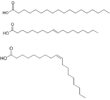
Delta (Δ) FA nomenclature is a simple mean of classification and identification of different FAs by specifying the chain length and number of double bonds, separated by a colon. In addition the number superscripted above the Δ represent the number of carbons from the carboxylic acid end to the first carbon in the double bond (Lehninger et al., 2008) (illustration 4). This simplified nomenclature, along with common names, will be recurrently used throughout this thesis.
A.
B.
C.
Illustration 3. Distinct types of fatty acid–containing compounds. A. The structure of phosphatidylcholine (glycerolphosphate-based lipid), a phospholipid based on a glycerol backbone
along with a phosphate group, a choline group and two stearic acids (C18:0); B. The structure of a triacylglycerol comprising a glycerol and three distinct FA, from top to bottom: palmitic acid (C16:0),
oleic acid (C18:1, Δ9) and alpha-linolenic acid (C18:3, Δ9, 12, 15); C. The structure of the animal wax
14
3.2. Fatty Acid Catabolism
Animal cells can obtain FAs from three distinct pathways: fats ingested in the diet, lipolysis of lipids stored in cells and de novo synthesis.
Triacylglycerols are formed by esterification of the three hydroxyl groups of glycerol by carboxyl groups of organic acids. They function as highly concentrated energy stores accumulated in cell depots, mainly in liver and adipose tissue. Their energy yield from complete oxidation is nearly 38 kJ g-1 whereas from carbohydrates and proteins is 17 kJ g-1. Such difference lies in the fact that FAs are much more reduced than carbohydrates or proteins (Berg et al., 2012).
Adipose tissue cells or adipocytes, are specialized cells that act as major site for triacylglycerol storage and synthesis. In mammals, adipose tissue can be found all over the body, particularly under the skin (subcutaneous) and surrounding internal organs (visceral). This tissue works mainly as an energy depot, where droplets of triacylglycerols tend to form large globules that can fill most of cell’s volume. The surface of these droplets is covered with perilipins that restrict the access to the lipids (Berg et al., 2012). Lipolysis and β-oxidation of free fatty acids (FFAs) is the main energy-yielding pathway for animal tissues. This process occurs in the mitochondria and is characterized by the degradation of FAs to generate acetyl-CoA. Access to the lipid energy reserves stored in adipose tissue involves three main stages: i) mobilization of FFAs; ii) activation and transport to the mitochondria; and iii) degradation into acetyl CoA (Berg et al., 2012; Lehninger et al., 2008).
Illustration 4. Example of fatty acids classification using the delta nomenclature, from top to bottom: stearate (C18:0), trans-oleate (C18:1, Δ9) and cis-oleate (C18:1, Δ9).
15
3.2.1. Mobilization of Free Fatty Acids
The first step consists in isolating the FAs stored as triacylglycerols or mobilizing FFAs. For that, lipases of adipose tissue are activated to hydrolyze triacylglycerols into glycerol and FAs in a process also known as lipolysis (illustration 5).
Adipocyte lipases are activated by hormones such as epinephrine or glucagon. These hormones are responsible for the trigger of seven transmembrane (7TM) receptors that induce adenylate cyclase (EC 4.6.1.1) activity. Increased levels of cyclic adenosine monophosphate (AMP) stimulate protein kinase A (EC 2.7.11.11) activity which, afterwards, activates cytosol hormone-sensitive lipases (EC 3.1.1.79) and perilipins by phosphorylation (Berg et al., 2012; Lehninger et al., 2008). Activated perilipins are the proteins responsible for: restructuring of the lipid droplet in a way that it’s easier for lipases to access triacylglycerols; the release of a coactivator for the adipose triglyceride lipase (ATGL, EC 3.1.1.3). ATGL hydrolyzes a FA forming diacylglycerol in the process, while hormone-sensitive lipase removes a second FA. Monoacylglycerol lipase (EC 3.1.1.23) completes the mobilization producing a last FFA and glycerol (Berg et al., 2012).
Released FFAs are not soluble in blood, so they bind into serum albumin, which will carry them to the energy-requiring tissues. On the other hand, the glycerol is absorbed by the liver where it’s transformed into glyceraldehyde 3-phosphate and can participate in either the glycolytic or gluconeogenic pathways (Berg et al., 2012).
3.2.2. Activation and Transport to the Mitochondria
Full animal FA oxidation occurs in the mitochondrial matrix. FA with chain lengths of 12 or fewer carbons do not need membrane transporters to enter in the mitochondria. Longer FFAs need to be activated and then carried by a carnitine based transport mechanism to reach the mitochondrial matrix (Lehninger et al., 2008).
Illustration 5. Hydrolysis of a trigliceride into glycerol and
FAs by adipocyte lipases. Adapted from Berg et al. (2012)
16
Activation occurs on the outer mitochondrial membrane where long-chain FFAs are linked to Coenzyme A (CoA) by a thioester bond, in a reaction catalyzed by acyl CoA synthetase (EC 6.2.1.3). This process is ATP-dependent and comprehends 2 steps. On the first one, the FA reacts with ATP forming acyl adenylate releasing pyrophosphate (illustration 6) (Berg et al., 2012; Lehninger et al., 2008).
On the second step, the thiol group of CoA displaces AMP, conducing to the formation of the thioester acyl CoA (illustration 7).
Both reactions are reversible and have an equilibrium constant close to 1. In order to pull these reactions in the forward direction, the pyrophosphate formed in the first reaction is rapidly hydrolyzed into two monophosphates by an inorganic pyrophosphatase (EC 3.6.1.1) (Berg et al., 2012; Lehninger et al., 2008).
FAs are activated on the outer mitochondrial membrane, yet they are oxidized in the mitochondrial matrix. Their transfer across the mitochondrial membrane involves carnitine. The acyl group present in the activated long-chain FAs binds to the hydroxyl group of carnitine to form acyl carnitine in a reaction catalyzed by carnitine acyltransferase I (EC 2.3.1.21) (Berg et al., 2012; Lehninger et al., 2008) (illustration 8).
Illustration 6. Formation of acyl adenylate and pyrophosphate in the first reaction of the FA activation process.
Illustration 7. The second activation step comprises the formation of the thioester acyl CoA. Adapted from Berg et al. (2012)
17
Acyl carnitine translocase mediates the entrance of the acyl carnitine ester to the mitochondrial matrix. There, the acyl group is transferred from acyl carnitine to intramitochondrial CoA, in a reaction catalyzed by carnitine acyltransferase II (EC 2.3.1.21), releasing free carnitine. Carnitine can, then, return to the cytosolic side of the mitochondrial membrane through the translocase in exchange for acyl carnitine to balance the process (Berg et al., 2012) (illustration 9).
Illustration 8. Transfer of the activated acyl into the mitochondrial matrix involves a previous reaction, on the outer mitochondrial membrane, with carnitine forming acyl carnitine.
Illustration 9. Summary of the acyl group transportation from cytosol to the mitochondrial matrix, a process mediated via acyl carnitine translocase.
Adapted from Berg et al. (2012)
18
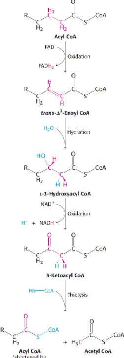
3.2.3. Fatty Acid Degradation – β-oxidation
Within the mitochondrial matrix, a four-step operation known as β-oxidation occurs. This cycling oxidation process takes place at the β carbon atom and includes a first oxidation by flavin adenine dinucleotide (FAD), hydration, oxidation by oxidized nicotinamide adenine dinucleotide (NAD+) and thiolysis by CoA (illustration 10) (Berg et al., 2012).
In the first step, acyl CoA is oxidized to produce a double bond between carbon atoms 2 and 3 in an oxidation process also known as dehydrogenation that is catalyzed by acyl CoA dehydrogenase (EC 1.3.8.7). The lost hydrogen atoms reduce the electron carrier FAD to its reduced form FADH2 (Berg et al., 2012;
Lehninger et al., 2008).
The resultant product, trans-Δ2-enoyl CoA, is then hydrated on the second step conducing to the formation of L-3-hydroxyacyl
CoA in a reaction catalyzed by enoyl CoA hydratase (EC 4.2.1.17) (Berg et al., 2012; Lehninger et al., 2008).
The third step involves another oxidation reaction, which converts the hydroxyl group at C-3 into a keto apart from generating reduced nicotinamide adenine dinucleotide (NADH) from NAD+. This oxidation is specifically catalyzed by L-3-hydroxyacyl CoA
dehydrogenase (EC 1.1.1.35) (Berg et al., 2012; Lehninger et al., 2008).
In the last step (thiolysis), the previously formed 3-ketoacyl CoA is cleaved by the thiol group of a free coenzyme A molecule, yielding acetyl CoA and an acyl CoA molecule shortened by two carbon atoms. This reaction is catalyzed by acyl CoA acetyltransferase (thiolase, EC 2.3.1.16) (Berg et al., 2012; Lehninger et al., 2008).
Illustration 10. β-oxidation involves 4 main steps: oxidation, hydration, oxidation
and thiolysis. Adapted from Berg et al. (2012)
19
Therefore, for each oxidation cycle, a determined acyl CoA is shortened by two carbon atoms and one molecule of FADH2,
NADH and acetyl CoA are formed. The last oxidation cycle of an acyl CoA with an even number of carbon atoms ends up producing two copies of acetyl CoA and one molecule of the electron carriers FADH2 and NADH (Lehninger et al.,
2008).
The acetyl CoA produced from β-oxidation enters the Citric Acid Cycle (Kreb’s Cycle), as well as those derived from glucose via glycolysis, where it’s further oxidized to CO2, while generating
more electron carriers such as NADH and FADH2. These, in turn, transfer their
energy to the mitochondrial respiratory (electron-transfer) chain, thus inducing the synthesis of more ATP. This can be represented as a three-stage process as shown on illustration 11 (Berg et al., 2012; Lehninger et al., 2008).
3.2.4. Degradation of Unsaturated Fatty Acids
Oxidation of UFAs requires up to two additional reactions compared to the previously described oxidation sequence. The double bonds present in these FAs are frequently in cis configuration, which prevents them from being hydrated on the second degradation reaction. This is because enoyl CoA hydratase, the enzyme responsible for catalyzing the addition of H20, operates only on trans double bonds. Besides, the presence
of a double bond between C-3 and C-4 prevents the formation of another double bond between C-2 and C-3. The solution to this problem comes from cis-Δ3-enoyl CoA
isomerase (EC 5.3.3.8), an enzyme capable of shifting the position and configuration of the cis-Δ3 double bond to a trans-Δ2 double bond. The substrate is now similar to those of the SFA oxidation sequence and β-oxidation can proceed (Berg et al., 2012; Lehninger et al., 2008) (illustration 12).
Illustration 11. After β-oxidation, the main resultant product, acetyl CoA, enters the Citric
Acid Cycle for e- production that can be
transported via NADH and FADH2 carriers to
enter the respiratory chain for ATP synthesis. Adapted from Lehninger et al. (2008)
20
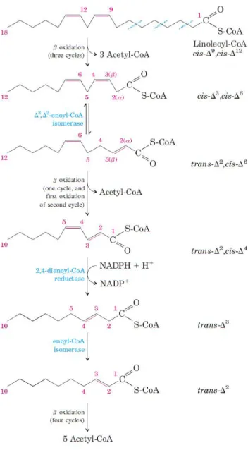
Polyunsaturated fatty acids (PUFAs), have more than one double bond and require an additional reaction to get fully oxidized.
At some point of the β-oxidation pathway, a stalemate occurs from the formation of a trans-Δ2, cis-Δ4-dienoyl CoA intermediate which is not a substrate for trans-enoyl CoA hydratase and therefore, cannot be directly hydrated. Nevertheless, 2,4-dienoyl CoA reductase (EC 1.3.1.34), a NADPH-dependent enzyme can convert this substrate to trans-Δ3-enoyl CoA. This new substrate can now be converted by cis-Δ3-enoyl CoA isomerase
to trans-Δ2-enoyl CoA and full oxidation can proceed normally (Berg et al., 2012) (illustration 13).
Illustration 12. The full oxidation of a monounsaturated fatty acid (MUFA) involves the action of an isomerase to change the double bond location and configuration.
21
3.3. Dietary Lipid Absorption
Most of ingested lipids come in the form of triacylglycerols and must be degraded so they can be absorbed in the intestinal epithelium (illustration 14). Dietary triacylglycerols are emulsified in the small intestine lumen by bile salts, stored in the gallbladder, and hydrolyzed into FFAs and monoacylglycerol by water-soluble intestinal lipases, secreted by the pancreas. These products cross the intestinal mucosa, where they are reconverted to triacylglycerols and are incorporated, along with cholesterol and apolipoproteins, into lipoprotein transport particles known as chylomicrons (Berg et al., 2012; Lehninger et al., 2008).
Illustration 13. The full oxidation of a polyunsaturated fatty acid involves the action of an isomerase and a reductase.
22
Chylomicrons are released into the lymphatic system and then into the blood, which transports them to muscle and adipose tissue. On the capillaries, apolipoprotein C-II present in chylomicrons activates lipoprotein lipase, which hydrolyses triacylglycerols to FAs and glycerol. The FAs can then be taken by the target tissue cells, where they can be oxidized for energy (myocyte) or reesterified for storage as triacylglycerols (adipocyte) (Berg et al., 2012; Lehninger et al., 2008).
Illustration 14. Absorption of dietary lipids in vertebrates. Adapted from Lehninger et al. (2008)
23
3.4. De Novo Synthesis
The excess dietary carbohydrates and proteins can be converted/recycled to FAs and then incorporated into triacylglycerols for storage in a process known as de novo synthesis. Generally, this endogenous lipogenic pathway for FA biosynthesis becomes unnecessary when the body’s lipid requirements are attained in the diet. Nevertheless, this synthesis is particularly important during embryonic development and lactation in mammary glands (Berg et al., 2012; Strable and Ntambi, 2010).
The final product of this synthesis is palmitate, a sixteen carbon SFA that is the precursor for the formation of other FAs in a task involving other enzymatic systems. De
novo synthesis occurs in the cytoplasm of hepatic, renal and adipose tissue cells besides
lactating mammary glands. This process can be divided into two distinct stages: activation of acetyl CoA and the set of reactions of the fatty acid complex (Berg et al., 2012).
3.4.1. Activation – acetyl CoA carboxylation
The end-product of FA degradation, acetyl-CoA, is now the precursor and source of carbons for the synthesis of any FA while the reductant role is played by NADPH molecules. Therefore, the first step of de novo synthesis is to obtain acetyl CoA and its further activation (Berg et al., 2012).
Acetyl CoA is a common organic intermediate product of oxidation of pyruvate, FAs, amino acids and ketone bodies. Nevertheless, the mitochondrial membrane is impermeable to acetyl CoA. Acetyl CoA must be conjugated with oxaloacetate to form citrate, in a reaction catalyzed by citrate synthase (EC 2.3.3.1). Citrate can then be freely transferred to the cytoplasm through the citrate carrier (CiC). These mitochondrial transporters play an important intermediary metabolic role, connecting carbohydrate catabolism and lipogenesis, by facilitating an exchange flow of citrate and malate between the mitochondrial matrix and the cytosol, respectively (illustration 15). In fact, CiC helps citrate get through the permeable inner mitochondrial membrane, which is then transported by passive diffusion from the mitochondrial outer membrane to the cytosol through voltage dependent anion channels. In the cytoplasm, citrate is then cleaved to acetyl CoA and oxaloacetate, in a reaction catalyzed by ATP citrate lyase (EC 2.3.3.8.) In addition, cytosolic citrate can provide, via malic enzyme or NADP malate dehydrogenase (EC 1.1.1.40), reducing equivalents as NADPH molecules that are required in further lipogenesis phases. Nevertheless, most of required NADPH for FA synthesis comes from the pentose phosphate pathway (Gnoni et al., 2009).
24
Carboxylation of acetyl CoA expresses the beginning of de novo synthesis, a reaction catalyzed by acetyl CoA carboxylase (ACC). This process is irreversible and results in the formation of the substrate malonyl CoA (illustration 16). ACC in animals is a single multifunctional polypeptide that has three functional domains (see illustration 17): the biotin carboxylase, which activates a carboxyl group derived from hydrogencarbonate in an ATP-dependent reaction; the biotin carrier protein, which accepts the activated carboxyl group binding it to a nitrogen in the biotin ring; and the transcarboxylase, which is responsible for the transfer of the activated carboxyl group from the carboxybiotin intermediate to acetyl CoA yielding malonyl CoA (Berg et al., 2012; Lehninger et al., 2008).
Illustration 15. The metabolic key role played by the CiC providing a linkage between glycolysis and lipogenesis. HMGCoA, 3-hydroxy-3-methylglutaryl-CoA; PyC, pyruvate carrier.
(a) Pyruvate dehydrogenase, (b) citrate synthase, (c) ATP-citrate lyase, (d) acetyl-CoA carboxylase, (e) fatty acid synthase, (f) 3-hydroxy-3-methylglutaryl-CoA reductase, (g) cytosolic malate dehydrogenase, (h) mitochondrial malate dehydrogenase, (i) malic enzyme.
Illustration 16. The first step of de novo synthesis comprises the carboxylation of acetyl CoA into malonyl CoA.
Adapted from Gnoni et al. (2009)
25
This reaction is the rate-limiting step in FA synthesis, with ACC playing a key role in controlling the balance between oxidation and synthesis in the metabolism of FAs. Inactivation of ACC occurs by phosphorylation in an ATP-dependent reaction catalyzed by AMPK that dissociates the polypeptide into monomeric subunits which causes loss of activity (see illustration 18). AMPK works has a fuel gauge since it’s activated by AMP and inhibited by ATP. Therefore, in circumstances of low cellular energy levels, cells tend to stop synthesizing new lipids. On the other hand, protein phosphatase 2A (EC 3.1.3.16) catalyzes the inverse reaction, the activation of ACC by dephosphorylation (Berg et al., 2012; Lehninger et al., 2008).
Illustration 17. The acetyl CoA carboxylase is a three subunit polypeptide that mediates the carboxylation of acetyl CoA to malonyl CoA.
26
The flow of precursors into malonyl CoA can also be regulated by hormones and substrates (illustration 19). Citrate stimulates allosterically ACC activity. High levels of citrate induce polymerization of inactive subunits that can partly reverse the inhibition produced by phosphorylation. On the other hand palmitoyl CoA, the main product of FA synthesis, inhibit ACC activity by causing the same effects as phosphorylation, dissociating the ACC polypeptide into inactive subunits. Palmitoyl CoA also limits FA synthesis by inhibiting CiC, preventing the entry of citrate from the mitochondria into the cytosol. Glucagon and epinephrine, under conditions of fasting and exercise, stimulate the mobilization of FAs from triacylglycerols in cell depots which will be used as urgent energy source. These hormones also inhibit FA synthesis by inhibiting acetyl CoA carboxylase and though the exact mechanisms are not yet entirely understood, it is recognized that they enhance the inhibition provoked by the activity of AMPK by phosphorylating ACC (Berg et al., 2012; Lehninger et al., 2008).
Illustration 18. Regulation of acetyl CoA carboxylase by phosphorylation/dephosphorilation.
Illustration 19. Control on the FA biosynthesis by hormonal triggers and other substrate factors. Adapted from Berg et al. (2012)