Development Of Antimicrobial Coatings
Based On Antimicrobial Peptides
Hélia Cristina Barros Fernandes
Integrated Master in Bioengineering Branch of Molecular Biotechnology
Supervisor: Drª Cláudia Monteiro Co-Supervisor: Drª Cristina Martins
The work described on this thesis was conducted at:
i3s/INEB – Instituto de Investigação e Inovação em Saúde da Universidade do
Porto/Instituto de Engenharia Biomédica
Unless you try To do something beyond What you have already mastered, You will never grow.
i
Acknowledgements
Gostaria de agradecer a todas as pessoas que me apoiaram e ajudaram das mais diversas formas durante esta jornada.
Em primeiro lugar gostaria de agradecer à minha orientadora, Cláudia Monteiro, por toda a ajuda e apoio durante o desenvolvimento da dissertação. Agradeço toda a partilha de conhecimento e técnicas, a supervisão, o esclarecimento de dúvidas e o incentivo ao meu trabalho autónomo. Agradeço imenso pela aceitação e integração na equipa, pela introdução no mundo da investigação e acima de tudo pela confiança em mim e na minha potencialidade.
Também quero agradecer à minha co-orientadora, Cristina Martins, por todos os concelhos, dicas e pelas diversas reuniões pelas quais passamos. A partilha de ideias e possíveis formas de contornarmos os percalços que foram aparecendo foram cruciais para a evolução do meu trabalho e para recorrer a mais técnicas, moldando o caminho a seguir.
Quero dirigir o meu agradecimento também à nossa equipa, Bioengineered Surfaces, por toda a partilha, apoio e por me fazerem sentir bem-vinda. O meu obrigada a todas pela disponibilidade e ajuda nos momentos em que os percalços apareceram, na escrita da dissertação e por me manterem sempre calma ao longo da fase final e crucial da dissertação. Rita Gomes, minha companheira de jornada, obrigada por todas as dicas, por toda a ajuda e apoio que me deste ao longo dos últimos anos. Foi um orgulho conhecer-te, fazer o mestrado e terminar esta longa jornada do teu lado. Mariana Barbosa e Inês Borges, obrigada pela ajuda e pelos dias passados na câmara de fluxo, em conjunto, para que eu pudesse completar da melhor forma o trabalho a que me tinha predisposto. Andreia T. Pereira, obrigada pelas dicas, pelos empréstimos e pelos arranjos que proporcionaste e que foram importantíssimos para resolver situações desaprazíveis.
Agradeço encarecidamente à Dalila Pedro e ao Ricardo Vidal por todas as formações, por toda a ajuda e disponibilidade ao longo do desenvolvimento da dissertação e por estarem sempre a uma chamada de distância. O meu obrigado também ao CEMUP – Centro de Materiais da Universidade do Porto, especialmente ao Engenheiro Carlos Sá pela ajuda com o XPS.
Agradeço, do fundo do meu coração, aos meus colegas dos Bombeiros Voluntários de Lordelo e à AFPS – Associação Formar para Salvar, por todo o apoio durante o nosso intensivo curso de TAS - Tripulante de Ambulância de Socorro. Dois meses com os fins de semana passados ao vosso lado foram essenciais para me manter terra-a-terra e distraída
ii
com outra das minhas grandes paixões, a ajuda ao outro. Todos os formadores da AFPS e colegas bombeiros foram me ajudando e apoiando, apesar de não o terem feito de forma intencional, compreendendo sempre a situação em que me encontrava. Foi um orgulho terminar essa caminhada do vosso lado e da mesma forma desenvolver da melhor forma a minha dissertação.
O meu agradecimento final e também um dos mais importantes vai para a minha família. Aos meus pais, Hélia e Paulo, muito obrigada por todos os sacrifícios que fizeram para que eu pudesse concretizar um dos nossos sonhos, ser formada. Obrigada pela forma como me criaram, sempre desejosa de ser melhor e lutadora para alcançar os meus sonhos e cumprir os meus objetivos. Obrigada por me darem sempre tudo o que era necessário para eu puder melhorar e por todo o apoio indiscriminado ao longo da minha vida. À minha irmã Carla, obrigada por estares sempre do meu lado, por serem quem és e acima de tudo, pelo apoio incondicional que me deste nos últimos anos. Ao meu namorado Rui, agradeço do fundo do coração pelas palavras carinhosas que sempre tiveste, pelos “acredita em ti como eu acredito”, “eu sei que vais conseguir” e o mais importante “estou sempre aqui para o que precisares”. Obrigada pelo ombro amigo, pela compreensão e por toda a confiança em mim.
A todos vós, esta conquista é também um pouco vossa. Partilho convosco o meu sucesso e agradeço todos os incentivos e apoios ao longo desta prazerosa jornada que chega da melhor forma ao seu término.
iii
Abstract
Bacterial infections, namely nosocomial infections occur very frequently after bacterial colonization of implanted devices/biomaterials. This type of infections, besides leading to loss of biomaterials therapeutic effect, are extremely difficult to treat being a huge treat public health. Staphylococcus epidermidis, a gram-positive bacterium, colonizer of the human skin and mucosal surfaces (Namvar et al., 2014), is considered the most common cause of biomaterial-associated infections, especially due to its ability to form a very resistant biofilm. Infections caused by S. epidermidis are amongst the most burdensome, expensive and difficult to treat (Brescó et al., 2017).
Bioengineered approaches appear as very promising tools to prevent or treat infection in a variety of scenarios especially as antibiotics face loss of efficacy due to resistance development. Different strategies can be adopted, being one of the most promising the use of biomaterials combined with antimicrobial peptides (AMPs). MSI-78, also known as Pexiganan is a promising AMP, already in phase III clinical trials for the treatment of infected diabetic foot ulcers and presents outstanding in vitro activities against multi-resistant pathogens, however this AMP still presents some drawbacks such as high production cost and cytotoxicity. To solve that problem, shorter, less toxic but still active derivatives of MSI-78 were previously developed in our team (Monteiro et al., 2015). The 17-mer derivatives were used in the development of two different systems to prevent or treat biomaterials-associated infections.
For the first system, MSI-78 (4-20)-chitosan film, aiming at the development of an antimicrobial coating for orthopaedic implants, MSI-78 (4-20) was immobilized on chitosan ultrathin films through a PEG linker. This system was characterized by determination of thickness (ellipsometry), hydrophilicity (contact angle measurement), surface composition (x-ray photoelectron spectroscopy) and antimicrobial activity.
For the second system MSI-78 (1-17)/MSI-78 (2-18)-PU films, aiming at the development of an antimicrobial coating for intravascular catheters, peptides were dissolved in the polymer solution and films produced by solvent casting. The release kinetics of these systems was evaluated using PBS and 1% Human Plasma and antimicrobial activity was also determined.
In the contact-killing system, MSI-78 (4-20)-chitosan film, ellipsometry results demonstrated a thickness increase after SM(PEG)8 addition, while peptide
iv
SM(PEG)8 addition, as expected, as the spacer is hydrophilic. The addition of peptide
revealed a slight contact angle increase when compared to the spacer sample. Regarding the antimicrobial activity of the surface, surfaces with peptide immobilized presented around 70% of bacterial death (72 % (Cys-AMP) and 76 % (AMP-Cys)).
Regarding the release-based systems, MSI-78 (1-17)/MSI-78 (2-18)-PU films, the release kinetics showed a gradual release without bursting effects. MSI-78 (1-17) films released more peptide than MSI-78 (2-18) films, both in PBS and 1% human plasma and in general release was increased in 1% human plasma.
The results of this research are quite promising and suggest that both, AMP contact-killing coatings or AMP release-based coatings can be a good strategy to prevent or treat biomaterial associated infections.
v
Table of Contents
Acknowledgements ... i
Abstract ……….iii
Table of Contents ... v
Figure List ………ix
Table List ..………...xii
Abbreviations and symbols ... xiii
Chapter 1 - General Introduction
... 1
1.1
Motivation and Aim ... 1
1.2
Dissertation Structure ... 2
Chapter 2 - Literature Review
... 3
2.1 Biomaterials ... 3
2.1.1 Biomaterials-associated infections ... 3
2.2
Coagulase-negative Staphylococcus (CoNS) ... 4
2.2.1 Staphylococcus epidermidis ... 6
2.3
Biofilms ... 7
2.3.1 Protein adsorption... 7 2.3.2 Biofilm formation ... 8 2.3.3 Effect of Biofilm ...102.4
Orthopaedic Devices ... 11
2.4.1 Chitosan ...122.4.2 Chitosan’s degree of acetylation ...14
2.5
Intravascular catheters ... 15
2.5.1 Polyurethane (PU) ...15
2.6
Antibacterial biomaterials - Strategies to prevent or treat
biomaterials-associated infections ... 18
2.6.1 Low adhesion or bacterial repulsion ...19
2.6.2 Bactericidal Activity ...20
2.6.3 Quorum quenching or enzymatic biofilm disruption ...21
vi
2.7
Antimicrobial peptides (AMPs)... 22
2.7.1 Modes of action ...24
2.7.2 MSI-78 (Pexiganan) ...24
2.7.2.1 MSI-78 Derivatives ...25
2.7.3 Use of antimicrobial peptides in biomaterials ...26
2.8
AMPs based Biomaterials (Contact-killing surfaces vs Release-based
systems)
……….27
2.8.1 Contact-killing Surfaces ...28
2.8.1.1 Solid supports and chemical coupling strategies ...29
2.8.1.2 Spacers ...30
2.8.1.3 Peptide concentration...30
2.8.1.4 Peptide orientation after immobilization ...31
2.8.1.5 Long-term stability ...31
2.8.2 Release-based systems...32
2.8.2.1 Influence of the polymers on AMP release ...33
Chapter 3 – Methods
... 35
Immobilization of MSI-78 (4-20) on chitosan ultrathin films
... 35
3.1
Purification of Chitosan ... 35
3.1.1 Dry chitosan ...35
3.1.2 Hydration ...35
3.1.3 Dissolution of chitosan ...36
3.1.4 Chitosan’s Neutralization and Precipitation ...36
3.1.5 Freeze-drying ...36
3.1.6 Milling ...36
3.1.7 Fourier-Transform InfraRed Spectroscopy (FTIR) ...37
3.2
MSI-78(4-20) chitosan films preparation ... 38
3.2.1 Gold plates preparation ...38
3.2.2 Chitosan solution 0.4% (w/v) in Acetic Acid 0.1M ...38
3.2.3 Preparation of chitosan ultrathin films by spin-coating ...38
3.2.4 Crosslinker Immobilization ...39
3.2.5 Peptide immobilization ...40
vii
3.3.1 Ellipsometry ...40
3.3.2 Contact Angle Measurement ...41
3.3.3 X-ray Photoelectron Spectroscopy ...41
3.4
Antimicrobial assays ... 42
3.4.1 Bacterial strains, media and growth conditions ...42
3.4.2 Surface antimicrobial activity characterization ...42
3.4.2.1 Sample preparation ...42
3.4.2.2 Sample incubation with bacteria ...42
3.5
Statistical Analysis ... 44
Immobilization of MSI-78 (1-17) and MSI-78 (2-18) on Polyurethane films
... 45
3.6
Polyurethane (PU) ... 45
3.7
AMPs ... 45
3.8
Film Preparation ... 46
3.8.1 Dissolution of AMP in THF ...46 3.8.2 Dissolution of PU pellets ...47 3.8.3 Solvent Casting ...47 3.8.4 Cut films ...473.9
Peptide release kinetics ... 48
3.10
Antimicrobial activity ... 49
3.10.1 Bacterial strains, media and growth conditions ...49
3.10.2 Surface antimicrobial activity characterization ...49
Chapter 4 – Results and Discussion
... 51
Immobilization of MSI-78 (4-20) on chitosan ultrathin films
... 51
4.1
Characterization of Chitosan ... 51
4.1.1 DA determination ...52
4.2
Surface Characterization ... 55
4.2.1 Ellipsometry ...55
4.2.2 Contact Angle Measurement ...56
4.2.3 X-ray Photoelectron Spectroscopy ...58
4.3
Antimicrobial activity characterization – Viability of adherent bacteria.... 62
Immobilization of MSI-78 (1-17) and MSI-78 (2-18) on Polyurethane films
... 65
4.4
Release kinetics ... 65
viii
Chapter 5 - Conclusions and Future Perspectives
... 69
Conclusions... 69
Future work ... 70
ix
Figure List
Figure 1: Main microorganisms that cause orthopaedic device-associated infections, based on Montanaro et al (Montanaro et al., 2011) ... 5
Figure 2: Incidence of microorganisms in central venous catheters-associated bloodstream infections, based on Patil et al. (Patil, Patil, Ramteerthkar, & Kulkarni, 2011) 5 Figure 3: Staphylococcus epidermidis. Photo credit: National Institute of Allergy and Infectious Diseases (NIAID) ... 6 Figure 4: Four stages of biofilm formation. Adapted from Salwiczek et al. (Salwiczek et al., 2014). ...10 Figure 5: Structure of Chitin and Chitosan based on Jayakumar, where the acetyl group from chitin is eliminated, leaving the amino group free (Jayakumar et al., 2011) ...13 Figure 6: Standard two-step reaction to form Polyurethane. Adapted from (Cauich-rodríguez et al., 2012). ...17
Figure 7: Schematic representation of the different strategies used to avoid establishment of infections on medical devices. Adapted from Campoccia et al. (Campoccia et al., 2013) ...18 Figure 8: Antimicrobial Peptides characteristics and modes of action. ...23
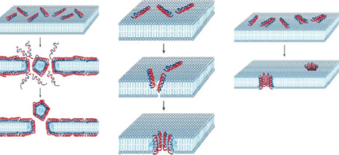
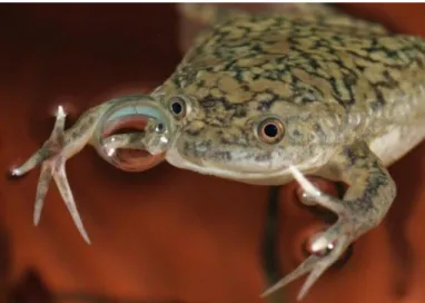
Figure 9: Peptide-membrane interaction is initially mediated by the positive charge of the peptide. The peptides fold into distinct secondary structures, separating the hydrophilic and hydrophobic domains. The amphiphilic peptides then induce disruption of the membrane through three different mechanisms: (A) the carpet model; (B) the barrel-stave model and (C) the toroidal pore model (F. Costa et al., 2011) ...24 Figure 10: Frog Xenopus laevis, also known as African Clawed Frog ...25
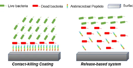
Figure 11: Different strategies to use AMPs on biomaterials. Contact-killing materials kill only by contact, while Release-based systems deliver AMPs locally, and direct contact between the microorganism and material is not required. ...27 Figure 12: Schematic representation of the steps involved in the production of MSI-78 (4-20) chitosan ultrathin films. First gold plates were washed and ultrathin films of chitosan were deposited by spincoating in the surface. Then, a PEG crosslinker and MSI-78 (4-20) were successively covalently immobilized. ...35
x
Figure 13: Brugnerotto equation used for DA determination using the two peaks located approximately at 1320 cm.1 and 1420 cm-1. ...37
Figure 14: Schematic representation of the chemical reaction used for MSI-78 (4-20) immobilization. Conjugation was performed using a PEG heterobiofuncional crosslinker, with N-hydroxysuccinimide (NHS) ester and maleimide groups that allow covalent conjugation with the amines of chitosan and the sulfhydryl group of the peptide. ...39
Figure 15: Schematic representation of the method used to test antimicrobial activity. ...43
Figure 16: After staining, samples were placed in a microscope slide and mounted as exemplified in the figure, using two gold plates to obtain the needed height to place and fixate the coverslip on top properly. ...43
Figure 17: Scheme of AMP-PU films production using Solvent Casting. ...45
Figure 18: Struture of 5(6)-Carboxytetramethylrhodamine - Sigma Aldrich catalogue, product number 21953. ...46 Figure 19: Polyurethane films were cut using a 13-mm metal puncher. ...48
Figure 20: Equation for calculation of corrected concentration of released AMP. ...48
Figure 21: Schematic representation of the method used to test the antimicrobial activity. ...50 Figure 22: Schematic representation of the protocol followed after 5-hour incubation. The coverslip was removed and the supernatant was retrieved, diluted and plated for CFU counts ...50 Figure 23: FTIR spectrum of Chitosan ...53
Figure 24: Peaks of the FTIR spectrum of chitosan used for DA determination ...54
Figure 25: Surface characterization of chitosan modified films with SM(PEG)8 and
Cys-MSI-78(4-20) / Cys-MSI-78(4-20)-Cys using ellipsometry (thickness of surface). Statistically significant differences are indicated with **** (p<0.0001). ...56
Figure 26: Surface characterization of chitosan modified films with SM(PEG)8 and Cys-MSI- 78(4-20)/ MSI- 78(4-20)-Cys using contact angle measurement. Statistically significant differences are indicated with **** (p<0.0001) ...57
Figure 27: Pictures taken after 3 min of contact between a drop of water and functionalized samples. ...58
xi
Figure 28: Equation for determination of the kinetic energy of photoelectrons in XPS ...58 Figure 29: XPS survey spectra of the different modified surfaces ...60
Figure 30: XPS high resolution spectra of carbon of the different modified surfaces. ...61
Figure 31: Antimicrobial activity of MSI-78(4-20) chitosan films. Quantification of bacterial adhesion using the Live/dead BacLight™ kit. Statistically significant differences are indicated with **** (p<0.0001). ...63 Figure 32: Representative images of bacterial adhesion to AU, chitosan and chitosan modified surfaces. Images were obtained by IFM with 400 x magnification. ...64 Figure 33: Release profile of MSI-78 (1-17) in (A) PBS and (B) 1 % Human Plasma66
Figure 34: Release profile of MSI-78 (2-18) in (A) PBS and (B) 1% Human Plasma 66
Figure 35: Antimicrobial activity of MSI – 78 (1-17) and MSI – 78 (2-18) – PU films regarding non-adherent viable bacteria. (A) in PBS and (B) in 1 % Human Plasma. ...68
Figure S1: Evaluation of SM(PEG)8 structure by NMR. NHS group (2.84 peak) is
present in the expected amounts however, maleimide group (6.69 peak), responsible for the bounding to the sulfhydryl group of the peptide, was present in levels around 50%. This quantification was performed by experts and required peak modulation, which is not presented here as it out of our expertise. ...73 Figure S2: Calibration curve of MSI-78 (1-17)-5(6)-TAMRA. The peptide was dissolved in 1% acetic acid. ...74 Figure S3: Calibration curve of MSI-78 (2-18)-5(6)-TAMRA. The peptide was dissolved in 1% acetic acid. ...74
xii
Table List
Table 1: List of 17-mer derivatives designed by Monteiro et al., including its name, size, amino acid sequence and Minimum inhibitory concentration for S. epidermidis (Monteiro et al., 2015) ...26
Table 2: Information about non-modified and cysteine-modified MSI-78 (4-20)...40
Table 3: Information about MSI-78 (1-17) and MSI-78 (2-18). ...46
Table 4: Information about fluorescently modified MSI-78 (1-17) and MSI-78(2-18). 46
Table 5: Values of the peaks used for DA determination ...52
Table 6: Atomic composition (%) of the different surfaces obtained from XPS element analysis...60 Table 7: Relative percentage (%) of the different Carbon bonds obtained from high-resolution XPS spectra. ...61
xiii
Abbreviations and symbols
5(6)-TAMRA – 5(6)-Carboxytetramethylrhodamine Aap – Accumulation-associated protein
AMP – Antimicrobial Peptide
ATR-FTIR – Attenuated Total Reflection – Fourier-Transform InfraRed Spectroscopy C – Carbon
CEMUP – Centro de Materiais da Universidade do Porto CFU – Colony Forming Units
CoNS – Coagulase-negative Staphylococcus DA – Deacetylation degree
ddH2O – deionized water
DMSO – Dimethyl sulfoxide ECM – Extracellular matrix
Embp – Extracellular matrix binding protein EPS – Extracellular Polymeric Substances GAG – Glucosaminoglycans
H2O2 – Hydrogen peroxide
H2SO4 – Sulfuric acid
IFM – Inverted Fluorescence Microscope IFN-γ – Interferon-γ
IL-12 – Interleukin-12 IR – InfraRed
KBr – Potassium bromide
MCP-1 – Monocyte chemoattractant protein – 1 MIC – Minimun Inhibitory Concentration
MSI-78 – Pexiganan
MSI-78 (1-17) – Pexiganan derivate with aminoacids from 1 to 17 MSI-78 (2-18) – Pexiganan derivate with aminoacids from 2 to 18 MSI-78 (4-20) – Pexiganan derivate with aminoacids from 4 to 20 MW – Molecular Weight
N – Nitrogen
xiv NaOH – Sodium Hydroxide
NHS – N-hydroxysuccinimide
NMR – Nuclear Magnetic Resonance O – Oxygen
OD – Optical Density
OCA – Optical Contact Angle PBS – Phosphate-buffered saline PEG – Polyethylene glycol PI – Propidium iodide PU – Polyurethane
rpm – rotations per minute RT – Room Temperature S – Sulphur
SD – Standard Deviation
SM(PEG)8 – Succinimidyl-(N-maleimidopropionamido)-octaethyleneglycol ester
TCEP – Tris(2-carboxyethyl)phosphine THF – Tetrahydrofuran
TSA – Tryptic soy agar TSB – Tryptic soy broth
1
Chapter 1 - General Introduction
1.1 Motivation and Aim
The use of Biomaterials to restore a function to damaged tissues or as medication systems is widely spread. So far, some challenges persist, such as biomaterials-associated infections, clotting problems that can result in thrombosis or difficult tissue integration. Ultimately, these problems can lead to biomaterials failure and in extreme cases, biomaterial substitution or removal. Biomaterials associated infections, besides being expensive and burdensome, are difficult to eradicate using ordinary treatments, namely antibiotics and biocides. Treatment inefficiency is mainly caused by specific antibiotic resistance and biofilm formation, a very relevant problem in biomaterials associated infections.
In the past years, research has been exploring different strategies to solve this problem, namely biomaterials surface modifications, development of delivery systems and new and efficient antimicrobial substances. Antimicrobial peptides (AMP) appear as promising antibiotic alternatives that can be combined with biomaterials to improve biomaterials performance. Thereby, AMP immobilization or incorporation into biomaterials are interesting strategies to prevent or treat biomaterials-associated infections.
The major goal of this dissertation is the development of antimicrobial coatings based on AMPs to reduce bacterial colonization and decrease the rate of biomaterials-associated infections. Three different AMPs designed by our team have been used, MSI-78 (4-20), MSI-MSI-78 (1-17) and MSI-MSI-78 (2-18) (Monteiro et al., 2015). These 17-mer MSI-MSI-78 derivatives are cost effective short AMPs, derived from the well-known MSI-78 (pexiganan), a 22-mer AMP. Despite the truncation of five amino acids, all these derivatives maintain antimicrobial efficacy against S. epidermidis being MSI-78 (4-20) the most efficient derivative, showing similar antimicrobial activity to MSI-78 and more selectivity towards bacterial cells.
Two approaches with different aims have been investigated: 1) Development of a contact killing coating based on AMP-chitosan films to be used as coating on orthopaedic implants, 2) Development of a release-based coating, a AMP-PU film for intravascular catheters.
1) Development of AMP-chitosan films based on the covalent immobilization of the AMP MSI-78 (4-20) on chitosan ultrathin films. Chitosan was chosen due its
2 osteogenic and antimicrobial properties. This project involved the following objectives: 1) Preparation of AMP-chitosan thin films using the free amine groups of chitosan. Covalent immobilization was done through a SM(PEG)8 spacer, a heterobiofuncional crosslinker with N-hydroxysuccinimide (NHS) ester and a maleimide group that allow conjugation with the amines of chitosan and the sulfhydryl group of Cys-MSI-78 (4-20)/ MSI-78 (4-20)-Cys. 2) Characterization of the antimicrobial activity of AMP-chitosan thin films by quantification of bacterial adhesion/viability on the coating.
2) Development of AMP-PU films based on the physical incorporation of the AMPs MSI-78 (1-17) and MSI-78 (2-18) on polyurethane (PU). Pellethane 2363-80 AE was selected for this study due to its application in the production of intravascular catheters, its versatile physical properties and hemocompatibility. This project involves the following objectives: 1) preparation of AMP-PU films by AMP incorporation during PU film production. 2) Characterization of AMP release kinetics and antimicrobial activity.
1.2 Dissertation Structure
In this dissertation, a literature review was performed, presenting the state of the art and the current knowledge and information about the subjects to be studied.
Biomaterials-associated infections, with focus on the main bacteria involved and its major virulence factor – biofilm production, are reviewed in the second chapter of the dissertation. These infections can affect different biomaterials, as orthopaedic implants and intravascular catheters. Characteristics of the two main polymers used, chitosan for orthopaedic implants and polyurethane for intravascular catheters are also presented.
Strategies to overcome biomaterials associated-infections are described and antimicrobial peptides (AMP) are reviewed in detail as they have been explored in this study to improve the antimicrobial properties of biomaterials. Delivery vs immobilization systems and the factors that influence their performance are presented.
Materials and Methods used in this work are described in Chapter 3. The obtained results are presented and discussed in Chapter 4. And finally, in Chapter 5, conclusions and future perspectives are presented.
3
Chapter 2 - Literature Review
2.1 Biomaterials
The definition of Biomaterials has been changing over the years along with the evolution of the field. Nowadays biomaterials are defined as “a substance that has been engineered to take a form which, alone or as part of a complex system, is used to direct, by control of interactions with components of living systems, the course of any therapeutic or diagnostic procedure, in human or veterinary medicine” (Williams, 2009).
Nowadays, the use of biomaterials that can restore a function to damaged tissues is widely spread. However, their application still involves some challenges, such as clotting problems that can lead to thrombosis (Jaffer, Fredenburgh, Hirsh, & Weitz, 2015; Uludag, 2014), deficient tissue integration (R. Agarwal & García, 2015) and biomaterials-associated infections (Neethirajan, Clond, & Vogt, 2014). A foreign body inside the organism does not have contact with efficient blood supply having therefore a restriction of the immune cells and antibodies to clear possible existing infections (Werner Zimmerli & Sendi, 2011), making them very vulnerable.
2.1.1 Biomaterials-associated infections
Bacterial, fungal and viral agents contribute to almost 2 million cases of hospital associated infections, the so called nosocomial infections, that have a high economic impact (Salwiczek et al., 2014) and lead to approximately one hundred thousand deaths per year (Green, Fulghum, & Nordhaus, 2011; Sun, Shahzad, Li, Wang, & Xu, 2014). Biomaterials-associated infections, are very often acquired in hospitals and medical facilities - during implant surgery or hospitalization (Busscher et al., 2012) – contributing to the high numbers of nosocomial infections. Device implantation/insertion actually facilitates infection, since microbes introduced into the surgical/insertion site may be capable of adhering and accumulating on the device’s surface (Brescó et al., 2017). As such, the majority of biomaterials-associated infections has its source on direct contact with healthcare settings in medical environments (Hocevar et al., 2012) however, other sources such as temporary bacteraemia with origin in other infections in the body may be also cause contamination
4 (Salwiczek et al., 2014). Moreover, despite causing infection, microbial attachment can reduce the sensitivity and efficacy of devices, modifying its therapeutic action (Hongbin Zhang & Chiao, 2015). Several publications review the details of pathogenesis, diagnosis, prevention and treatment of biomaterials-associated infections, being biofilm formation the main mechanism through which infection establishment occurs (Vergidis & Patel, 2013; Werner Zimmerli & Moser, 2012). In humans, around 80 percent of all nosocomial infections are due to biofilm formation (Römling & Balsalobre, 2012; Singh & Gupta, 2015; van Kleef, Robotham, Jit, Deeny, & Edmunds, 2013) and their establishment can occur in devices such as orthopaedic implants and intravascular catheters (Francolini & Donelli, 2010).
Typical treatment methods for contaminated devices include device replacement and prolonged antibiotic therapy that leads to additional health care costs due to long hospitalization periods and increased mortality (Banerjee, Pangule, & Kane, 2011; Jansen, Kirstinsson, Jansen, Peters, & Pulverer, 1992).
There are some approaches to reduce device-associated infections based on antimicrobial materials via incorporation of antibiotics or biocides. However, difficulties in controlling antibiotics release may lead to sub-lethal concentrations that can be responsible for promoting bacterial resistance and formation of a protective environment to evade antibiotics action. As microbial colonization is the prelude to device-associated infections (Hall-Stoodley, Costerton, & Stoodley, 2004; K. Lewis, 2001), the development of antimicrobial biomaterials that can prevent the early stages of bacterial colonization is being the focus to fight biomaterial-associated infections (Cloutier, Mantovani, & Rosei, 2015).
2.2 Coagulase-negative Staphylococcus (CoNS)
The human skin is the biggest organ in the body and acts as a physical barrier, protecting the organism from the outside environment, being colonized by a multitude and diversity of microorganisms (Grice & Segre, 2011). Staphylococcus, namely Coagulase-negative Staphylococcus (CoNS), are gram-positive bacteria and represent a regular part of the skin microbiota of humans and animals, as described by Becker (Becker, Heilmann, & Peters, 2014).
CoNS, historically regarded as non-pathogenic strains, are highly opportunistic pathogens, (Shin et al., 2011) and the most frequent cause of nosocomial infections (Wisplinghoff et al., 2003). Their infection comprises either local or bloodstream-related
5 entities and is highly associated with implanted or inserted medical devices. It is important to comprehend CoNS infection mode to successfully prevent infections (von Eiff, Jansen, Kohnen, & Becker, 2005), especially with the increasing use of medical devices (Weinstein et al., 1997).
The majority of CoNS infections is caused by a specific Staphylococcus strain – S. epidermidis, both in the case of orthopaedic devices and intravascular catheters-associated infections – Figures 1 and 2. This strain has become the most important and well-studied model organism for device-associated infections as it is a very common cause of bacteraemia (Sievert et al., 2013).
Figure 1: Main microorganisms that cause orthopaedic device-associated infections, based on Montanaro et al
(Montanaro et al., 2011)
Figure 2: Incidence of microorganisms in central venous catheters-associated bloodstream infections, based
on Patil et al. (Patil, Patil, Ramteerthkar, & Kulkarni, 2011)
S. epidermidis 39% S. aureus 32% P. aeruginosa 6.10% E. faecalis 2.40% S. warneri 4.90% S. hominis 2.40% E.coli 2.40% Others 11.10% Staphylococcus epidermidis 45% Staphylococcus haemolyticus 15% Staphylococcus aureus 15% Klebsiella pneumoniae 10% Escherichia coli 5% Candida albicans 5%
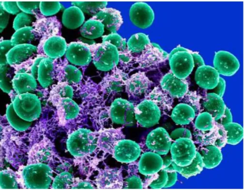
6 2.2.1 Staphylococcus epidermidis
Staphylococcus epidermidis (Figure 3) is a Gram-positive and coagulase-negative bacteria, commensal of the human skin, mucosal surfaces (Namvar et al., 2014) and also a colonizer of epithelial cells (Michael Otto, 2009) being more prevalent in moist areas, such as the axillae, inguinal and perineal areas, conjunctiva and toe webs (Becker et al., 2014). Considering the tremendous abundance of this strain on the human skin, it is not surprising that it is considered the most common cause of device-associated infections (Mack, Horstkotte, Rohde, & Knobloch, 2006) as it can be easily introduced during implantation/insertion, even though its regarded as harmless commensal inhabitant of the skin (M. Otto, 2008).
S. epidermidis is considered as the most frequent cause of nosocomial infections (Cerca et al., 2005; Rogers, Fey, & Rupp, 2009; Von Eiff, Peters, & Heilmann, 2002), that leads to biomaterials-associate infections, especially due to its ability to form biofilms (F. Gomes, Teixeira, & Oliveira, 2014). Devices infection most likely occurs during device insertion, after material surface contamination that permits the inoculation of a small number of bacteria from the patient’s skin or mucous membranes (Becker et al., 2014; Ramasamy & Lee, 2016). These infections caused by S. epidermidis are amongst the most burdensome, expensive and difficult to treat (Brescó et al., 2017).
7
2.3 Biofilms
Bacteria survive in physiological environments by assembling and forming communities embedded into a self-produced extracellular matrix by adhering to each other and/or to surfaces and interfaces (Costerton & Lewandowski, 1995). This phenomenon is known as biofilm formation (Salwiczek et al., 2014) and is considered as one of the major virulence factors (Vuong & Otto, 2002). In addition to microbial components, for example secreted macromolecules, biofilms may contain non-cellular materials, such as mineral crystals, particles formed due to corrosion and even blood components (Singh & Gupta, 2015). This biofilm environment protects individual cells from hostile factors, such as immunological responses, nutrient limitations and even antibacterial agents (Ramasamy & Lee, 2016).
The ability of S. epidermidis to form biofilms is regarded as the most important non-specific mechanism of resistance (M. Otto, 2008) and often leads to persistent infections (Costerton, 1999) as bacteria in biofilms resist antibiotic therapy (Arciola, Campoccia, & Montanaro, 2002; Monzón, Oteiza, Leiva, Lamata, & Amorena, 2002).
2.3.1 Protein adsorption
In the context of biomaterial-associated infections, biofilms are the result of the interaction between microorganisms, devices surfaces and host proteins present. Biomaterials and medical devices, upon implantation into living tissues or in contact with blood, induce tissue responses (Anderson, 2001), protein adsorption (Bhakta, Evans, Benavidez, & Garcia, 2015), immune recognition and immunological response (Gristina, Shibata, Giridhar, Kreger, & Myrvik, 1994; Moyano, Liu, Peer, & Rotello, 2016) that can lead to the reduction of efficacy and may result in harmful side effects. Within seconds after implantation, protein adsorption occurs on the biomaterials surface and can induce foreign body reactions (Ratner, 2001), being a concern for the design and application of biomaterials (Nakanishi, Sakiyama, & Imamura, 2001). This biological response can also result in the device’s isolation, due to the production of a fibrous avascular capsule, reducing its effectiveness (Meyers & Grinstaff, 2012).
Protein adsorption from serum-containing media is identified as a mild immobilization process with great potential due to the maintenance and preservation of the
8 native structure of the elements, widely studied for a long time for the production of inert surfaces (Amirgoulova et al., 2004; Ostuni, Chapman, Holmlin, Takayama, & Whitesides, 2001). It is a process driven mainly by hydrophobic, electrostatic and van der Walls interactions (Bhakta et al., 2015) and is a fast and simple way to attach proteins to surfaces. The rate and the strengths of the initial interactions between proteins and the surface determine the conformation, stability and the activity of those proteins. Besides, this issue is crucial in determining the biocompatibility of materials (Barnthip, Parhi, Golas, & Vogler, 2009; Roach, Farrar, & Perry, 2005).
Initially, highly mobile proteins adsorb to the surface but are replaced, with time, by other proteins with more affinity, the well-known “Vroman effect” (Jung et al., 2003; Noh & Vogler, 2007; Vroman & Adams, 1969). Some proteins that adhere to the biomaterial surface are fibronectin, fibrin, among others like albumin (Francolini & Donelli, 2010).
Protein adsorption is considered the true interface, the most important factor, of the interaction between biomaterials and body fluids or even tissues (Wei et al., 2014) and the prevention of nonspecific protein adsorption on the surface is crucial to improve the biomaterials’ biocompatibility (Wei et al., 2014). Adsorbed proteins form a layer that provides a prone environment for biofilm formation and microbial colonization of the surface. This phenomenon, which is initially dependent on the biomaterial physical and chemical properties and on the composition of the surrounding fluids, is mediated by different types of interactions and allows bacterial adhesion and colonization. (Banerjee et al., 2011).
2.3.2 Biofilm formation
Most of the bacterial species are well-known biofilm-producers (Jouenne, 2006). Biofilms on indwelling devices can be composed of both gram-positive and gram-negative bacteria. The gram-positive bacteria most common are Staphylococcus epidermis, Staphylococcus aureus, Streptococcus viridans and Enterococcus faecalis, while among gram-negative bacteria Klebsiella pneumoniae, Escherichia coli, Proteus mirabilis and Pseudomonas aeruginosa (Rodney M. Donlan, 2001) can be found. These bacterial strains can form biofilms on both abiotic and biotic surfaces, including different types of medical devices, such as prosthesis and catheters (Steven L. Percival et al., 2012; Seth et al., 2012).
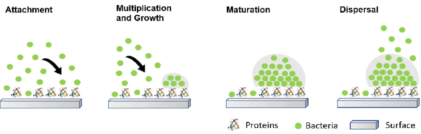
The formation of biofilms on biomaterials surfaces is well characterized and can be divided into four different stages as can be seen in Figure 4.
9 Stage 1 – attachment
The attachment process involves two different phases: the reversible and irreversible attachment. The first consists in the identification of the surface by free-floating microorganisms and is characterized by non-specific cellular association bacteria-surface, by van der walls forces, electrostatic forces, hydrophobic interactions, and even polar or ionic interactions (R M Donlan, 2001; Von Eiff et al., 2002). The second phase, irreversible attachment, occurs due to specific adhesions and is mediated by a preformed film of biomolecules and proteins from plasma components, that provide specific binding sites for bacterial surface proteins - adhesins (Gottenbos, Busscher, Van Der Mei, & Nieuwenhuis, 2002). In the case of S. epidermidis, different adhesins like polysaccharide intercellular adhesin (PIA), accumulation-associated protein (Aap) and extracellular matrix binding protein (Embp) mediate aggregation and biofilm accumulation (Schommer et al., 2011).
Stage 2 – multiplication and growth
The bacteria newly attached produce extracellular polymeric substances (EPS), that induce colonization of the surface and form a conditioning film (Singha, Locklin, & Handa, 2017). This stage comprises the formation of microcolonies after local multiplication and assembly into an aggregate. The formed microcolony depends on the secreted and surface-adsorbed biomolecules (Michael Otto, 2009).
Stage 3 - maturation
Recruitment of more microorganisms, bacterial adaptation, biofilm maturation and convergence of microcolonies leads to biofilm development (Singh & Gupta, 2015). This stage comprises the formation of a structured and thick layer containing channels, filled with fluids, to ensure oxygen and nutrient delivery to bacterial cells located in the deeper layers of the biofilm (Costerton, Stewart, & Greenberg, 1999).
Stage 4 – dispersal
After biofilm structuring, certain environmental cues and self-stress signals promote the detachment of single bacteria or agglomerates from the biofilm (Bester, Wolfaardt, Joubert, Garny, & Saftic, 2005) that return to planktonic model. In this mode, microbes are translocated or disseminated through the bloodstream and may attach to a new location, and so, there is a higher risk of infection spreading (Stoodley et al., 2005).
10 Figure 4: Four stages of biofilm formation. Adapted from Salwiczek et al. (Salwiczek et al., 2014).
2.3.3 Effect of Biofilm
Changes in the physiology of the biofilm environment influences virulence and pathogenicity of microorganisms (Singh & Gupta, 2015). Biofilm formation is accompanied by significant changes in gene and protein expression as well as metabolic activity (Dötsch et al., 2012; Sanchez et al., 2013).
The major hallmarks of biofilms in vivo are the presence of aggregated bacteria that tolerate the host defense and high concentrations of antimicrobial agents for even longer periods of time due to adaptive and genetic changes and physical protection by the matrix (Bjarnsholt, Alhede, & Alhede, 2014; Cloutier et al., 2015). Biofilm structure also resists the adaptive and innate immune system responses of the host by avoiding its action in the bacteria (Jensen, Givskov, Bjarnsholt, & Moser, 2010).
The development of resistance towards antibiotics, can be attributed to four possible characteristics of the biofilm (Stewart & William Costerton, 2001), including:
1) the matrix and layer of cells in the biofilm which create a physical barrier to slow down or cause incomplete penetration of the antibiotics and antimicrobial agents (Francolini & Donelli, 2010; Lynch & Robertson, 2008);
2) formation of a resistant phenotype, persister cells, that remain in a transient and dormant state and can cause recurrent infections (Kim Lewis, 2007). These metabolically less active bacteria facilitate resistance towards antimicrobial agents (Corbin, Pitts, Parker, & Stewart, 2011), surviving in a dormant state for long-lasting periods, evading antibiotics and host immune responses for up to several years before awakening in more virulent modes (Costerton et al., 1999);
11 3) the metabolic activity in the outer biofilm can create acidic or anoxic areas that can
contribute to antibiotic degradation (McConoughey et al., 2014);
4) Moreover, as transport by diffusion is also limited, matrix components can create a gradient of antibiotic exposure. In this case, the population within the biofilm is exposed to sub lethal doses of antibiotics and can consequently develop resistance (Jefferson, Goldmann, & Pier, 2005).
2.4 Orthopaedic Devices
Orthopaedic implants are mainly used for bone fixation or joint replacement (Kurtz et al., 2008) and have particular features such as high biocompatibility and resistance to corrosion (Koseki et al., 2014). Internal fixation devices used after a fracture are temporary and removed after bone healing (W. Zimmerli, 2014). Prosthetics joints replace damaged articulations, alleviate pain and improve function (Jämsen et al., 2013), usually due to osteoarthritis (joint degeneration), that is characterized by pain, loss of function and deformity (B. G. X. Zhang, Myers, Wallace, Brandt, & Choong, 2014). Over the past decades, there is an increasing need of medical devices in orthopaedics due to trauma situations, osteoporotic diseases, bone cancer, joint and spinal diseases, among others (Lin et al., 2017).
Different materials can be used for orthopaedic devices’ production, from metals to ceramics and polymers. Polymers are being more used due to its properties and functionalization capacities. Besides, the type of prosthetic has to be considered when choosing the material to have the desirable morphology, topography and specific characteristics for the proposed function (Getzlaf et al., 2016).
Bone tissue infections represent some of the worst possible complications in orthopaedics (Moriarty et al., 2016), especially after device implantation (Dibartola, Swearingen, Granger, Stoodley, & Dusane, 2017). Since 2001, in United States of America, around 2.6 million orthopaedic devices were implanted and approximately 4.3% of those became infected. Direct medical costs associated with these infections exceed $3 billion annually and are expected to increase as more implants are being implanted each year (Yin & Luan, 2016).
Improvement of orthopaedic devices to inhibit bacterial colonization while promoting desirable tissue responses (Lin et al., 2017) is of uttermost importance.
12 2.4.1 Chitosan
Chitin and its derivatives, especially chitosan, are known for their excellent properties and are widely studied as orthopaedic devices coatings. These materials are biocompatible, biodegradable, nontoxic and possess inherent antimicrobial and antibacterial activities (Gupta & Edwards, 2009).
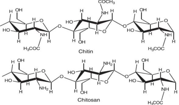
Chitin is a biological material, a linear polymer and is composed of N-acetyl-D-glucosamine residues liked by β(14)-bonds (Figure 5). It is widely available, inexpensive and obtained from the skeleton of invertebrates, from shells of crustaceans or from the cell wall of fungi (Jayakumar, Prabaharan, Sudheesh Kumar, Nair, & Tamura, 2011).
Chitosan is produced by deacetylating chitin and is a linear natural biopolymer composed of random units of 2-acetamido-2-deoxy-β-ᴅ-glucan (N-acetyl-ᴅ-glucosamine) and 2-amino-2-deoxy-β-ᴅ-glucan (ᴅ-glucosamine), linked by β(14) bonds (Jayakumar, Nagahama, Furuike, & Tamura, 2008). The structure of chitosan is presented in Figure 5, where the acetyl group from chitin is eliminated leaving a free amino group.
Chitosan has similar structure to Glycosaminoglycans, also known as GAG, present in the extracellular matrix (ECM). Depolymerization of chitosan can be performed by enzymes as glucosaminidases, lipases and lysozyme and its monomeric product, glucosamine is metabolized or excreted from the body, explaining chitosan’s biodegradability and biocompatibility (Logithkumar et al., 2016). Chitosan is also known for satisfying most of the properties needed for its use for tissue engineering applications (Jiang, James, Kumbar, & Laurencin, 2014; Niranjan et al., 2013; Saranya, Saravanan, Moorthi, Ramyakrishna, & Selvamurugan, 2011).
Chitosan can be easily processed into different types of matrices, from hydrogels (Nagahama, Kashiki, et al., 2008; Nagahama, Nwe, et al., 2008; Tamura, Furuike, Nair, & Jayakumar, 2011), membranes (Madhumathi et al., 2009; Marreco, Da Luz Moreira, Genari, & Moraes, 2004; Yusof, Wee, Lim, & Khor, 2003), nanofibers (Shalumon et al., 2009, 2010), beads (Jayakumar, 2006), particles (Anitha et al., 2009, 2011; Prabaharan, 2008), scaffolds (Maeda, Jayakumar, Nagahama, Furuike, & Tamura, 2008; Peter et al., 2009) to sponges (Muramatsu, Masuda, Yoshihara, & Fujisawa, 2003; Portero, Teijeiro-Osorio, Alonso, & Remuñán-López, 2007).
Chitosan is the only natural cationic polysaccharide that is protonated at pH bellow 6.5, its pKa, being therefore able to interact with negatively charged compounds, which can be advantageous for instance for production of biomaterials combining chitosan and a negatively charged polymer (Campana, Casettari, Ciandrini, Illum, & Baffone, 2017).
13 Chitosan pKa also influences its solubility in aqueous solutions leading to a limited solubility at neutral pH, which is advantageous for use at physiological pH (Logithkumar et al., 2016). Chitosan antimicrobial activity is well characterized and as a film, chitosan displays antimicrobial activity against a wide range of microorganisms and efficiently reduces or even prevents bacterial adhesion and biofilm formation when coated onto surfaces (Carlson, Taffs, Davison, & Stewart, 2008; Martinez et al., 2010).
The molecular weight of chitosan may influence its antimicrobial activity. Lower molecular weight is associated with reduced microorganisms growth and multiplication (Goy, Britto, & Assis, 2009). Size and conformation, namely, the mobility and ionic interaction of small chains is easier than big ones, facilitating an extended conformation and an effective binding to the membrane surface (Vishu Kumar, Varadaraj, Gowda, & Tharanathan, 2005). So, lower molecular weight chitosan is associated to higher antimicrobial activity.
Even though chitosan chemical and biological properties are easily adjustable (Kong, Chen, Xing, & Park, 2010; Rinaudo, 2006), in certain applications these properties are not sufficient to obtain the desired results. In that case, functionalization of chitosan structure through chemical modifications of the amine and hydroxyl groups offer a solution for the limitations (Logithkumar et al., 2016).
Due to its tunable properties, antimicrobial properties and easy functionalization, chitosan is a promising material for the production of coatings for medical devices.
Figure 5: Structure of Chitin and Chitosan based on Jayakumar, where the acetyl group from chitin is eliminated,
14 2.4.2 Chitosan’s degree of acetylation
The degree of acetylation (DA) of chitosan is referred to the fraction of N-acetyl glucosamine to glucosamine units in the chain (Croisier & Jérôme, 2013). The deacetylation process of chitin to obtain chitosan provides available amine groups, increasing chitosan’s reactivity and solubility when compared to chitin (Gupta & Edwards, 2009).
This parameter influences charge density, crystallinity and solubility and can be determined by different techniques as FTIR (Kasaai, 2009), Raman (Zaja̧c, Hanuza, Wandas, & Dymińska, 2015) and NMR spectroscopy (Thevarajah, Bulanadi, Wagner, Gaborieau, & Castignolles, 2016).
Chitosan’s antimicrobial activity is increased as the degree of acetylation decreases (Andres, Giraud, Gerente, & Le Cloirec, 2007; G J Tsai, Zhang, & Shieh, 2004). Studies with chitin and chitosan with different DA were made, analysing its activity against fungi, gram-positive and gram-negative bacteria. In all cases, the antimicrobial activity is increased with decreased DA (Andres et al., 2007; Guo Jane Tsai, Su, Chen, & Pan, 2002).
The degree of acetylation of chitosan also influences degradation, with a higher DA being related to quicker degradation (Kean & Thanou, 2010; Şenel & McClure, 2004; Tomihata & Ikada, 1997; Hua Zhang & Neau, 2002). On the other side, a lower DA is associated with better cell responses. Amaral el at tested the influence of chitosan’s DA in the cellular response and osteogenic differentiation of rat bone marrow stromal cells (rBMSCs). Using low DA (4%) chitosan, rBMSCs were able to spread, proliferate and differentiate better, reaching higher differentiation levels when compared to high DA chitosan (Amaral, Lamghari, Sousa, Sampaio, & Barbosa, 2005).
Besides, it is hypothesized that DA can affect the layer of adsorbed proteins, which in turn affects cell behaviour, including attachment, spreading, among others (Amaral, Lamghari, et al., 2005). Lower DA is known for favouring cell adhesion (Chatelet, Damour, & Domard, 2001; Prasitsilp, Jenwithisuk, Kongsuwan, Damrongchai, & Watts, 2000).
An explanation for the effect of lower DA in the properties presented above is the presence of -OH and more -NH2 groups available to react (Andres et al., 2007). So, lower DA is usually associated to higher antimicrobial effect, improved cell behaviour and low biodegradation rate.
15
2.5 Intravascular catheters
The use of intravascular catheters is related to management of hospitalized patients, either as a delivery system of blood products (transfusions), fluids, nutritional solutions and medications (chemotherapy, among others) or as an access in dialysis treatment (R. M. Donlan, 2008; Hewlett & Rupp, 2012; S. L. Percival & Kite, 2007).
Materials such as plastic poly(vinylchloride), polyurethanes, silicones and even latex rubbers have been used in the production of catheters due to its superior malleability (Johnson, Kuskowski, & Wilt, 2006). These materials have also been developed to include most of the characteristics desirable in a catheter, such as, high tensile strength, soft, pliable and inherent chemical resistance and biocompatibility, without discarding the flow requirements (Singha et al., 2017).
Catheters are highly susceptible to microbial accumulation, either by single bacteria or clustered cells that form biofilm, causing infections (Stickler, 2014). Microorganisms are introduced at the catheter insertion site and can adhere to both the inner and outer surface of the catheters to form a biofilm (Donelli & Vuotto, 2014; Rodney M. Donlan, 2011).
Infections in catheters are the leading cause of nosocomial infections and are associated with significant morbidity or mortality (L. Zhang & Rickard, 2017). Around 80 000 catheters get infected among patients in United States intensive care units per year (Mermel, Mermel, & Hudson, 2000), but 250 000 are estimated when considering data from hospitals (Maki, Kluger, & Crnich, 2006). When these infections are left untreated, they can be disseminated through the bloodstream, leading to sepsis (septicemia) or even death in extreme cases (Steven L. Percival, Suleman, & Donelli, 2015; Singha et al., 2017).
2.5.1 Polyurethane (PU)
Polyurethane is a widely used synthetic polymer in biomedical applications, originated from petroleum products (Zuber et al., 2015), due to its adequate biocompatibility and mechanical properties (Akduman, Özgüney, & Kumbasar, 2016; Lee, Hong, & Lee, 2007).
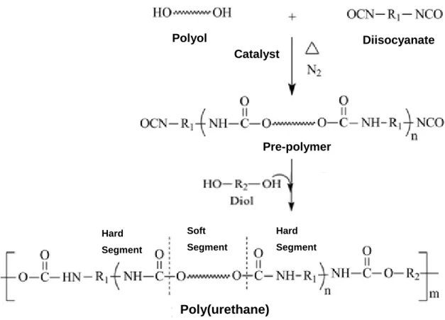
PU is synthesized through a two-step reaction (Figure 6). First, the isocyanate group from the diisocyanate compound reacts with the hydroxyl group of the polyol soft segment,
16 by a urethane linkage (Mahmood Zia, Ahmad, Anjum, Zuber, & Naveed Anjum, 2015; Rahimi & Mashak, 2013; Sattar, Kausar, & Siddiq, 2014), forming the prepolymer. In the second step, the chain extender diol (with low MW) is used to link the prepolymer segments, yielding a high MW polymer (Cauich-rodríguez, Chan-chan, Hernandez-sánchez, & Cervantes-uc, 2012) Polyurethane can possess other moieties in the structure, such as urea, aromatic, ester and ether (Chattopadhyay & Raju, 2007; Chattopadhyay & Webster, 2009).
This copolymer possesses linear block polymers with soft amorphous sections and hard crystalline components allowing a subtle control of structure and properties (Kucińska-Lipka, Gubańska, & Janik, 2014), by varying its structural components (Mahmood Zia, Ashair Iqbal, et al., 2015). The first sections permit the stretching while the latter hold the structure together (Gupta & Edwards, 2009). The content of the hard segments influence the degree of phase separation (Janik, Pałys, & Petrovic, 2003; Król & Pilch-Pitera, 2003), affecting the physical and mechanical properties (Ioan, Grigorescu, & Stanciu, 2002) and also the degradation rate (Tang, Labow, & Santerre, 2001).
The PU used in this research is Pellethane 2363 80 AE, a poly(ether-urethane), that results from the reaction of poly(tetramethylene oxide), methylene diphenyl diisocyanate and 1,4-butanediol. Aromatic isocyanates, as the methylene diphenyl diisocyanate, are known for being highly reactive. These groups can be altered by some factor, such as light or temperature, and are responsible for PU stiffness. In this case, the polyol used was poly(tetramethylene oxide) and the diol responsible for the chain extension was 1,4-butanediol.
The major properties that make PU a great material besides the presented above are its durability, elasticity, resistance to fatigue and even compliance, besides acceptance and tolerance by the human body (Guan, Gao, Feng, & Shen, 2001). Besides, PU can have a wide range of mechanical and physical properties, from stable to degradable and from hydrophobic to hydrophilic, depending on their composition and procedure of synthesis (Kucińska-Lipka, Gubańska, & Janik, 2013; Kucińska-Lipka et al., 2014). Even tough PU possesses low microbial colonization rate, good compatibility with blood and bioresorbability (Q. Chen, Liang, & Thouas, 2013) when compared to other materials, the possibility of infections, thrombosis and inflammation related to their use are still a problem. This problem has a big impact due to the high necessity of catheters use, especially in patients with a compromised immune system and in need of prolonged drug delivery or fluids removal (Cotogni & Pittiruti, 2014).
17 Polyurethane is a material that can be used as shaped, soft, stretchable and elastic fibers or even films. PU has diverse applications, either in coatings (Mumtaz, Zuber, Zia, Jamil, & Hussain, 2013), foams, elastomers (Mahmood Zia, Ahmad, et al., 2015) and composites, especially due to their great abrasion resistance, toughness, flexibility at low temperatures, mechanical strength and also resistance to chemicals and corrosion (Tate, Akinola, & Kabakov, 2009). Since PU started being used in the catheters production, their thickness has been minimized while increasing its flow, patients’ comfort has been improved and their properties to prevent thrombosis have been upgraded (Zdrahala & Zdrahala, 1999).
These advantageous features of PU permitted its widespread use in implantable devices from vascular grafts (Detta et al., 2010), lead insulators of pacemakers (Lelah, Michael and Stuart, 1986), intravenous or urinary catheters (Bach et al., 1994), cartilage replacements (Chetty et al., 2008; Eglin, Grad, Gogolewski, & Alini, 2010) to artificial heart valves to medical applications (Gupta & Edwards, 2009; Huynh et al., 2010; Zdrahala & Zdrahala, 1999), among others (Macocinschi, Filip, & Vl, 2011).
Figure 6: Standard two-step reaction to form Polyurethane. Adapted from (Cauich-rodríguez et al., 2012). Catalyst Polyol Diisocyanate Pre-polymer Hard Segment Hard Segment Soft Segment
Poly(urethane)
18
2.6 Antibacterial biomaterials - Strategies to prevent or treat biomaterials-associated
infections
The treatment of biomaterial-associated infections is not desirable as it is a complicated process. In most cases, the best solution to eradicate biofilm-associated infections is the removal of the infected device (Boucher et al., 2009). However, in some cases such as implantable prosthesis and cardiac implants, removal is extremely difficult (Fey, 2010). Then, the use of bioengineered approaches that would prevent biofilm formation is rising as the best way to evade biomaterial-associated infections. Ultimately, improving biomaterials properties is an effective and promising strategy to prevent the mortality associated with biofilm infections (Singh & Gupta, 2015).
Biomaterials can be functionalized to increase devices’ biocompatibility, functionality and reduce microbial contamination in order to prevent biofilm-associated infections (Jaffer et al., 2015; Prasad, 2014) or can also be used to deliver medical substances for prevention, treatment or reduction of infections.
There is an increasing need for more strategies and products to reduce the susceptibility of medical devices to bacterial colonization and biofilm formation and to treat other biofilm-associated infections. Campoccia et al. (Campoccia, Montanaro, & Arciola, 2013), reviewed some of the strategies for the design of antibacterial materials, that are also summarized in the Figure 7.
Figure 7: Schematic representation of the different strategies used to avoid establishment of infections on
19 2.6.1 Low adhesion or bacterial repulsion
This strategy does not kill microorganisms but instead prevents its attachment on surfaces, avoiding biofilm formation (Campoccia et al., 2013; Tenke et al., 2012). Bacterial behaviour and adhesion can vary according to the chemical and physical properties and functional groups present in the surface (Campoccia et al., 2013). Influencing bacterial behaviour or altering the surface arises as a promising strategy to prevent infection by preventing biofilm formation.
The design of surfaces to avoid bacterial adhesion should consider the species of bacteria commonly known to colonize the materials and their characteristics in order to increase efficacy. (Campoccia et al., 2013). The first step for biofilm formation is bacterial adhesion, as such, if adhesion is impaired, infection will not occur.
After implantation, a layer of proteins deposits in the surface of biomaterials and is considered the true interface with bacteria as it provides a prone environment for bacterial attachment. Protein adsorption on a surface can be reduced by modification of the interaction potential or by decreasing the rate of adsorption using barriers for the interaction, with the latter achieved by, for example, polymer grafting and introduction of repulsive forces (Poncin-Epaillard et al., 2012). If the materials are exposed to high concentrations of proteins, it is possible to lower bacterial adhesion by conditioning the surfaces using pre-adsorbed molecules or host adhesins, increasing surfaces hydrophobicity or hydrophilicity. Heparin is one example of a molecule used to reduce bacterial adhesion to catheters and artificial lenses (Nagaoka & Kawakami, 1995). In this case, hydrophilicity is increased by the formation of a highly-hydrated layer in the interface between bacteria and the surface. Heparin in solution has proven to interfere with S. epidermidis adhesion, inhibiting the binding of bacterial adhesins to fibronectin (Arciola et al., 2003; Bustanji et al., 2003).
Another option is the use of compounds that possess hydrophilic and hydrophobic moieties to reduce bacteria’s ability to bind to the surface of prosthetics (Rodrigues, 2011), with the widely known example of polyethylene glycol (PEG). As PEG’s antifouling properties against proteins have been widely reported in the literature (Bearinger et al., 2003; Liu, Jastromb, & Bhatia, 2002; Xia, May, McArthur, & Castner, 2002) and bacterial attachment usually occurs through a layer of adsorbed proteins, it is speculated that using a PEG coating it is possible to impair protein adsorption and so, avoid bacterial attachment (Banerjee et al., 2011). Nowadays, many researchers use PEG-coated surfaces to resist bacterial adhesion (Kenan et al., 2006; Kingshott, Wei, Bagge-Ravn, Gadegaard, & Gram, 2003; Ostuni, Chapman, Liang, et al., 2001; Saldarriaga Fernández, van der Mei,
20 Lochhead, Grainger, & Busscher, 2007). However, different studies have contradictory results as it was suggested that a small quantity of microbes could adhere to these so called “anti-adhesive” surfaces and after adhesion, modifications could occur and create a conditioning film that would permit the binding of more microbes (Bryers, 2008).
The morphology and topography of a surface can also be modified to reduce adhesiveness to bacteria even if there is not an enhanced bactericidal activity (Montanaro, Campoccia, & Arciola, 2008).
2.6.2 Bactericidal Activity
This strategy aims to kill the microbes instead of reducing its deposition, protecting the patients from infections, decreasing its associated hospital care costs. These agents can either be embedded in the polymer and released to kill bacteria or can be bond to the surface of materials and kill microbes upon contact (Singha et al., 2017).
Materials that can exert antibacterial action without modifications or functionalization can be described as intrinsically antibacterial. Some examples of these materials are metals as silver, copper and zinc and polymers as chitosan and bioactive glasses. These metals are usually associated with toxicity and so, their use must be considered according to the objective. Polymers do not pose that problem. Chitosan is one example of a intrinsically bioactive polymer derived from chitin, known for its intrinsic antibacterial and antifungal activities, usually enhanced at low pH (Muzzarelli et al., 1990). Different derivatives of chitosan were developed to exalt the antibacterial properties of chitosan.
Bioactive antibacterial coatings have been developed to achieve anti-infective properties at the interface between the biomaterial and the tissues, without affecting the characteristics of the bulk material. Bulk materials have been optimised to respond in the best way possible to the physico-mechanical and biocompatibility requirements of the destination within the physiological environment, but it is crucial to find a balance between bactericidal effects and biocompatibility properties of the material itself. This strategy relies on different approaches such as: functionalization of the materials surface with antimicrobial molecules, polymer coatings with bactericidal functional groups; polymer coatings releasing bactericidal molecules; thin films to deliver antimicrobial molecules or production of bactericidal surfaces or coatings with nanostructured materials.
21 Another option is the production of biomaterials delivering antimicrobials, achieved by incorporating or coating biomaterials with antibiotics or other bactericidal substances. These bactericidal substances can be mixed with the ingredients during production, absorbed in porous or permeable biomaterial, bounded to the surface coatings, among others. The release can therefore be due to diffusion, degradation of the matrix, hydrolysis of covalent bonds and so on. Antibiotic delivery systems have been implemented on medical devices, such as catheters and orthopaedic implants, and present good results regarding their efficacy, against non-resistant bacteria. But there are associated concerns as the repetitive use of these biomaterials can promote resistance to the antibiotics used and systemic toxicity in patients with antibiotic-loaded orthopaedic prosthesis (Campoccia, Montanaro, Speziale, & Arciola, 2010). Besides, in such materials, the antibiotic release has an initial burst and then decreases to low levels, becoming subinhibitory which may favour biofilm formation instead of inhibiting it (Dunne, 1990; Hoffman et al., 2005; Qian Wang et al., 2010).
Bacteriophage therapy is a host specific and bactericidal therapy to treat biofilm formation. Lytic bacteriophagic viruses can be used alone (Carson, Gorman, & Gilmore, 2010) or combined with antibiotic drugs (Yilmaz et al., 2013) and cause a rapid destruction of the bacterial cell (Burrowes, Harper, Anderson, McConville, & Enright, 2011).
Antimicrobial peptides are also attractive candidates for the development of promising coatings to substitute the use of antibiotics, as a bactericidal therapy.
2.6.3 Quorum quenching or enzymatic biofilm disruption
A different strategy using substances with antibiofilm activity, either coated on surfaces or released by coatings or nanoparticles is also possible (Arciola, Montanaro, & Costerton, 2011). These substances include molecules as enzymes capable of degrading specific polymeric substances in the biofilm (e.g. Dispersin B), bactericidal substances capable of eliminating even metabolically quiescent bacterial cells (e.g. some AMPs), molecules that interfere with the Quorum sensing system (e.g. furanones) and even molecules that downregulate expression of biofilm extracellular polymeric substances (e.g. N-acetylcysteine).