DOI: http://dx.doi.org/10.18363/rbo.v75.2018.e1091 Original Article/Dental Prothesis/Microbiology
Evaluation of bacterial contamination on irreversible
hydrocolloid impressions before and after disinfection
Laís Falcão Borgo,1 Francisco de Assis Bozzetti,1 Julieti Fernandes Flor,1 Paula Sampaio de Mello,1 Thaís Dias Lemos Kaiser11School of Dentistry, São Francisco de Assis University, Santa Teresa, ES, Brazil
• Conflicts of interest: none declared.
AbstrAct
Objective: to evaluate microbial contamination and characterization of irreversible hydrocolloid impressions, as well as the antimicrobial efficacy of 1% sodium
hypochlorite and 2% chlorhexidine solutions in decontamination for 5 and 10 minutes. Material and Methods: we made upper arch impressions from 50 den-tistry students of the Escola Superior São Francisco de Assis, the sample from each impression was collected with a sterile cotton swab and inoculated in BHI broth, being transferred to solid blood agar for evaluation of the contamination. Subsequently, the impressions were washed or disinfected with 1% hypochlorite or 2% chlorhexidine, a new collection was made to evaluate the degree of decontamination by spraying. Results: data regarding the number of CFU before and after the decontamination procedures were tabulated, we compared the results using the Wilcoxon signed rank test. Thus, we verified that the microbial load was not reduced after washing the impression with sterile water. In contrast, decontamination using both chemical agents proved to be effective, for 5 and 10 minutes. Moreover, from the characterization of the microorganism test, we noticed that most bacteria are common within the oral microbiota. However, we also found Staphylococcus aureus in 5 impressions (8.33%), a pathogenic bacterium that is not common in the oral microbiota. Conclusion: decontamination procedures are effective to remove microorganisms present in impressions.
Keywords: Disinfectants; Dental Impression Materials; Disinfection.
Introduction
T
here is a considerable risk of transmission ofinfec-tious diseases within dental clinics, every day the dentist is exposed to a variety of microorganisms
present in the oral cavity, which are often pathogenic.1 The
transmission of microorganisms between patients, pro-fessionals, auxiliaries and prosthesis laboratories is called
cross-contamination.2
Several microorganisms may be transmitted by cross-con-tamination, many of them pathogenic to humans, such as: Staphylococcus aureus, Streptococcus spp., Candida albicans and even Enterobacteria. Viral particles such as the Hu-man Immunodeficiency Virus (HIV) and Hepatitis B and
C viruses may also be transmitted.3 Many of these
microor-ganisms can survive in different areas of the office through
spills and aerosols, even with little moisture.2,4
Raising awareness about the risks of cross-contamination during appointments is important in all dental practice ac-tivities. However, some studies point to the lack of care from some dental surgeons regarding biosafety and the disinfec-tion/sterilization procedures, especially when performing
non-invasive procedures, such as in impressions.5-7
Dental impressions consist of registering the anatomy of the mouth to be evaluated. During this procedure there is direct contact of the material with saliva, bacterial plaque and sometimes blood of the patient. The most used imprint material is alginate (irreversible hydrocolloid). When used for an imprint procedure, it collects many microorganisms since it is a hydrophilic material. For this reason, it becomes
a means of contamination in dental offices and laboratories.8
Given this context, this study examined bacterial
con-tamination and the characterization of microorganisms existing in irreversible hydrocolloid impressions before and after disinfection through collection with sterile swabs. In addition, we evaluated the effectiveness of different disin-fectant agents on the decontamination of these impressions.
Material and Methods
The project was sent to and approved by the Research Ethics Committee no. 47149015.0.0000.5070, CEP/Hospital Meridional S/A (Cariacica, ES, Brazil). The volunteers who participated in the study had to sign an Informed Consent Form.
This is a microbiological laboratory study, of descriptive and comparative nature, the objective was to evaluate the contamination of irreversible hydrocolloid impressions be-fore and after a disinfection procedure. For such, we made impressions of the upper arch of 50 volunteer students en-rolled in the undergraduate course of Dentistry of the Escola Superior São Francisco de Assis (ESFA) in Santa Teresa, Es-pírito Santo, Brazil.
Impression Procedure
We performed the impression procedure on 50 students of the undergraduate course of Dentistry of the ESFA. The imprints were made in the upper arch of the participants using irreversible hydrocolloid (sodium alginate), which contained chlorhexidine in its composition, AVAGEL (Den-tisply, Milford, DE, USA). The impression material was han-dled according to the instructions of the manufacturer and inserted into a plastic tray (Maquira, Maringá, PR, Brazil). The trays were previously sterilized in an autoclave
accord-gins to touch the hard tissue and gingiva. After the period of jellification, the tray was removed from the mouth.
Material Collection and Laboratory Analysis
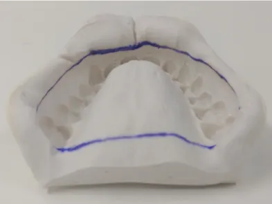
After the imprinting procedure, we immediately collect-ed the material to be analyzcollect-ed using a sterile swab in a pre-determined region of the alginate impression, as shown in Figure 1.
Each swab was then inoculated in Brain Heart Infusion (BHI) broth (OXOID, Pinheiros, São Paulo, Brazil).
We followed the disinfection procedures recommended
by the Brazilian Ministry of Health (2000),9 the impression
was washed in sterile water by spraying, each impression was sprayed with the disinfectant solutions of 1% sodium hypochlorite or 2% chlorhexidine. The impressions were stored in plastic bags for 5 or 10 minutes.
After this period, the impression was washed in sterile water again and another collection using a sterile swab was performed. The swabs were inoculated in new BHI broth to continue the microbiological analyses. We also washed the impressions using only sterile water as the control for the
hypochlorite for 10 minutes.
• Group 2: 10 Impressions disinfected with 1% sodium hypochlorite for 5 minutes.
• Group 3: 10 Impressions disinfected with 2% chlorhex-idine for 10 minutes.
• Group 4: 10 Impressions disinfected with 2% chlorhex-idine for 5 minutes.
• Group 5: 10 Impressions washed only in sterile water for a minute (control group).
Each tube of BHI broth was incubated for 24h at 37°C. After this period, we transferred 100 µL of broth of each sample to a solid blood agar medium and spread by surface scattering technique using a Drigalski spreader. Then, the plates were incubated for 48h at 37°C to promote microbial growth.
After growth on the plates, we manually counted the Col-ony-forming Units (CFU). We considered as the maximum value the cases in which the count exceeded 300 CFU. Iso-lated colonies were characterized through Gram stain us-ing optical microscopy increased by 100x, the colonies were identified and classified using Gram stain. For Gram-posi-tive bacteria we used the following tests: Catalase, DNase, hemolytic profile, PYR, bacitracin sensitivity, sulfa-tri-methoprim susceptibility and growth on mitis salivarius agar. For Gram-negative bacteria: test series of glucose and oxidase fermentation. Regarding fungal growth, the identi-fication was performed using only morphological analysis using the Gram stain.
We tabulated the data regarding CFU before and after the disinfection procedure. Due to being nominal qualitative data, we performed the comparisons using the Wilcoxon signed rank test.
Results
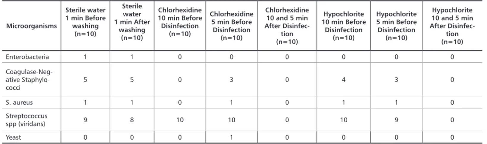
We obtained 50 impressions total, they were analyzed for the number of CFU on two different moments: before and after the disinfection procedures. Table 1 shows the median of the results for the count of colonies in the five experimen-tal groups, as well as the comparison, for each group, before and after disinfection.
Figure 1. Region of the impression where we performed the collection
Table 1. CFU count on the impressions before and after the disinfection procedure
Microorganisms Sterile water 1 min Before washing (n=10) Sterile water 1 min After washing (n=10) Chlorhexidine 10 min Before Disinfection (n=10) Chlorhexidine 5 min Before Disinfection (n=10) Chlorhexidine 10 and 5 min After Disinfec-tion (n=10) Hypochlorite 10 min Before Disinfection (n=10) Hypochlorite 5 min Before Disinfection (n=10) Hypochlorite 10 and 5 min After Disinfection (n=10) Enterobacteria 1 1 0 0 0 0 0 0 Coagulase-Nega-tive Staphylococci 5 5 0 3 0 4 3 0 S. aureus 1 1 0 1 0 1 1 0 Streptococcus spp (viridans) 9 8 10 10 0 10 9 0 Yeast 0 0 0 1 0 0 0 0
We can notice that washing with sterile water (positive control) was not effective to reduce the microbial load in ir-reversible hydrocolloid impressions. On the other hand, all other experimental groups presented satisfactory behavior, regardless of the time of exposure to the disinfectant agent.
Table 2 summarizes the major microorganism groups present in the impressions.
Microorganisms Sterile water 1 min Before washing (n=10) Sterile water 1 min After washing (n=10) Chlorhexidine 10 min Before Disinfection (n=10) Chlorhexidine 5 min Before Disinfection (n=10) Chlorhexidine 10 and 5 min After Disinfec-tion (n=10) Hypochlorite 10 min Before Disinfection (n=10) Hypochlorite 5 min Before Disinfection (n=10) Hypochlorite 10 and 5 min After Disinfec-tion (n=10) Enterobacteria 1 1 0 0 0 0 0 0 Coagulase-Neg-ative Staphylo-cocci 5 5 0 3 0 4 3 0 S. aureus 1 1 0 1 0 1 1 0 Streptococcus spp (viridans) 9 8 10 10 0 10 9 0 Yeast 0 0 0 1 0 0 0 0
Table 2. Characterization of contaminant microorganisms on the alginate impressions before and after the disinfection procedure
From 50 impressions made, we obtained 60 plates for analysis of microorganisms. This is explained by the bacte-rial growth before and after washing with sterile water. This did not occur in the disinfectant groups because there was growth only prior to disinfection. Thus, there was a predom-inance of Gram-positive bacteria in 58 plates (96.66%). For the most part, biochemical tests identified microorganisms that are common in the oral microbiota, such as Strepto-coccus sp of the viridians group in 37 impressions (61.66%); negative-coagulase Staphylococcus in 20 (33.33%); howev-er, we also found Staphylococcus aureus in 5 impressions (8.33%), this is a pathogenic and unusual bacterium in the oral microbiota.
Discussion
Some irreversible hydrocolloids feature disinfectant agents in their composition, such as chlorhexidine.
Accord-ing to Moreira and Cruz,10 these agents are effective on
an-timicrobial action when present in irreversible hydrocolloid powder. However, the results showed that the chlorhexidine in the formulation used did not cause antimicrobial action, since after washing using only sterile water, over 300 CFU were found when counting the colonies (Table 1). The ineffi-ciency of the agent in this case can be explained by its aggre-gation to the irreversible hydrocolloid during jellification, or
due to a low concentration in the alginate formulation.11
Other studies found similar results, impressions that were
washed only using water presented bacterial growth.12-15
Washing the impressions using water is a usual procedure, however, using antibacterial substances is still needed for a
more effective disinfection.16
The results of this study showed that after the disinfec-tion procedure, the impressions were free of contamina-tion. Therefore, the disinfectant agents used – 1% sodium hypochlorite and 2% chlorhexidine – were effective in the decontamination of the impressions through the spraying
technique. Esteves et al.,11 evaluated the direct
antimicro-bial action of these agents against S. mutans and S. aureus
in vitro, they observed that the substances were effective in
inhibiting microbial growth. Furthermore, Linhares et al.,17
found that 1% sodium hypochlorite was effective in disin-fecting impressions, also by spraying for 10 minutes. Al-though the sodium hypochlorite solution is recommended for disinfection, some care is needed in its handling, since it is a hazardous material that can cause irritation on the skin, mucous membrane and eyes, as well as the corrosion
of metallic materials.18
Regarding the disinfection time, we observed that it was effective for both 5 and 10 minutes (Table 1). The advantage from a reduced disinfection time is the optimization of the work routine. In addition, a longer time in this procedure can lead to some significant dimensional changes on the
irreversible hydrocolloid, as stated by Nassar et al.,19 and
Anusavice8 in their research. Our results corroborate with
Okazaki et al.,20 whom also found good results of
disinfec-tion using 1% sodium hypochlorite for only 5 minutes. The oral cavity houses around 500 to 700 species of mi-croorganisms. This occurs since several sites (habitats) with different environmental conditions that favor microbial growth exist, including the teeth, gingival sulcus, tongue, cheek, hard and soft palate and tonsils. Many of these mi-cro-organisms can be transmitted during dental procedures
and may be associated with diseases.21
Similarly, Egusa et al.,22 showed the existence of different
microorganisms in alginate impressions. From 30 samples investigated in selective agar medium, we detected among the isolates 56.7% Staphylococcus sp., 30% Candida albi-cans sp, 26.7% Methicillin-resistant Staphylococcus aureus (MRSA) and 6.7% Pseudomonas aeruginosa. This shows that
In another research, Egusa et al.,23 found that the
fre-quency of isolation of Streptococcus, Staphylococcus, Can-dida, MRSA and P. aeruginosa were 100%, 55.6%, 25.9%, 25.9% and 5.6%, respectively.
The isolation of S. aureus in some of the impressions shows the presence of pathogenic bacteria that may be present on
the oral mucosa transitionally. According to Saramanayake3
several enzymes and toxins are produced by Staphylococcus aureus. The most important are the coagulase and entero-toxin, the main diseases caused by S. aureus are osteomyeli-tis, endocardiosteomyeli-tis, sepsis and pneumonia.
The importance of disinfection procedures is emphasized by the presence of this microbial diversity in the impressions (Table 2) since some bacteria can survive for long periods of
time on surfaces. Žilinskas et al.,24 investigated the survival
of fungi and bacteria in plaster models that were prepared with 1 mL bacterial suspension and stored in a sterile con-tainer for evaluation between 1 hour and 120 hours. The re-sults found that S. aureus remained viable for four days.
Our results show that the impressions must be systemat-ically disinfected, so the material contaminated with blood and saliva does not become a source of cross-infection. Thus, disinfection and sterilization procedures must be adopted
This study presented limitations regarding the popula-tion and sample collecpopula-tion area. Larger populapopula-tion numbers and a larger collection area could show greater bacterial variability.
Conclusion
Our results evidence the microbial contamination in the handling of these impressions, and that the microbial load was not reduced after the impression was washed only with water (control group). Thus, a treatment with disinfection agents is still required, since this procedure is not effective in eliminating the microorganisms in the impressions. Despite the predominance of oral bacteria, the presence of S. aureus in some impressions points to the possibility of pathogenic microorganisms being found.
The microbial load was reduced after the impressions were disinfected with 2% chlorhexidine or 1% sodium hy-pochlorite, proving that these disinfection procedures are effective to remove microorganisms present in the alginate impressions. Furthermore, the disinfection of the impres-sions for 5 minutes was as efficient as it was for 10 minutes. A reduced disinfection time can optimize the work routine in the dental office.
13. Haralur SB, Al-Dowah OS, Gana NS, Al-Hytham A. Effect of alginate chem-ical disinfection on bacterial count over gypsum cast. The journal of advanced prosthodontics. 2012;4(2):84-8.
14. Correia-Sousa J, Tabaio AM, Silva A, Pereira T, Sampaio-Maia B, Vascon-celos M. The effect of water and sodium hypochlorite disinfection on alginate impressions. Revista Portuguesa de Estomatologia, Medicina Dentária e Cirurgia Maxilofacial. 2013;54(1):8-12.
15. Pakdin M, Soufiabadi S, Shahrakipour M. Evaluation of Contamination Re-duction on Gypsum Casts from Alginate Impressions Disinfected with Four Dif-ferent Materials. Global Journal of Health Science. 2016;8(12):127.
16. Badrian H, Ghasemi E, Khalighinejad N, Hosseini N. The effect of three dif-ferent disinfection materials on alginate impression by spray method. ISRN den-tistry. 2012;2012:1-5.
17. Linhares SMDS, Gallito MA, Rozário HHD, Silva ACPD, Maciel RMDV. Desinfecção de moldagens na clínica integrada da Faculdade de Odontologia de Campos. Rev Flum Odontol. 2010;10(34):36-42.
18. Barsano, P. R. Biossegurança: ações fundamentais para promoção da saúde. 1. Ed. São Paulo: Ed. Érica, 2014. p. 120.
19. Nassar U, Aziz T, Flores-mir C. Dimensional stability of irreversible hydro-colloid impression materials as a function of pouring time: a systematic review. The Journal of prosthetic dentistry. 2011;106(2):126-33.
20. Okazaki LK, Cato CH, Teramoto L, Costa ACBP, Fava M, Becker JBM, et al. Comparison of disinfection protocol of irreversible hydrocolloid (algi-nate) impressions through plastic and metallic trays. Brazilian Dental Science. 2014;17(3):54-9.
21. Lorenzo JL, Mayer MPA. Componentes Bacterianos da Microflora Bucal. In: Lorenzo JL. Microbiologia para o estudande de odontologia. 1. ed. São Paulo: Atheneu; 2004. 274 p.
22. Egusa H, Watamoto T, Matsumoto T, Abe K, Kobayashi M, Akashi Y, et al. An Analysis of the persistent presence of opportunistic pathogens. The International journal. 2008;21(1):62-8.
References
1. Krieger D, Bueno R, Gabardo MCL. Perspectivas de biossegurança em odonto-logia. Rev Gestão Saúde. 2010;1(2):1-10.
2. Silva ASF, Risso M, Ribeiro MC. Biossegurança em odontologia e ambientes de saúde. 2. ed. Sao Paulo: ed. Icone; 2009. P. 13-19.
3. Samaranayake L. Essential Microbiology for destistry. 4. Ed. Rio de Janeiro: Elsevier; 2012. 392 p.
4. Hymer R, Almeida TF. Riscos Biológicos em Odontologia: Uma Revisão da Literatura. Revista Bahiana de Odontologia. 2015;6(1):34-46.
5. Fernandes JKB, Barros KSM, Thomaz EBAF. Avaliação da adesão ás normas de biossegurança em clinicas de odontologia por estudantes de graduação. Revista de Pesquisa em Saúde. 2012;13(3):42-6.
6. Fernandez CS, Mello EBD, Alencar MJSD, Albrecht N. Conhecimento dos dentistas sobre contaminação das hepatites B e C na rotina odontológica. Rev Bras Odontol. 2013;70(2):192-5.
7. Bezerra ALD, de Sousa MNA, Feitosa ADNA, de Assis EV, Barros CMB, de Abreu Carolino EC. Biossegurança na odontologia. ABCS health sci. 2014;39(1):29-33.
8. Shen C. Materiais de moldagem. In: Anusavice JK. Phillips Materiais Dentári-os. 12. ed. Rio de Janeiro: Elsevier; 2013. 165-175 p.
9. Ministério Da Saúde. Secretaria de Políticas da Saúde, Coordenação Nacional DST e AIDS. Controle de infecção e a prática odontológica em tempos de AIDS: manual de condutas - Brasília. 2000. 118 p.
10. Moreira ACA, Cruz JFW. Efetividade da clorexidina incorporada a hidro-colóide irreversível. Rev Ci Méd Biol. 2005;4(2):113-7.
11. Esteves RA, Sousa EGD, Celestino Júnior AF, Maranhão KM, Pedrosa SS, Gauch LMR. Análise da eficácia antimicrobiana dos alginatos autodesinfetantes. RGO. 2007;55(1):23-8.
12. Meira DM, Collares T, Van der Sand ST, Collares FM, Leitune VCB, Sam-uel SMW. Influência do Tempo na Desinfecção de Alginato Contaminado com Staphylococcus Aureus em Ácido Peracético ou Gluataraldeído. Rev Fac Odontol. 2011;52(1/3):11-4.
Submitted: 01/15/2018 / Accepted for publication: 03/25/2018
Corresponding Author Laís Falcão Borgo
E-mail: laisfalcaoborgo@gmail.com
Mini Curriculum and Author’s Contribution
1. Thais Dias Lemos Kaiser – DDS and MSc. Contribution: conception and design of the study; standardization of the methodology; data interpretation; preparation of the manuscript; writing the manuscript; critical review and final approval.
2. Paula Sampaio de Melo – DDS and MSc. Contribution: preparation of the manuscript; critical review and statistic.
3. Laís Falcão Borgo – Undergraduate student. Contribution: conception of the study; preparation of culture media; sample collection; inoculation and Peak; identi-fication tests of microorganisms; analysis of results; data interpretation and preparation of the manuscript.
4. Julieti Fernandes Flor – Undergraduate student. Contribution: preparation of culture media; sample collection; inoculation and Peak; identification tests of microo ganisms; analysis of results.
5. Francisco de Assis Bozzetti – Undergraduate student. Contribution: preparation of culture media; sample collection; inoculation and Peak; identification tests of microorganisms; analysis of results.
23. Egusa H, Watamoto T, Matsumoto T, Abe K, Kobayashi M, Akashi Y, et al. Clinical evaluation of the efficacy of removing microorganisms to disinfect patient-derived dental impressions. International Journal of Prosthodontics. 2008;21(6):531-8.
24. Zilinskas J, Junevicius J, Ramonaitė A, Pavilonis A, Gleiznys A, Saka-lauskienė J. Viability changes: Microbiological analysis of dental casts. Med Sci Monit. 2014;20:932-7.