Universidade de Aveiro 2017
Departamento de Ciências Médicas
MARIANA CAMPOS
MARQUES
Respostas Celulares à Infeção Viral: Proteostase e
Imunidade Inata
Cellular Responses to Viral Infection: Proteostasis
And Innate Immunity
Universidade de Aveiro 2017
Departamento de Ciências Médicas
MARIANA CAMPOS
MARQUES
Respostas Celulares à Infeção Viral: Proteostase e
Imunidade Inata
Cellular Responses to Viral Infection: Proteostasis
And Innate Immunity
Tese apresentada à Universidade de Aveiro para cumprimento dos requisitos necessários à obtenção do grau de Mestre em Biomedicina Molecular,
realizada sob a orientação científica da Doutora Daniela Maria Oliveira Gandra Ribeiro, Professora Auxiliar Convidada do Departamento de Ciências Médicas da Universidade de Aveiro e Investigadora Principal do grupo “Organelle Dynamics in Infection and Disease” do Instituto de Investigação em Biomedicina (iBiMED), Universidade de Aveiro.
Thesis submitted at University of Aveiro to fulfil the requirements to obtain the Master degree in Molecular Biomedicine, held under the scientific guidance of Dr. Daniela Maria Oliveira Gandra Ribeiro, Principal Investigator at Organelle Dynamics in Infection and Disease group at Institute for Research in
o júri
presidente Doutora Ana Gabriela Henriques
Professora Auxiliar Convidada do Departamento de Ciências Médicas da Universidade de Aveiro
Doutora Daniela Maria Oliveira Gandra Ribeiro
Professora Auxiliar Convidada do Departamento de Ciências Médicas da Universidade de Aveiro
Doutora Rute Conceição do Nascimento Veríssimo Afonso
Bolseira de pós-doutoramento do Instituto Gulbenkian de Ciência e Médica Interna de Ano Comum do CHO- Centro Hospitalar Oeste
agradecimentos
Um grande obrigado a todos os que me acompanharam ao longo desta etapa
e que, de uma maneira ou de outra, me ajudaram a chegar ao fim.
À Dra. Daniela Ribeiro, por me ter apoiado a dar os primeiros passos na
investigação, por ter orientado o desenvolver deste projeto, por toda a
experiência que me transmitiu, por todos os conselhos, e por me contagiar
com o seu entusiasmo pelo mundo da virologia.
A todos os que partilharam e têm vindo a partilhar comigo a experiência de
trabalhar no ODID Lab, especialmente por me terem integrado tão bem
dentro e fora laboratório. Foi e tem sido um prazer fazer parte deste grupo.
À Isabel, obrigada por toda a ajuda, por partilhares os teus conhecimentos
comigo, pela companhia e por enriqueceres o meu vocabulário com as tuas
expressões malucas.
À Beatriz, por estares sempre pronta a ajudar-me e por animares os meus dias
de trabalho sempre com uma banda sonora.
À Joana, ao Ricardo, à Catarina e ao Rui pela companhia e por partilharem
comigo o que é iniciar um projeto num grupo novo.
Um especial e enorme agradecimento à Rita, por todas as horas boas e menos
boas. Por acreditar em mim e no meu trabalho, por tudo o que me ensinaste e
por me ajudares a ultrapassar os mil e um problemas que foram surgindo,
mesmo com um oceano pelo meio. Por teres partilhado comigo alguns
segredos de Aveiro, pelos passeios e pelos petiscos, sobretudo pela tua
amizade.
À Carolina e à Marisa por me terem aturado mais do que deviam, sempre
prontas a aliviar os ânimos. Por toda a vossa ajuda no decorrer das
experiências, a analisar os resultados e a encontrar soluções.
À Dra. Ana Soares, por me ter ajudado em todas das situações em que me
ajudou a perceber “o que é que não tinha corrido bem desta vez” e por ter
aceite participar neste projeto com entusiasmo.
À Dra. Maria João Amorim e ao seu grupo no Instituto Gulbenkian de Ciência,
por me terem acolhido e ajudado a planear e desenvolver as experiências da
infeção.
Ao pessoal do mestrado e a todas as pessoas que conheci em Aveiro, obrigada
por terem descoberto esta cidade comigo, pelas horas de maluqueira e
desespero. Foram sem dúvida imprescindíveis nessa etapa!
Ao Daniel, pela força, pela companhia e por me dares ânimo para ultrapassar
os momentos menos bons. Obrigada por acreditares em mim e nos meus
“primers”.
À minha família. Aos meus pais, ao meu irmão, aos meus avós, aos meus tios,
um obrigado muito especial. Por sempre se mostraram presentes e
interessados no meu trabalho, e por terem feito o que podiam para me ajudar
a terminar esta etapa com sucesso.
Also, I want to sincerely thank to Daniela and Prof. Dr. Wolfram Brune for
giving me the opportunity to spend some time in Hamburg. It was definitely a
great experience that allowed me to meet wonderful people that shared with
me their experience in the lab and also a lot of curiosities from their different
cultures and interests.
Lastly, I want to thank to my roommates and to the people I met there, for
making me miss Hamburg the way I do.
palavras-chave
Infeção viral, Vírus da Influenza A, Citomegalovírus, Dinâmica de Organelos, Peroxissomas, Mitocôndrias, Proteostase, Imunidade Inataresumo
Os vírus são agentes infeciosos oportunistas. Os diferentes passos de um ciclo de vida viral, incluindo a entrada do vírus na célula, a replicação do seugenoma e a formação de novas partículas virais requerem interações com os diferentes componentes celulares do hospedeiro, nomeadamente com organelos. Neste projeto, propomos estudar dois tipos diferentes de vírus que afetam dois mecanismos distintos de sobrevivência celular: a influência do Citomegalovírus de humano (HCMV) na resposta imunitária inata e o efeito do Vírus da Influenza A (IAV) na proteostase.
O HCMV pode estar associado com consequências graves para a saúde da população, uma vez que tem a capacidade para estabelecer uma infeção latente e persistente no hospedeiro. Este vírus codifica para a vMIA, uma proteína anti-apoptótica que se localiza nos peroxissomas e nas mitocôndrias, induzindo a sua fragmentação e inibindo a resposta antiviral celular que é estabelecida em ambos. Com isto, sugerimos mapear os domínios da vMIA responsáveis pelas alterações na morfologia dos organelos e na inibição da resposta imune. Os nossos resultados revelaram que a sequência de aminoácidos 115-130 poderá ser importante para a fragmentação dos
organelos. Também descobrimos que a proteína m38.5 do Citomegalovírus de ratinho (MCMV), análoga à vMIA, parece localizar nos peroxissomas, induzir a sua fragmentação e claramente inibir a resposta antiviral dependente deste organelo. Estes resultados sugerem que este vírus poderá ser útil para complementar os nossos resultados com experiências animais ou no contexto de infeção viral.
O IAV é o agente causativo da maioria das epidemias anuais em humanos. Durante a infeção com IAV, ocorre acumulação de proteínas com conformação errada e a formação de locais especializados de replicação viral, resultando na formação de agregados insolúveis ou inclusões. Neste estudo, propusemos determinar se a infeção com IAV conduz à acumulação de proteína com pré-disponibilidade para formar agressomas. Os nossos resultados, embora preliminares, sugerem que existe formação destas estruturas durante a infeção viral, previamente à libertação do genoma viral no citoplasma.
keywords
Viral infection, Influenza A Virus, Cytomegalovirus, Organelle Dynamics, Peroxisomes, Mitochondria, Proteostasis, Innate Immunityabstract
Viruses are small opportunistic infectious agents. Virus entry, replication and assembly are dynamic and coordinated processes that require precise interactions with host components, often with cellular organelles. Hence, we proposed to study two different viruses affecting two distinct cellularsurveillance mechanisms: Human Cytomegalovirus (HCMV) and Influenza A Virus (IAV) influence on the innate immune response and proteostasis, respectively.
HCMV might be associated with additional long-term health consequences in human due to its ability to establish a lifelong persistent latent infection. HCMV encodes vMIA, an anti-apoptotic protein known to co-localize at peroxisomes and mitochondria, induce their fragmentation and inhibit the downstream cellular antiviral response that is established at both organelles. In the present work, we aimed to characterize the role of vMIA in the peroxisomal-MAVS dependent antiviral response. We proposed to map the vMIA domains responsible for the organelles’ morphology changes and innate immune
response inhibition. Our results revealed that the 115-130 amino acid sequence might be important for the organelles’ fragmentation. We also found that m38.5, an analogue of vMIA in murine CMV (MCMV) seems to localize at
peroxisomes, induce the organelle’s fragmentation and clearly inhibit the peroxisome-dependent antiviral immune response. These results suggest that this virus may be useful to complement our results with experiments performed in animals or in the context of a viral infection.
IAV is the causative agent for most of the annual epidemic in humans. During IAV infection, it occurs the accumulation of unfolded proteins and the formation of specialized sites of viral replication, resulting in the formation of insoluble aggregates or inclusions. In this study, we proposed to determine whether and how IAV infection leads to aggresomal-prone proteins accumulation. Our preliminary results suggest aggresomes formation during viral infection, previous to the vRNP release in to the cytoplasm.
List of abbreviations
ATF4/6 Activating Transcription Factor 4/6 ATF6f ATF6 fragment
ATP Adenosine Triphosphate
Bid BH3 interacting-domain death agonist BIP Binding Immunoglobulin Protein c/EBP cAMP response Element-Binding Protein cAMP cyclic Adenosine Monophosphate
CARD caspase activation and recruitment domains CARDIF CARD adaptor inducing IFN-β
cGAMP cyclic GMP-AMP synthase
cGAMP cyclic Guanosine Monophosphate–Adenosine Monophosphate cGAS cGAMP Synthase
CHOP C/EBP Homologous Protein CLRs C-type Lectin Receptors
CMV Cytomegalovirus
cRNA complementary RNA
DAMPs Damage-Associated Molecular Patterns
DLP/DRP1 Dynamin-Like Protein/Dynamin-Related-Protein GTPase DNA Deoxyribonucleic Acid
dsDNA double stranded DNA
EGFR Epidermal Growth Factor Receptor eIF2α eukaryotic Translation Initiator Factor 2α eIF4 eukaryotic Initiation Factor 4
ER Endoplasmic Reticulum ERAD ER-associated degradation
FADD FAS-associated Death Domain Protein FasR FAZ receptor
FDA Food and Drug Administration FIS1 Fission Protein 1
gB/gH glycoprotein B/H
GDAP1 Ganglioside-Induced Differentiation-Associated Protein GTP Guanosine-5'-Triphosphate
HA Hemagglutinin
HCMV Human Cytomegalovirus HDAC6 Histone Deacetylase 6 HHV-5 Human Herpesvirus 5
HIV Human Immunodeficiency Virus HSF1 Heat Shock Factor 1
HSPs Heat Shock Proteins IAV Influenza A virus
ICTV International Committee on Taxonomy of Viruses IFN type-I Interferons
IRE1 Inositol-Requiring Enzyme 1 IRF Interferon Regulatory Factor ISGF Interferon-Stimulated Gene Factor ISGs IFN-Stimulated Genes
JAK-STAT Janus Kinase-Signal Transducers and Activators of Transcription JNK c-Jun N-terminal kinase
kbp kilo base pairs kDA kilo Daltons
LE Late Endosome
LGP2 Laboratory of Genetics and Physiology MAMs Mitochondria-Associated Membranes MAVS Mitochondrial Anti-Viral Signalling MCMV Murine Cytomegalovirus
MDA5 Melanoma Differentiation-Associated Gene-5 MDVs Mitochondria-Derived-Vesicles
MFF Mitochondrial Fission Factor
MOMP Mitochondrial Outer Membrane Permeabilization
mRNA messenger RNA
MTOC Microtubule Organizing Centre
NA Neuraminidase
NF-κB Nuclear Factor kappa B NLR NOD-Like Receptors
NLS Nuclear Localization Sequences
NOD Nucleotide-binding Oligomerization Domain
NP Nucleoprotein
NPC Nuclear Pore Complex NS1A/NS2 Non-Structural Protein
ORF Open Reading Frame
PA Polymerase Acidic Protein
PAMPs Pathogen-Associated Molecular Patterns PB1/2 Polymerase Basic Protein 1/2
PERK PKR-like ER kinase
Pex Peroxin
PKR Protein Kinase RNA
PMP Peroxisomal Membrane Protein PRR Pattern-Recognition Receptors
RD Repressor Domain
RdRP RNA-dependent RNA polymerase RIDD Regulated IRE1-dependent mRNA decay RIG-I Retinoic Acid-Inducible Gene-I
RIPK1 Receptor Interacting Protein Kinase-1 RLRs RIG-I-like Receptors
RNA Ribonucleic Acid ROS Reactive Oxygen Species ssRNA single strand RNA
sXBP1 spliced XBP1 t-Bid truncated Bid TLRs Toll-like Receptors TNF Tumor Necrosis Factor
TRADD TNFR-associated Death Domain Protein TRAF TNF Receptor-Associated Factors TRAIL TNF-Related Apoptosis-Inducing Ligand TRAILR TRAIL Receptor
tRNAiMet transfer RNA methionine initiator
UPR Unfolded Protein Response UPS Ubiquitin-Proteasome System
vICA viral Inhibitor of Caspase-8 Activation VISA Virus-Induced Signaling Adaptor
vMIA viral Mitochondria-Localized Inhibitor Of Apoptosis
vRNA viral RNA
vRNPs viral Ribonucleoprotein complexes WHO World Health Organization XBP1 X Box-Binding Protein 1
i
Index
CHAPTER I Human Cytomegalovirus and Innate Immunity 1
1.1 Human Cytomegalovirus 3
1.2 Epidemiology 3
1.3 Genome and morphology 4
1.4 Life cycle 4
Treatment 6
1.5 Antiviral innate immune sensing 6
Tool-like receptors 6
Cyclic GMP-AMP synthase (cGAS) 7
RIG-I-like receptors (RLRs) 7
1.6 RIG-I-MAVS signaling 8
1.7 Virus-induced apoptosis 10
1.8 vMIA’s dependent CMV evasion from antiviral cellular responses 11
Organelle’s morphology and RIG-I-MAVS signaling 11
Apoptosis 13
CHAPTER II Influenza A Virus and Proteostasis 15
2.1 Influenza A virus 17
Epidemiology 17
2.2 Genome and morphology 17
2.3 Replication cycle 18
2.4 Treatment 21
2.5 Proteostasis and Quality Control Machinery 21
2.6 Protein aggregation and aggresome formation 23
2.7 Unfolded protein response 25
Influenza A virus infection and UPRER inducing 27
CHAPTER III Aims of the study 29
Human Cytomegalovirus and Innate Immunity 31
Influenza A Virus and Quality Control Machinery 31
CHAPTER IV Materials and Methods 33
4.1Materials 35
4.1.1 Cell lines 35
4.1.2 Cell Culture Solutions 35
4.1.3 Bacterial strains 35
4.1.4 Bacterial Media 35
4.1.5 Viruses 36
ii
4.1.7 Vectors 36
4.1.8 Primers and Oligonucleotides 36
4.1.9 Transfection Reagents 37
4.1.10 Markers and Loading Dyes 37
4.1.11 Enzymes 37
4.1.12 Kits 37
4.1.13 Antibodies 38
4.1.14 Solutions and Buffers 38
4.1.15 Databases and Software 39
4.2Methods 40
4.2.1 Cell Culture 40
4.2.2 Transformation of competent bacteria 40
4.2.3 Transient Mammalian Cell Transfection Methods 42
4.2.4 Immunocytochemistry 42
4.2.5 Reverse transcriptase - quantitative Polymerase Chain Reaction (PCR) 43
4.2.6 Infection 45
4.2.7 Immunobloting 45
4.2.8 Quantification methods 46
4.2.9 Clustered Regularly Interspaced Short Palindromic Repeats/CRISPR associated system 9
(CRISPR/Cas9) for gene knockout 47
4.2.10 Statistical Analysis 48
CHAPTER V Results and Discussion 49
5.1 Human Cytomegalovirus and Innate Immunity 51
Mapping the vMIA domains responsible for organelles’ morphology change 51 Mapping the vMIA domains responsible for the inhibition of innate immune response 54
Creation of a PEX19 KO cell line 55
Study of the vMIA analogue in MCMV 56
5.2 Influenza A Virus and Quality Control Machinery 59
CONCLUDING REMARKS 63
Human Cytomegalovirus and Innate Immunity 65
Influenza A Virus and Quality Control Machinery 65
Publications resulting from this work 67
REFERENCES 69
List of figures
Figure 1 Schematic representation of HCMV structure. ... 4
Figure 2 Schematic representation of HCMV life cycle in a human cell.. ... 5
Figure 3 Organelle-Specific MAVS Signaling. ... 9
Figure 4 Representation of vMIA anti-apoptotic domains. ... 11
Figure 5 Representation of mitochondrial (A) and peroxisomal (B) growth and division in mammalian cells. ... 12
iii
Figure 7 Schematic representation of IAV virion. ... 18
Figure 8 IAV life cycle ... 20
Figure 9 A schematic representation of the cellular quality control machinery involved in the maintenance of proteostasis. ... 22
Figure 10 IAV hijacks aggresome processing machinery during virus entry. ... 24
Figure 11 Schematic representation of UPR pathway... 26
Figure 12 Reverse transcription PCR cycle of cDNA synthesis. ... 43
Figure 13 PCR cycle used to amplify GAPDH gene. ... 44
Figure 14 RT-qPCR cycling protocol. ... 45
Figure 15 Structural and functional characterization of vMIA and different sequence-deletion vMIA mutants. ... 52
Figure 16 Peroxisomal morphology and vMIA mutants localization in MAVS PEX cells.. ... 53
Figure 17 Deletion mutants of vMIA inhibit the peroxisome-dependent innate immunity signaling. ... 54
Figure 18 Complementary characterization of vMIA and its different mutants.55 Figure 19 Schematic and shorten representation of CRISPR/Cas9 system. ... 56
Figure 20 CRISPR/Cas9 PEX19 KO cell lines. ... 56
Figure 21 Peroxisomal morphology and m38.5 localization within transfected MAVS PEX cells.57 Figure 22 m38.5 inhibits MAVS PEX innate immunity signaling. ... 58
Figure 23 Aggresomal formation in HeLa HSPB1-GFP cells infected with ΔNS1 IAV. ... 60
Figure 24 Caracterization of insoluble protein fraction upon infection at different time points, normalized to the total fraction. ... 61
1
CHAPTER I
Human Cytomegalovirus and Innate Immunity
3
Human Cytomegalovirus and Innate Immunity
1.1 Human Cytomegalovirus
According to the International Comittee on Taxanomy of Viruses (ICTV, 2011), cytomegalovirus (CMV) belongs to the subfamily Betaherpesvirinae, family Herpesviridae and order Herpesvirales. It takes part of the Group I of Baltimore’s Classification of double stranded desoxyribonucleic acid (dsDNA)1. Several
species of CMV have been identified and classified for different mammals, being humans and monkeys its natural hosts.
Cytomegaloviruses are widely distributed in nature and are characterized generally by slow growth, restricted species specificity, and by inducing a typical cytopathic effect on infected cells involving specific nuclear and cytoplasmic inclusions as well as cell enlargement. Human herpesvirus 5 (HHV-5), commonly named as Human cytomegalovirus (HCMV), is common in the human population and is becoming increasingly apparent that it might be associated with additional long-term health consequences due to its ability to establish a lifelong persistent latent infection2. Murine CMV (MCMV),
a natural mouse pathogen, shares a high degree of sequence homology and biology with HCMV, and is a widely used model to study HCMV infection3.
1.2 Epidemiology
HCMV is a highly widespread pathogen that infects people of all ages, with higher seroprevalence in the elderly, being more common in developing countries and in communities with lower socioeconomic status4.
Upon primary infection, the virus is intermittently shed in multiple body fluids, hence, transmission can occur via saliva, sexual contact, blood transfusion and solid-organ or hematopoietic stem cell transplantation5. Recurrent infection occurs with reactivation of latent virus in response to
immunosuppression, or reinfection with a different strain in a seropositive individual for CMV.
Considering an immunocompetent host, primary infection is almost always benign with minimal or no clinical manifestation, yet it can result in horizontal or vertical transmission6. Occasionally, healthy
individuals develop a self-limited mononucleosis syndrome, sore throat, glandular fever and a mild hepatitis. Howecer, HCMV infection turns into a leading cause of illness and life-threatening in immunocompromised subsets of the population, including patients who are undergoing hemodialysis or receiving immunosuppressive drugs, as well as patients with cancer, HIV-infected, or organ transplant recipients7,8. In this setting, CMV serves as a major opportunistic pathogen, being a lifelong
burden to immune dysfunction.
Also, HCMV has been described as one of the major causes of congenital disorder, including severe and permanent neurological injury in newborns. Vertical transmission can occur transplacentally, during childbirth, or through breastfeeding9.
4
1.3 Genome and morphology
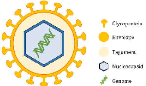
HCMV has a spherical to pleomorphic structure, and is the largest of the eight known human herpesviruses, with 150-200 nm in diameter and a 230 kilo base pairs (kbp) non-segmented genome encoding for over than 200 conventional open reading frames (ORFs).
The HCMV virion (Figure 1) consists of a core containing a long non-segmented linear dsDNA surrounded by a symmetric icosahedral capsid. These components are enclosed into a lipid bilayer envelope derived from the host cell endoplasmic reticulum (ER)-Golgi compartments, that contains at least 20 virus-encoded glycoprotein complexes involved in cell attachment and penetration. Between the envelope and the capsid is an amorphous, proteinaceous asymmetric matrix designated the tegument holding few cellular and viral ribonucleic acid (RNA) and the majority of the virion proteins, which can either have a structural role, a modulatory function of the host cell response to infection or be important for the virion (dis)assembly10. In addition to viral DNA, HCMV virions also carry mRNAs
into the host cells11.
Figure 1 Schematic representation of HCMV structure. The dsDNA genome is surrounded by a symmetric icosahedral capsid and enclosed into a lipid bilayer spiked with at least 20-virus encoded glycoprotein complexes, as gB, gH and gL that mediate virus entry in human cells. In between, there is the tegument, a proteinaceous matrix.
1.4 Life cycle
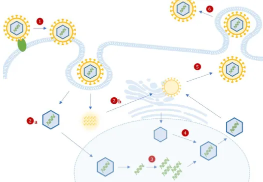
Virus Entry: Attachment and fusion of infectious particles with the host cell membrane requires interaction of several viral glycoproteins (e.g. gB and gH12) and cell-surface proteoglycans and receptors
(e.g. epidermal growth factor receptor (EGFR) and β1 integrins). HCMV can enter the cells either through endocytosis or direct fusion of the envelope with the cellular membrane (Figure 2, step 1).
After internalization, nucleocapsids, virion mRNA and tegument proteins are released into the cytoplasm (Figure 2, step 2a and 2b), where virion mRNAs are translated. Tegument proteins bound to the capsid are believed to interact with the host microtubule machinery to transport viral capsids to into nucleus, where the genome is released (Figure 2, step 3) and where viral transcription, genome replication and encapsidation occurs13.
Viral Gene Expression and Replication: HCMV starts to express its genes using the cellular transcriptional machinery. Productive replication leads to the temporal-coordinated synthesis of three classes of proteins, each regulating different aspects of the infectious cycle. Tegument proteins by
5
incoming virions tightly inhibit the initial steps of immune response and initiate the time-dependent cascade of viral genome expression13.
Immediate early genes (0–4 hours after infection) have been shown to be responsible for the early cytopathic effect, to protect the virus against innate host immunity and be involved in the regulation of transcription. Early viral genes (4–48 hours after infection) are involved in viral DNA replication and further transcriptional regulation. Furthermore, late genes are expressed during the remainder of infection up to viral egress and typically code for structural proteins14.
When latent, viral genomes take the form of closed circular episome in tandem with the host cell DNA and retain the capacity to replicate using the host cell replication machinery, although being expressed only a small subset of viral genes15.
Virion assembly and release: Late gene expression drives capsid assembly in nuclear viral factories and nuclear egress to the cytoplasm (Figure 2, step 4). Capsids are then associated with tegument proteins and trafficked to the viral assembly complex, comprised by host’s endoplasmic reticulum, Golgi apparatus and endosomal machinery, to acquire a tegument layer and an envelope13. Furthermore,
cellular and viral RNAs are packaged into virions in proportion to their intracellular concentration11
(Figure 2, step 5).
Figure 2 Schematic representation of HCMV life cycle in a human cell. HCMV attaches to the cell via interactions between viral glycoproteins (e.g., gB and gH) and specific surface receptors (e.g. EGFR), and is incorporated either through direct fusion or the endocytic pathway (1), followed by the release of nucleocapsids (2a), viral proteins and viral mRNA (2b) into the cytoplasm. These mRNA are translated and the nucleocapsids are translocated into the nucleus, where viral DNA is released (3), and initiates the expression of IE genes. Viral replication and maturation involves the encapsulation of replicated viral DNA as capsids (4), which are then transported to the cytoplasm. Secondary envelopment occurs in at the ER-Golgi intermediate compartment (5). This is followed by
6
Enveloped infectious particles along with non-infectious dense bodies are next released into the extracellular space by exocytosis (Figure 2, step 6). HCMV is a lytically replicating virus, thus after it causes massive cell enlargement, its life cycle culminates with the destruction of the infected cell.
Treatment
The most common treatment for patients with weakened immune system who have HCMV infection symptoms is prophylactic antiviral medication. Currently, all licensed anti-HCMV drugs are nucleoside analogs and all share the same fundamental mechanism of action, namely the inhibition of viral DNA polymerase, and consequently viral replication, being ineffective against a latent virus. In clinical practice, these drugs are frequently used for broader indications related to the treatment and prevention of HCMV infection in immunocompromised hosts9.
Cytogam®, Cytomegalovirus Immune Globulin Intravenous is an immunoglobulin G containing a consistent number of antibodies to HCMV. Alone or in combination with an antiviral agent, this medication has been approved by Food and Drug Administration (FDA) for the prophylaxis of HCMV disease in high-risk patients having an organ transplant and reduce the risk of HCMV-related diseases and death in some of the highest-risk patients9,17.
1.5 Antiviral innate immune sensing
The innate immune system on eukaryotic organisms holds very well-defined defense mechanisms against evading pathogens in early phases of infection. The innate antiviral immunity is activated with the detection of evolutionarily conserved structures termed pathogen-associated molecular patterns (PAMPs), by a set of host’s germline-encoded pattern-recognition receptors (PRRs). Endogenous products produced during cell damage or tissue destruction upon infection also stimulate PRRs18,19.
According on their localization, PRRs may be classified into membrane-bound PRRs, that include Toll-like receptors (TLRs) and C-type lectin receptors (CLRs); and cytoplasmic PRRs, including nucleotide-binding oligomerization domain (NOD)-like receptors (NLRs), retinoic acid-inducible gene-I (RIG-I)-like receptors (RLRs), as well as cytosolic viral DNA sensors such as cyclic guanosine monophosphate– adenosine monophosphate (cGAMP) synthase (cGAS)18. The recognition of viral PAMPs, that mainly
consists of viral nucleic acids, such as 5’ triphosphate terminal RNA, is possible due to endosomal TLR, cytosolic DNA sensors and cytosolic RLRs20.
Tool-like receptors
TLRs are considered the primary sensors of pathogens19. TLRs are type I membrane glycoproteins and
consist of one extracellular and one cytoplasmic domains, required for PAMP recognition and downstream signaling, respectively. Following TLR activation by PAMPs, a variety of adaptor molecules are activated and induce a signaling cascade that culminates in the activation of transcription factors that will regulate the expression of interferon (IFN), cytokines and chemokines20.
7
To date, 10 TLR family members have been identified in humans. TLR1, 2, 4, 5 and 6 are primarily expressed on the cell surface, whether TLR3, 7, 8 and 9 are exclusively expressed within endocytic compartments20.
HCMV virions were shown to trigger inflammatory cytokine responses via envelope gB and gH recognition by TLR2, in a mechanism dependent of nuclear factor kappa B (NF-κB) activation21, and
were proven to induce TLR4 signaling components and downstream IFN expression22. On the other
hand, endosomal TLR3 and 9 were demonstrated as essential components in the innate immune defense to MCMV infection23.
Cyclic GMP-AMP synthase (cGAS)
cGAS is a cytosolic DNA sensor belonging to the nucleotidyltransferase family. Once bound to DNA, cGAS catalyzes cGAMP synthesis, which in turn binds to and activates the ER protein stimulator of interferon genes (STING). This protein is a critical signaling molecule of the innate immune response against DNA viruses, once it further activates the antiviral type I IFN signaling pathway.
It is known that UL122, which encodes the immediate-early 2 86 kilo Dalton (kDa) protein (IE86), strongly abolished cGAMP-mediated type I interferon (IFN) promoter activation, as it facilitates proteasome-dependent degradation of STING24. Also, the HCMV tegument protein UL82 was identified
as a negative regulator of STING-dependent antiviral responses25. Furthermore, STING was described as
necessary for the first phase of type I IFN production that limits early CMV replication, proven that the cGAS-STING pathway has a pivotal role in the initial detection of CMV infection26.
RIG-I-like receptors (RLRs)
RLR family are expressed in most cell and tissue types and consists in three molecules: retinoic acid-inducible gene (RIG-I), melanoma differentiation-associated gene-5 (MDA5) and laboratory of genetics and physiology 2 (LGP2). These sensors recognize the RNA from RNA viruses in the cytoplasm of infected cells and induce inflammatory cytokines and type I interferons.
Structurally, all three members contain an intermediate DExD/H-box RNA helicase domain which is involved in recognition and binding to pathogen nucleotides, as well as adenosine triphosphate (ATP) hydrolysis-involved conformational changes; and a C-terminal repressor domain (RD)27,28. RIG-I and
MDA5 also contain two N-terminal tandem caspase activation and recruitment domains (CARDs) that are essential for downstream signaling cascade29,30. The RD of RIG-I maintains the receptor in a closed,
stable and inactive conformation that constrains the activation of the downstream signaling, being necessary its activation via conformational changes to expose its RIG-I CARD to further initiate antiviral signaling. Opposing to RIG-I, MDA5 has the CARD domains permanently exposed20.
Although RIG-I and MDA-5 have specificities for different ligands, effective sensing of PAMPs rapidly induces host immune responses via the activation of intracellular signaling cascades that ultimately leads to the induction of IFN and IFN-stimulated genes (ISGs), as well as pro-inflammatory cytokines19,31,
8
which subsequently may function as direct antiviral effectors, preventing viral genome replication, viral particle assembly, or virion release from infected cells, and shape the adaptive immune response18.
1.6 RIG-I-MAVS signaling
Mitochondrial anti-viral signaling (MAVS), also known as IFN-β promoter stimulator (IPS-1), CARD adaptor inducing IFN-β (CARDIF), and virus-induced signaling adaptor (VISA), is localized in the outer mitochondrial membrane32, peroxisomes33 and mitochondria-associated membranes (MAM)32. Both
peroxisomal and mitochondrial MAVS have specific signaling pathways which result in different but complementing activities20 and are required for antiviral responses with either temporal or functional
differences, which suggest that they may be recruiting distinct subsets of adaptor proteins. The different kinetics of ISG expression induction by peroxisomal and mitochondrial MAVS suggest that more than one mechanism of RLR-induced ISG expression may operate in virus-infected cells29.
Peroxisomes and mitochondria are ubiquitous organelles present in eukaryotic cells, with remarkably dynamic and high plasticity, capable of moving throughout the cell in a motor protein-dependent manner along cytoskeletal tracks. They continuously adapt their abundance, morphology, distribution, enzyme content and activity, according to the metabolic needs, or physiological changes in their cellular environment upon external stimuli. Peroxisomal and mitochondrial abundance varies according to the cell type and is regulated by organelle formation, half-life, and autophagy-mediated degradation34,35.
Mitochondria are double membrane-bound organelles containing their own genomes and transcription/translation systems. It can adopt a variety of different shapes that can range from small, spherical compartments of 0.1 to 1µm to elongated tubulo-reticular networks up to 10 μm34.
Peroxisomes are bordered by a single-membrane that surrounds a granular matrix, are devoid of DNA or protein synthesis machinery and are spread throughout the cytoplasm of most eukaryotic cells36.
Similarly, its shape and size vary greatly in different tissues, ranging from a spherical to rod-like form and from 0.1 to 0.5 μm in diameter, but they can also appear as elongated tubular organelles and small, tubulo-reticular networks with up to 5 μm, which are frequently associated with lipid droplets34,37.
Besides cooperating in the establishment of an effective cellular antiviral response, peroxisomes and mitochondria cooperate coordinately in managing diverse metabolic processes in mammals, as maintenance of cellular reactive oxygen species (ROS) homeostasis, fatty acids oxidation, and serve as signaling platforms that modulate diverse physiological and pathological processes including inflammation and cell fate transitions34.
Fluctuations in organelle abundance can be expected to have significant effects on their functional output, and to adjust organelle quantity in response to changing environmental and developmental stimuli, cells coordinate the formation of new organelles and their subsequent degradation once they are excessive or non-functional.
9
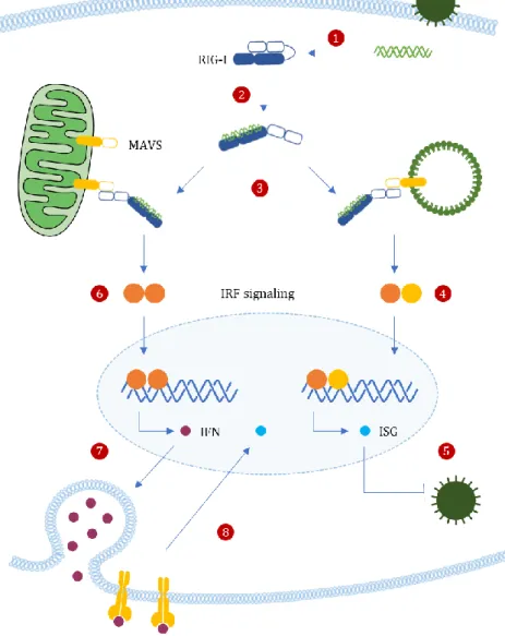
Upon infection, the recognition and binding of exogenous 5’-ppp panhandle dsRNA structures to RD leads to a conformational switch of RIG-I, which releases the autorepressed CARDs18 (Figure 3, step 1).
Activated RIG-I recruits its downstream adaptor MAVS (Figure 3, step 2). MAVS N-terminal contains a CARD-like domain and a proline-rich region allows MAVS to bind with upstream signaling molecules such as RIG-I and MDA5, through homotypic CARD–CARD-mediated interactions. MAVS activation induces the formation of a detergent-resistant prion fibre-like active aggregates that may involve the CARD domains of several MAVS38.
Figure 3 Organelle-Specific MAVS Signaling. Upon infection, detection of exogenous dsRNA structures by RIG-I (1) releases the autorepressed CARDs, leading to its activation (2). Activated RIG-I recruits and bind to mitochondrial and peroxisomal MAVS, through homotypic CARD–CARD-mediated interactions (3). Peroxisomal MAVS was shown is essential for the rapid expression of antiviral genes (ISGs) that will block early antiviral effects, via IRF1 and IRF3 (5). On the other hand, mitochondrial MAVS seems to act with slower kinetics, inducing delayed but sustained responses (6); it promotes type I IFN-dependent ISG expressions, via IRF2, 3 and 6 (7). Once secreted, IFNs bind to specific cell surface type I IFN receptors, leading to the activation of the JAK–STAT pathway, thus generating an amplifying loop leading to RIG-I accumulation during infection and additional ISGs transcription,
10
Peroxisomal MAVS was shown to be involved in early rapid, however transient responses, mitochondrial MAVS seems to act with slower kinetics, inducing delayed but long-lasting responses20,33. Perhaps the
peroxisomal pathway establishes a first outburst of antiviral effector proteins to temporarily block viral replication, while the mitochondrial pathway can induce a stronger and sustained antiviral state although delayed, to clear out the infection33.
Moreover, peroxisomal MAVS leads to the induction of ISGs via the transcription factors interferon regulatory factor 1 (IRF1) and IRF3, independent of type I IFN production33, whereas mitochondrial
MAVS promote type I IFN-dependent ISG expressions, via IRF2, 3 and 620,29(Figure 3). The main
advantage of peroxisomal early response is the absence of pro-inflammatory cytokines secretion that would most definitely cause unnecessary cell damage. However, if this fails to control viral infection, the mitochondrial pathway comes into action, stimulating a more powerful and persistent immune response, in an attempt to prevent further viral spread39.
Once secreted, IFNs bind to specific cell surface type I IFN receptors, leading to the expression of the interferon-stimulated gene factor 3 (ISGF3) through the activation of the Janus kinase-signal transducers and activators of transcritpion (JAK–STAT) pathway. ISGF3 then translocates to the nucleus and coordinates the transcription of hundreds of ISGs including RIG-I, thus generating an amplifying loop leading to RIG-I accumulation during infection and additional ISGs transcription, involved in the generation of the antiviral state18,29 (Figure 3, step 8).
1.7 Virus-induced apoptosis
Viruses are capable of exploit and reprogram the host metabolism to replicate, what may lead to the host’s cell death20. Apoptosis, both extrinsic and intrinsic pathways, schematized in Figure 6, can occur
in response to cellular stress induced by viral infection3.
The extrinsic pathway is a major mechanism of host immune clearance of virally infected cells. Death receptors, as tumor necrosis factor (TNF)-related apoptosis-inducing ligand (TRAIL) receptor (TRAILR) and FAS receptor (FasR), can activate initiator caspases, namely caspase-8 and -10, through dimerization mediated by adaptor proteins, such as FAS-associated Death Domain Protein (FADD), TNFR-associated Death Domain Protein (TRADD) and Receptor Interacting Protein Kinase-1 (RIPK1). The intrinsic pathway of apoptosis requires mitochondrial outer membrane permeabilization (MOMP), with subsequent release of mitochondrial interspaced proteins, as cytochrome c, that promotes the activation of apoptotic caspases. Bcl-2 family proteins function by regulating the integrity of the mitochondrial outer membrane, and comprise three functional subgroups: BH3-only proteins that act as stress sensors and initiate apoptosis; the effector proteins Bax and Bak that mediate MOMP; and pro-survival Bcl-2 proteins that maintain mitochondrial membrane integrity. Bcl-2 proteins have the capacity to bind to Bax and Bak and thus prevent their activation. BH3-only proteins initiate apoptosis by binding to Bcl-2 proteins and thereby releasing Bax and Bak, or by interaction with Bax and Bak and
11
directly catalyze their activation. In healthy cells, Bax and Bak exist as inert monomers, but as apoptosis proceeds the proteins undergo conformational changes resulting in the formation of large homo-oligomers that permeabilize the mitochondrial membrane3,40.
The extrinsic cell death pathway can intersect with the intrinsic signaling through the caspase-8-mediated cleavage of BH3 interacting-domain death agonist (Bid) to truncated Bid (t-Bid), which translocates to the mitochondria and interacts with Bax and Bak, to induce MOMP.
1.8 vMIA’s dependent CMV evasion from antiviral cellular responses
Goldmacher et al41 demonstrated that CMV infection provides resistance to apoptosis through the viral
mitochondria-localized inhibitor of apoptosis (vMIA) protein. This protein is the product of UL37 exon 1 (pUL37x1) and, besides being an important regulator of viral response to stress42, it has been
additionally shown to induce the release of ER calcium stores43. vMIA has two domains that are
necessary and sufficient for its anti-apoptotic function (Figure 4) encoded by Tyr5-Leu34 and Asp118 and Arg147 segments41,44. Furthermore, the mitochondrial localization signaling is located within the
amino acid 2-30 sequence44,45, and one or several amino acids within the 135-141 segment of vMIA are
essential for its interaction with Bax and further apoptosis inhibition46,47.
Figure 4 Representation of vMIA anti-apoptotic domains. Adapted from Hayajneh, 200144.
Organelle’s morphology and RIG-I-MAVS signaling
Upon HCMV infection, vMIA was shown to promote viral replication by efficiently increasing mitochondrial biogenesis in fibroblasts48. vMIA is also localized at peroxisomes, interacts with MAVS
and specifically diminishes the peroxisomal MAVS-dependent production of ISGs31.
Mitochondria are continuously remodeled through cycles of fusion and fission34, whereas peroxisomes
have distinct biogenesis pathways. Unlike mitochondria, mature peroxisomes cannot fuse with one another, thus new peroxisomes must arise from division of pre-existing peroxisomes49 or de novo
formation50. A well-defined sequence of morphological changes, including elongation, constriction, and
fission, is most likely the major proliferation process35.
Both organelles share key components of their fission/division machinery mammals51, including a
dynamin-like protein/dynamin-related-protein GTPase (DLP/DRP1), and its membrane adaptors mitochondrial fission protein 1 (FIS1), mitochondrial fission factor (MFF), and ganglioside-induced differentiation-associated protein (GDAP1). Overexpression or downregulation of their function has been shown to induce its fragmentation or elongation, respectively35.
12
DLP/DRP1 is a predominantly cytosolic protein known to function in elongation and fission, not required for organelle constriction. Recruitment of DLP/DRP1 to peroxisomal or mitochondrial division sites depends on FIS1, MFF, and GDAP1 recruitment to organelle membranes. Additionally, peroxisome membrane elongation requires members of the Pex11p family of peroxisomal membrane proteins (PMP) that initiates membrane remodeling and the formation of a tubular membrane extension on one side of the peroxisome34,52. This process is schematically represented in Figure 5.
Figure 5 Representation of mitochondrial (A) and peroxisomal (B) growth and division in mammalian cells. A well-defined sequence of morphological changes, including growth/elongation, constriction and fission contributes to organelles proliferation. In Mefs of wild-type cells, mitochondria are characterized by a network of extended tubules (up to 10 μm) distributed roughly throughout the cytoplasm (A1). Replicative mitochondrial fission is initiated by recruitment of cytosolic DLP/DRP1 to the constriction sites, by the adaptor proteins MFF and FIS1, located in the outer mitochondrial membrane (A2). Fragmented mitochondrial appeared mostly as sphere or oval shaped (A3). On the other hand, the activation of Pex11 at pre-existing peroxisomes initiates membrane remodeling and the formation of a tubular membrane extension on one side of the peroxisome (B1). Subsequently, the extension grows and acquires specific set of proteins, as Pex11p and Fis1 (B2). Pex11p and Mff-DLP1 complex concentrate at the sites of constriction (B3). In Mefs, peroxisomes appear as elongated tubular organelles and small tubule-reticular networks (up to 5 μm) and, when fragmented, for instance as a consequence of DLP/DRP1
13
The peroxisomal de novo formation might involve the budding and fusion of pre-peroxisomal vesicles derived from the ER or the mitochondrial membrane. It can be explained by a semi-autonomous model of peroxisome formation, whereby the ER and mitochondria supply existing peroxisomes with essential membrane lipids and proteins, including Pex19, Pex3 or Pex16, to allow its growth and division34,50.
In addition, vMIA had already been shown to induce mitochondrial fragmentation as a necessary step for the inhibition of the mitochondrial-dependent signaling pathway53,54. It also induces peroxisomal
fragmentation, a mechanism is not essential for vMIA to specifically inhibit signaling downstream the peroxisomal MAVS. Thus, vMIA appears to act at both organelles via distinct mechanisms31.
Apoptosis
With a slow replication cycle, HCMV depends on the sustained cell viability and, to prevent the premature death of infected cells, the virus is known to block apoptotic signaling pathways and subvert the host antiviral response31.
Several viral immediate early gene products with antiapoptotic properties have been identified in HCMV, including, UL36, UL37 and UL38, represented in figure 6. The UL36-38 immediate-early locus is highly conserved among HCMV strains and it is required for its replication, once it encodes for proteins that inhibit the ability of an infected cell to activate cell-degrading caspases44.
The UL36 gene product is known as vICA (or pUL36), which stands for viral inhibitor of caspase-8 activation. vICA blocks the extrinsic cell death pathway by binding to procaspase-8 and blocking its cleavage and subsequently activation3.
Furthermore, HCMV deficient for UL37x1 gene, that encodes for vMIA, has a severe growth defect as a result of strong induction of apoptosis in infected cells55,56.
vMIA is known to inhibit apoptosis through inactivation of Bax. It binds and sequesters Bax at mitochondria’s outer membrane in form of high-molecular weight and inactive oligomers that lacks capacity to induce MOMP46. Whether vMIA is capable of inhibiting apoptosis though Bak is still
controversial46,57,58.
Overall, vMIA prevents the formation of the mitochondrial permeability transition pore and inhibit the release of cytochrome c and pro-apoptotic factors into the cytoplasm, thereby preventing the activation of downstream executioner caspases31. The functional properties of vMIA’s localization resemble those
of Bcl-2 family anti-apoptotic proteins. However, vMIA does not possess homology to any BH domains and its function is independent of t-BID low concentrations55.
Lastly, UL38 has recently been shown to encode a cytosolic protein pUL38 that suppresses the ER-stress response, by inducing the expression of chaperones through activating transcription factor 4 (ATF4) and suppression of pro-apoptotic c-Jun N-terminal kinase (JNK) activity3.
14
MCMV encodes distinct inhibitors of Bax and Bak, m38.5 and m41.1, respectively. m38.5 protein is encoded at analogous position as HCMV vMIA within the respective viral genomes. Although they share little sequence homology, both seem to localize to mitochondria and inhibit Bax in an analogous manner, therefore preventing apoptosis. m38.5 was proposed to be a functional ortholog of vMIA in MCMV59,60.
A second MCMV-derived inhibitor, m41.1, associates with Bak at the mitochondrial membrane and acts as a viral inhibitor of Bak oligomerization (vIBO) from HCMV. Optimal replication of MCMV depends upon m38.5 and m41.1, whose combined activities maintain mitochondrial integrity88.
Figure 6 Inhibition of apoptosis by CMV. vMIA and vIBO inhibit MOMP and release of pro-apoptotic factors, as cytochrome c by interacting with BAX and BAK, respectively. MCMV encodes two specific inhibitors, m38.5 and m41.1, HCMV has only pUL37x1, whether the pUL37x1 protein is BAX-specific or inhibits both BAK and BAX is still controversial. The extrinsic apoptosis pathway initiated by death receptors is blocked by vICA, which is encoded by HCMV UL36 and MCMV M36 gene, respectively. APAF1: apoptotic protease activating factor 1; FasL: Fas ligand; FADD: Fas-associated death domain protein; RIPK1: receptor interacting protein kinase-1; TNFα: tumor necrosis factor α; TNFR: TNF receptor; TRADD: TNFR-associated death domain protein; t-BID: truncated BH3-interacting
15
CHAPTER II
Influenza A Virus and Proteostasis
17
Influenza A Virus and Proteostasis
2.1 Influenza A virus
Influenza viruses are among the most common viruses causing high morbidity and mortality. Influenza A virus (IAV) has been the causative agent for most of the annual epidemic in humans and the major pandemics of influenza in the last century61,62.
According to ICTV (2011), IAV belongs to the Influenzavirus A genus of the Orthomyxoviridae family that comprises enveloped viruses with segmented, negative-sense single-stranded RNA (ssRNA) genome62,
capable of bind sialic acid in mucoproteins. IAV is designed as a type V virus concerning the Baltimore’s classification system1. Up to this date, 16 different HA (H1 to H16) and 9 NA (N1 to N9) subtypes have
been identified62, being H1, H2 and H3 the virus subtypes identified in humans.
Epidemiology
Influenza viruses continuously undergo antigenic evolution, either by antigenic drift or antigenic shift. Antigenic drift involves the accumulation of point mutations within the viral RNA genome, particularly in genes that code for antigenic sites. These mutations and the emerging virus strain variants gain selective advantages and evade preexisting immunity, being generally responsible for winter epidemic outbreaks. On the other hand, antigenic shift implies genetic re assortment of RNA segments from different virus strain or different viruses. The new virus subtype arises presenting a novel phenotype with pandemic potential, since there is no immunity to the new virus subtype in the population, allowing the virus to spread rapidly and cause high morbidity and mortality61.
Both seasonal and pandemic influenza can afflict people of all ages, and most cases will result in self-limited illness in which the person recovers fully without treatment. Seasonal influenza is an acute respiratory disease that is characterized by the sudden onset of high fever, cough, headache and inflammation of the upper respiratory tree and trachea. In the elderly, in infants, and in people with chronic diseases, typical seasonal influenza is associated with especially high risk of developing severe complications within hours, as hemorrhagic bronchitis, pneumonia, and ultimately death in as little as 48 hours after the onset of symptoms61. Pandemic outbreaks cause most of its severe or fatal disease in
younger people, either in chronic patients and healthy individuals, and caused many more cases of viral pneumonia than is normally seen with seasonal influenza.
2.2 Genome and morphology
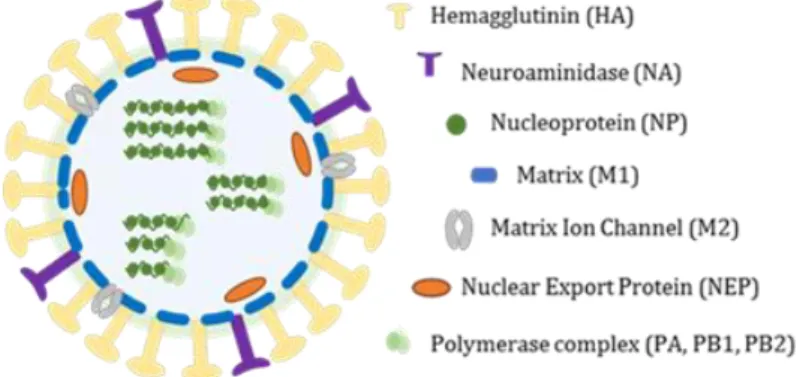
The genome of IAV consists of eight single-stranded, negative-sense linear RNA segments (-) ssRNA, encoding for 12 to 14 proteins depending on the strain most of which are necessary for efficient virus replication in host cells and for virion formation. Genome total size is about 3,5kb and the segments size range from 890 to 2341 nucleotides.
18
The three largest RNA segments encode the three viral RNA-dependent RNA polymerase (RdRP) proteins: polymerase acidic protein (PA), polymerase basic protein 1 (PB1) and PB2. The three intermediate-size RNA segments encode HA, NA and nucleoprotein (NP). The larger of the remaining two segments encodes M1 and M2 matrix proteins, and the smaller one encodes two nonstructural proteins, NS1A and NS261,63. To express spliced forms of viral proteins, as M2 and NS2, the virus uses the
host cell’s splicing machinery, while prevents the host cell from using it to process host cell messenger RNA (mRNAs), through NS1 interaction with small nuclear RNAs64.
Each of the eight RNA segments is separately enclosed in the virion in the form of ribonucleoprotein complexes (vRNPs), wrapped in a helical conformation with NP (one subunit binds ~20 nucleotides of vRNA and the vRdRp in both 3’ and 5’ ends62.
The influenza virions are known to display many forms, sometimes taking an irregular shape. They are generally spherical or elliptical in shape, ranging from approximately 80 to 120 nm in diameter, and occasional elongated or filamentous, reaching more than 20 µm in length. Regardless of their shape, all virions incorporate an organized set of eight RNPs62 (Figure 7).
The IAV genome is covered by an envelope coat made up of a lipid bilayer, derived from the host cellular membrane acquired during the budding, that is known to contain both cholesterol-enriched lipid rafts and non-raft lipids. The outer layer of the lipid envelope is spiked with numerous membrane-spanning viral glycoproteins, HA, NA and M2. The type I transmembrane HA is the most abundant envelope protein (~80%), followed by NA (~17%). M2, a highly selective type III transmembrane ion channel, is a very minor component, with only 16 to 20 molecules per virion64. The peripheral membrane protein,
M1, which is one of the most abundant viral proteins in the virion, binds to the lipid envelope to maintain virion morphology62.
Figure 7 Schematic representation of IAV virion. Eigh (-) ssRNA segments are inclosed into an lipid bilayer envelope spicked with viral glycoproteins.
2.3 Replication cycle
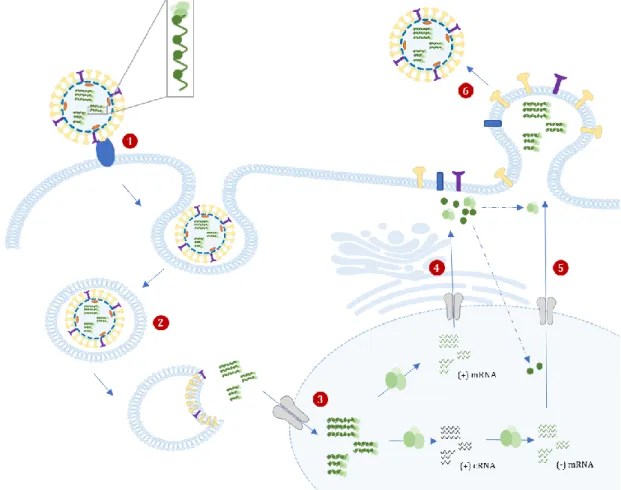
Attachment: The replication cycle begins when the viral surface HA binds to the sialic acid residues from glycoproteins or glycolipids on the host cells membrane64,65 (Figure 8, step 1). The specificity of HA
19
binding between species depends on the nature of the glycosidic linkage between the terminal sialic acid and the penultimate galactose residue on the receptor. Human influenza viruses preferentially bind to sialic acids attached to galactose in an α (2,6) configuration64.
Entry into host cell: After successful binding, virion internalization occurs essentially by receptor-mediated endocytosis and the virus is transported into the cell in an endocytic vesicle (Figure 8, step 2). The low pH in the endosome triggers conformational changes in the HA protein, which leads the fusion between the viral envelope and the endosomal membrane63,64, with the formation of a fusion pore
through which the viral genetic material is released. It also stimulates the proton flow into the virus via M2, which then weakens the interaction between the M1 and the vRNPs, promoting their dissociation. Nuclear transport: After being released into the cytoplasm, the vRNP are transported to the nucleus (Figure 8, step 3) by cellular nuclear import machinery recognition of the nuclear localization sequences (NLS) present in ribonucleoproteins63, where it undergoes transcription and replication processes.
Transcription and Replication: The (-) vRNA strand is used for the synthesis of capped, polyadenylated mRNAs, a readable form that it is further converted into proteins through process termed cap-snatching; and full-length positive sense (+) complementary RNA (cRNA), that will serve as template to produce more (-) vRNA strands to be then packed into the new virions. Both processes are carried out by the viral RdRp enzyme and, due to its short proteasome, the virus hijacks the host transcription machinery for its own purposes.
During the cap-snatching process, short oligomers from host pre-mRNA are recognized and bound by the viral PB2 subunit66, cleaved at the 5’ end by the PA endonuclease domain67 and then used to prime
mRNA synthesis via PB1 subunit68,69. The viral genome is thereby transcribed using host capped mRNA
segments as primers for initiation of viral mRNA synthesis, which ultimately leads to the synthesis of capped translatable viral mRNAs63. On the other hand, viral genome replication involves unprimed
synthesis of an exact full-length copy of the (-) vRNA into (+) cRNA, which lacks both the 5’ capped primer and 3’ polyadenylation tail and can be used as templates for further (-) vRNA synthesis further used in the assembly of vRNA complexes69,70.
NP molecules are required for both steps of replication and are deposited on the cRNA and vRNA during RNA synthesis. Both NP and the RNA polymerase components are complexed with newly synthesized vRNA to form vRNPs69.
Nucleus export of vRNPs and Translation: vRNPs are exported to the cytoplasm, either for translation into viral proteins and further assembly of new virus particles.
Synthesis and folding of viral core proteins occur entirely in the cytosol, taking advantage of host cell factors to perform viral mRNA translation70. The synthesis of viral envelope proteins HA, M2 and NA
also starts in the cytosol but are further folded and processed in the host ER and the Golgi apparatus where they undergo post-translational modifications, as glycosylation63. Subsequently, the proteins are
20
additionally modified and transported through the trans-Golgi network to the plasma membrane of the cell (Figure 8, step 4).
M1 interact with HA and NA, forming patches with a high density of HA and NA. Subsequently, newly formed RNPs interact actively with the M1 lining at these patches, which prevents re-entry of RNPs into the nucleus and direct them towards the assembly site on the apical membrane of polarized epithelial cells. This ensures that progeny viruses are releases back to the airways. The viral proteins accumulate in the cholesterol rich membrane region named lipid rafts, believed to be the site of virion formation. Virion assembly: The packaging of vRNPs favors the formation of infectious virus particles with all eight RNA segments required for efficient infection70 (Figure 8, step 5). After the budding of all viral proteins
and vRNP complexes, viral NA cleaves the sialic acid residues on cellular surface glycoproteins or glycolipids, which is bind to HA during the process. Doing that, recently formed virions are released from the host cell's surface (Figure 8, step 6) and start to spread and infection further cells throughout the respiratory tract64.
Figure 8 IAV life cycle. The cycle begins when the viral surface HA binds to the sialic acid residues on host cells membrane (1). IAVs is predominantly internalized by endocytosis and the virus is transported into the cell in an endosome (2). The low pH inside the endosome induces the formation of a fusion pore through which the viral genetic material is released and imported to the nucleus (3), where it undergoes transcription and replication processes. Viral core proteins synthesis occurs entirely in the cytosol, whether viral envelope proteins synthesis suffer further processing in the ER and Golgi apparatus (4), and both accumulate at the host membrane. After the budding of all viral proteins and vRNP complexes (5), recently formed virions are released from the host cell's
21
As an acute lytic viral infection, the entire process seriously disrupts the normal physiology of the infected cell and causes the destruction of its membrane, and consequently cell death and desquamation of the respiratory epithelium. However, cell lysis does not occur until the cell has produced many thousands of new virus particles during the latent phase of infection.
2.4 Treatment
Effective measures against influenza A diseases include prevention of infection by either administration of antiviral drugs or vaccination. Antiviral drugs can have both therapeutic and prophylactic effects, but to prevent disease they must be administered continuously at times of high influenza activity61.
Oseltamivir (Tamiflu®) is a selective NA inhibitor that induces the aggregation of viral particles on the host cell surface, preventing the release and spreading of the new progeny viruses71. Amantadine, a M2
ion channel blocker, slows the dissociation of M1 from the RNPs and the viral membrane, inhibiting subsequent steps in the viral life cycle72.
Nevertheless, vaccination is still the primary strategy for prevention and control of influenza virus, and both inactivated and attenuated vaccines are effective. Once virus subtypes are distinguishable serologically, and the continuous viral antigenic drift of IAV makes once effective vaccines ineffective after a few years' time, having the requirement for regular updates of the composition of the influenza vaccine and annual revaccination is thus recommended for those at high risk61.
2.5 Proteostasis and Quality Control Machinery
To be functional, most proteins go through a succession of folding intermediate states and adopt a defined three-dimensional native structure. A protein is correctly folded if it has attained its native conformation after required co- or post-translational modifications.
During the folding process, partially folded proteins, as folding intermediates and misfolded conformers, expose some hydrophobic domains that are typically hidden in the native structure, which can lead to nonproductive associations and are prone to trigger protein aggregation73. Besides, the proteins in the
native configuration can undergo unfolding, specially under stress conditions74.
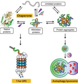
Misfolded proteins can interfere with normal cellular functions and be potentially toxic. Thus, cellular proteostasis maintenance is imperative to ensure successful development, healthy aging, resistance to environmental stresses and to minimize homeostasis perturbation by pathogens such as viruses. To suppress the formation of protein aggregates, cells have evolved an elaborated quality-control machinery (Figure 9) that can adapt to the severity of protein damage and acts through ensuring the fidelity of transcription and translation, and induction of stress responses75–78.
Distinct surveillance mechanisms that respond to misfolded and unfolded proteins have been characterized in the cytoplasm, in the ER and in the mitochondria. For instance, various molecular chaperones and/or chaperonins have evolved to assist post-translationally folding of newly synthesized
22
proteins and refolding of proteins damaged by stress and cellular injuries. For what is known, they occur ubiquitously in all cellular compartments that sustain protein synthesis and folding reactions. Also, while small monomeric proteins can fold in their absence in vivo, medium- to large-sized multidomain proteins critically require chaperones to undergo a correct fold79.
Most cellular proteins are folded directly after translation in the cytosol with the assistance of chaperones, foldases and lectines80 These molecules, mainly the heat shock proteins (HSPs) HSP60 and
HSP7078, bind to and stabilize exposed hydrophobic residues, prevent incorrect intra– and
intermolecular interactions between partially folded or unfolded polypeptides, prevent aggregation and promote the refolding of denatured model substrates and the proper formation of noncovalent interactions that lead to the desired folded state81.
Figure 9 A schematic representation of the cellular quality control machinery involved in the maintenance of proteostasis. Molecular chaperones support the folding of nascent polypeptides and refolding of proteins damaged by stress and cellular injuries. Additionally, they prevent misfolded or unfolded protein from aggregating and escort terminally misfolded protein for UPS degradation. The autophagy-lysosomal pathway aids to remove protein aggregates formed by the misfolded proteins that have escaped from the surveillance of chaperones and the
UPS. Adapted from Huabo et al, 200982.
Cellular proteins that are unable to fold properly, among non-functional protein fragments and no longer useful proteins, are targeted for degradation by a proteolytic mechanism, termed the ubiquitin– proteasome system (UPS). This system is found in cytosol and nucleus, and mediates degradation of cytosolic, nuclear, secretory and transmembrane proteins. Misfolded secretory and transmembrane proteins are first retained in the lumen or the membrane of the ER, retro translocated back to the cytosol and delivered to the proteasome83,84. The UPS involves at first the tagging of misfolded proteins with a
polyubiquitin chain, follow by recognition of the polyubiquitylated tag and degradation by the proteasome.
Furthermore, the autophagy-lysosomal pathway helps to remove protein aggregates formed by the aggregation-prone proteins that have escaped from the surveillance of chaperones and the UPS, and