Article
J. Braz. Chem. Soc., Vol. 27, No. 1, 2-9, 2016.
Printed in Brazil - ©2016 Sociedade Brasileira de Química 0103 - 5053 $6.00+0.00
A
*e-mail: [email protected], [email protected]
In vitro
Anti-HMPV Activity of New Synthetic Phenytoin Derivatives
Gabriella Mendes,a Geisa H. Aspesi,b Ana L. A. Arruda,b Maria T. V. Romanos*,a and Carlos K. Z. Andrade*,c
aLaboratório Experimental de Drogas Antivirais e Citotóxicas (LEDAC), Departamento de Virologia
do Instituto de Microbiologia Paulo Góes, Universidade Federal do Rio de Janeiro, UFRJ, CP 6804, 21941-590 Rio de Janeiro-RJ, Brazil
bDepartamento de Ciências Biológicas e da Saúde, Universidade Católica Dom Bosco, UCDB,
Avenida Tamandaré 6000, 79117-900 Campo Grande-MS, Brazil
cLaboratório de Química Metodológica e Orgânica Sintética, LaQMOS, Instituto de Química,
Universidade de Brasília, CP 4478, 70910-970 Brasília-DF, Brazil
New derivatives of synthetic 5,5-diphenylhydantoin (phenytoin) were prepared by N-alkylation with 1,3-dibromopropane. Subsequent treatment with sodium azide led to the respective azide. Reaction of the azide with phenylacetylene and 2-hydroxy-3-butyne and oxidation of the resulting alcohol with MnO2 resulted in three triazolic compounds that were evaluated in vitro for their antiviral activity against human metapneumovirus (HMPV). 5,5-Diphenyl-3-[3-(4-phenyl-1H-1,2,3-triazol-1-yl)propyl]imidazolidine-2,4-dione was the most active of the three compounds tested, with selectivity index of 129.87, even higher than ribavirin, the control substance. The three compounds showed activity in the early stages of viral replication presenting virucidal activity and binding to cellular receptors, preventing the adsorption of viral particles. These compounds showed higher activity in both experiments, inhibiting 98.3% of infection as virucidal and 98.9% when interacting with cellular receptors. Furthermore, they showed 73.8% of activity during the penetration of HMPV particles into cells. The derivative 3-{3-[4-(1-hydroxyethyl)-1H-1,2,3-triazol-1-yl]propyl}-5,5-diphenylimidazolidine-2,4-dione presented a mild anti-HMPV activity, with selectivity index of 2.74. 3-[3-(4-acetyl-1H-1,2,3-triazol-1-yl)propyl]-5,5-diphenylimidazolidine-2,4-dione inhibited less than 50% of HMPV replication.
Keywords: phenytoin, 1,2,3-triazoles, metapneumovirus, antiviral assay, real-time RT-PCR
Introduction
Human metapneumovirus (HMPV) is a negative-sense, single-stranded RNA virus first isolated in 2001 by van
den Hoogen and colleagues1,2 in previously virus-negative
nasopharyngeal aspirates from children with respiratory tract infections. It has been recognized as a common cause of respiratory infections in children, immunocompromised
and elderly populations.3
HMPV causes occasional upper respiratory tract infections, although lower respiratory tract infections can result in bronchiolitis, pneumonitis and asthma
exacerbations.4,5
In Brazil, the burden of HMPV infections has been
demonstrated by several scientists.6-11 Currently, there is no
antiviral therapy available for prevention or treatment of HMPV infection, with the exception of ribavirin, although
its activity is still questionable.12
Many hydantoin (imidazolidine-2,4-dione) derivatives have pharmacological activities (anticonvulsant, antifungal,
antibacterial and antiparasitic),13,14 related to the substituents
in positions 3 or 5 of the imidazolic-like ring.
Phenytoin (5,5-diphenyl-2,4-imidazolidindione or 5,5-diphenylhydantoin) belongs to a group of anti-epileptic drugs with clinical use in cases of partial and generalized seizures. It displays various effects such as inhibition of sodium, potassium and calcium channels in the neurons membrane, changes in local concentrations of neurotransmitters such as GABA, acetylcholine and noradrenaline and decreased neuronal excitation in
general.13 Furthermore, a number of phenytoin derivatives
interesting pharmacological properties, including
anticonvulsivant,15 antimicrobial16 and anti-arrhythmic.17
Due to its properties, this molecule was chosen as lead for the obtention of new 1,2,3-triazole derivatives in this work. 1,2,3-Triazole is an important nucleus, which can be easily obtained by a copper catalyzed alkyne-azide cycloaddition reaction (vide infra). It is present in many compounds with various pharmacological activities,
including antiviral.18 Thus, we envisaged that new
molecules containing both phenytoin and 1,2,3-triazole moieties could show enhanced activities compared to the isolated moieties.
Experimental
Chemistry
Commercially available reagents and solvents were purchased from Merck, Aldrich Chemical Co., Reagen, Fluka, Chemicals Group, Vetec and Ecibra and used without further purification. All compounds obtained in this work were obtained as solids and purified by recrystallization. Melting points were determined in a Köfler block and are uncorrected. The infrared spectra (IR) were recorded in a Bomem Hartmann & Brawn MB-100 spectrometer. Nuclear
magnetic resonance (NMR) 1H NMRspectra (300 MHz) and
13C NMR (75.46 MHz) were recorded in a Varian Mercury
plus (7.05 T) spectrometer. The NMR experiments were
referenced to tetramethylsilane (TMS) (d 0.0) as internal
standard for 1H, and CDCl
3 (d77.0) and dimethylsulfoxide
(DMSO) (d 39.7) for 13C, respectively. The letters s, t, q
and m are used to indicate a singlet, triplet, quadruplet and multiplet, respectively. High resolution electrospray (ESI) mass spectra (MS) were obtained on a Micro TOF-Bruker Daltonics instrument and IR spectra were recorded on a Varian 640-IR spectrometer. Compounds were analyzed
by IR, 1H NMR, 13C NMR and high resolution ESI mass
spectra giving data consistent with the proposed structures.
2-Hydroxy-1,2-diphenylethanone (3)19
A solution of potassium cyanide (6.63 g; 0.10 mol) in distilled water (50 mL) was added to a solution of benzaldehyde (50.0 g; 47.5 mL; 0.47 mol) in 96% ethanol (65 mL). The mixture was refluxed for 30 min and then cooled in an ice bath. The crude product was filtered, washed with cold distilled water and recrystallized from hot ethanol. White crystals were obtained (27.5 g; 0.13 mol,
55%); mp 136-138 ºC; IR (KBr) ν / cm-1 3414, 3377,
1678, 1263, 1092, 755, 595; 1H NMR (300 MHz, CDCl
3)
d 7.90-7.94 (m, 2H), 7.48-7.55 (m, 2H), 7.24-7.41 (m, 6H),
5.96 (s, 1H), 4.58 (broad s, 1H); 13C NMR (75.46 MHz,
CDCl3) d 198.8, 138.9, 133.8, 133.3, 129.1, 129.0, 128.6,
128.5, 127.7, 76.1.
Benzil (4)19
A mixture of 2-hydroxy-1,2-diphenylethanone (3)
(27.5 g; 0.13 mol) and concentrated nitric acid (140 mL) was refluxed for 2 h, until nitrogen oxide gas evolution stopped. The reaction mixture was poured into cold distilled water (400 mL), stirred and allowed to freeze for 24 h. After vacuum filtration, the crystals were washed with cold distilled water to completely remove the nitric acid. The product recrystallized from hot ethanol (66 mL) forming crystals as pale yellow needles (26.16 g; 0.12 mol; 96%);
mp 94-97 °C; IR (KBr) ν / cm-1 3551, 3477, 3416, 1676,
1657, 1211, 998, 875, 794, 718, 642; 1H NMR (300 MHz,
CDCl3) d 7.96-8.00 (m, 4H), 7.63-7.70 (m, 2H), 7.48-7.54
(m, 4H); 13C NMR (75.46 MHz, CDCl
3) d 194.5, 134.8,
132.7, 129.7, 128.9.
5,5-Diphenylimidazolidine-2,4-dione (phenytoin) (1)19
Potassium hydroxide 70% (8.1 mL), urea (2.30 g,
0.04 mol) and benzil 4 (4.20 g, 0.02 mol) were added
to ethanol (45 mL) in a round-bottom flask. The dough formed was refluxed for 3 h and became clear by heating. Then cold distilled water was added (100 mL) and the diphenylacetylenediurein precipitate was discarded. A solution of 50% sulfuric acid was added to the filtrate, with stirring, until acidic pH. The precipitate so formed was filtered under vacuum and washed with cold distilled water. The crystals were recrystallized from a solution of hot distilled water (4.5 mL) and sodium hydroxide (1.88 g, 0.05 mol) and treated with activated charcoal (0.3 g). After
filtration on Celite®, a solution of 50% sulfuric acid was
added to the filtrate until precipitation of the product as a beige colored solid, which was filtered and dried in oven at 100 ºC, yielding a beige powder (3.28 g, 0.013 mol, 65%);
mp 297-300 ºC; IR (KBr) ν / cm-1 3273, 3208, 1773, 1741,
1718, 1494, 1449, 1401, 1235, 1015, 787, 641; 1H NMR
(300 MHz, DMSO-d6) d 11.13 (s, 1H), 9.33 (s, 1H),
7.31-7.44 (m, 10H); 13C NMR (75.46 MHz, DMSO-d
6) d
174.9, 156.1, 139.9, 128.6, 128.1, 126.7, 70.3.
3-(3-Bromopropyl)-5,5-diphenylimidazolidine-2,4-dione (5)
DBU (1,8-diazabicycloundec-7-ene, 3.80 mL; 25.0 mmol) was added dropwise to a solution of
5,5-diphenylimidazolidine-2,4-dione 1 (6.30 g; 25.0 mmol)
in CH2Cl2 (30 mL). After stirring for 30 minutes at room
temperature, 1,3-dibromopropane (5.1 mL; 50.0 mmol) was added and the reaction mixture refluxed for 3 h. Then
CH2Cl2 (50 mL) was added to the reaction mixture and the
brine (20 mL), dried over Na2SO4 and concentrated under
vacuum. The crude residue was purified by recrystallization
(EtOH) yielding a white solid (6.50 g; 17.4 mmol; 69%);
mp 140-144 °C; IR (KBr) ν / cm-1 3454, 3406, 3220,
3099, 1772, 1706, 1494, 1417, 1255, 789, 700, 693, 518;
1H NMR (300 MHz, CDCl
3) d 7.36 (s, 10H), 7.14 (s, 1H),
3.72 (t, 2H, J 6.9), 3.34 (t, 2H, J 6.7), 2.16-2.25 (m, 2H);
13C NMR (75.46 MHz, CDCl
3) d173.2, 156.7, 138.9, 128.7,
128.5, 126.6, 70.0, 37.6, 31.0, 29.5; HRMS m/z, calc. for
C18H18BrN2O2 [M + H]+: 373.0552; found: 373.0549.
3-(3-Azidopropyl)-5,5-diphenylimidazolidine-2,4-dione (6)
Sodium azide (5.10 g; 78.6 mmol) was added to a mixture of 3-(3-bromopropyl)-5,5-diphenylimidazolidine-2,4-dione
5 (9.80 g; 26.0 mmol) in ethanol (80 mL) and distilled water
(16 mL). The white mixture was stirred under reflux for 35 h. Ethanol was evaporated and the aqueous phase extracted with
CH2Cl2 (80 mL). The organic layer was washed with distilled
water (2 × 40 mL), brine (40 mL), dried over Na2SO4, and
concentrated under vacuum. The yellowish oil was dissolved
in ethyl acetate, treated with charcoaland filtered through
Celite®. After solvent evaporation and purification by
recrystallization (ethanol) a pale yellow solid was obtained
(8.80 g; 24.3 mmol; 93%); mp 118-120 °C; IR (KBr) ν / cm-1
3448, 3413, 3222, 3171, 3094, 2099, 2079, 2053, 1770, 1708,
1495, 1245, 759, 534; 1H NMR (300 MHz, CDCl
3) d 7.47
(s, 1H), 7.36 (s, 10H), 3.65 (t, 2H, J 6.8), 3.30 (t, 2H, J 6.6),
1.85-1.94 (m, 2H); 13C NMR (75.46 MHz, CDCl
3) d 173.3,
156.9, 138.9, 128.6, 128.4, 126.6, 69.9, 48.7, 36.4, 27.3;
HRMS m/z, calc. for C18H17N5O2Na [M + Na]+: 358.1285;
found: 358.1285.
5,5-Diphenyl-3-[3-(4-phenyl-1H -1,2,3-triazol-1-yl)propyl]-imidazolidine-2,4-dione (7)
Phenylacetylene (1.7 mL; 15 mmol) was added to a
mixture of CuSO4.5H2O (3.70 g; 15.0 mmol), sodium
ascorbate (4.50 g; 22.5 mmol) and
3-(3-azidopropyl)-5,5-diphenylimidazolidine-2,4-dione 6 (5.00 g; 15.0 mmol) in
a mixture of distilled water (50 mL) and CH2Cl2 (25 mL).
The orange mixture was stirred under reflux for 24 h. To
the reaction mixture were added CH2Cl2 (100 mL) and
distilled water (100 mL). The organic layer was washed with distilled water (50 mL), brine (50 mL), dried over
Na2SO4 and concentrated under vacuum. The pale yellow
solid was purified by recrystallization (hexane/ethyl acetate 1:1) yielding a beige solid (5.50 g; 12.5 mmol; 83%); mp
190-193 °C; IR (KBr) ν / cm-1 3477, 3409, 3273, 3181,
3116, 3087, 1769, 1712, 1493, 1446, 1424, 1383, 1347,
1222, 1042, 792, 738, 516; 1H NMR (300 MHz, CDCl
3)
d 7.92 (s, 1H), 7.76-7.80 (m, 5H), 7.43 (s, 1H), 7.31-7.41
(m, 10H), 4.36 (t, 2H, J 6.8), 3.65 (t, 2H, J 6.5), 2.26-2.35
(m, 2H); 13C NMR (75.46 MHz, CDCl
3) d 173.5, 156.6,
147.6, 138.8, 130.4, 128.9, 128.7, 128.6, 128.1, 126.7,
125.7, 120.4, 70.2, 47.4, 35.9, 29.1; HRMS m/z, calc. for
C26H23N5O2Na [M + Na]+: 460.1749; found: 460.1755.
3-{3-[4-(1-Hydroxyethyl)-1H -1,2,3-triazol-1-yl]propyl}-5,5-diphenylimidazolidine-2,4-dione(8)
2-Hydroxy-3-butyne (1.60 mL; 20.0 mmol) was added
to a mixture of CuSO4.5H2O (3.70 g; 15.0 mmol), sodium
ascorbate (4.50 g; 22.5 mmol) and
3-(3-azidopropyl)-5,5-diphenylimidazolidine-2,4-dione 7 (5.00 g; 15.0 mmol) in
a mixture of distilled water (50 mL) and CH2Cl2 (25 mL).
The orange mixture was stirred under reflux for 24 h. Then,
CH2Cl2 (100 mL) and distilled water (100 mL) were added
to the reaction mixture. The organic layer was washed with
distilled water (50 mL), brine (50 mL), dried over Na2SO4
and concentrated under vacuum. The brownoil obtained
was dissolved in ethyl acetate, treated with charcoal
and filtered through Celite®. After solvent evaporation
and purification by recrystallization (CH2Cl2/hexane),
a white solid was recovered (4.00 g; 9.92 mmol; 66%);
mp 78-81 °C; IR (KBr) ν / cm-1 3548-3238, 3464, 3418,
3171, 2974, 2958, 2926, 1766, 1788, 1634, 1617, 1491,
1447, 1308, 1150, 1077, 759, 529; 1H NMR (300 MHz;
CDCl3) d 7.59 (s, 1H), 7.35-7.38 (m, 10H), 6.69 (s, 1H),
5.05 (q, 1H, J 6.7), 4.32 (t, 2H, J 6.9), 3.63 (t, 2H, J 6.6),
2.24-2.33 (m, 2H), 1.67 (s, 1H), 1.57 (d, 3H, J 6.7);
13C NMR (75.46 MHz; DMSO-d
6) d 173.4, 155.3, 152.7,
139.6, 129.8, 127.8, 125.7, 120.2, 69.2, 62.6, 46.6, 40.3,
24.7, 6.8; HRMS m/z, calc. for C22H23N5O3Na [M + Na]+:
428.1700; found: 428.1699.
3 - [ 3 - ( 4 - A c e t y l - 1H 1 , 2 , 3 t r i a zo l 1 y l ) p r o py l ] 5 , 5 -diphenylimidazolidine-2,4-dione(9)
MnO2 (0.87 g; 10.0 mmol) was added to a mixture of
3-{3-[4-(1-hydroxyethyl)-1H
-1,2,3-triazol-1-yl]propyl}-5,5-diphenylimidazolidine-2,4-dione 8 (0.40 g; 1.00 mmol)
in CH2Cl2 (20 mL). The reaction mixture was stirred at
room temperature for 24 h. Then, it was filtered through
Celite®and concentrated under vacuum. The pale yellow
semi-solid was purified by recrystallization (CH2Cl2)
yielding a white solid (0.37 g; 0.92 mmol, 92%); mp
151-152 °C; IR (KBr) ν / cm-1 3456, 3351, 3149, 1780,
1673 1713, 1493, 1528, 1449, 1236, 1207, 1035, 768,
623 cm-1; 1 H NMR (300 MHz, DMSO-d
6) d 9.72 (s, 1H),
8.77 (s, 1H), 7.33-7.44 (m, 10H), 4.44 (t, 2H, J 7.0), 3.49
(t, 2H, J 7.1), 2.55 (s, 3H), 2.10-2.20 (m, 2H); 13C NMR
(75.46 MHz, DMSO-d6) d 191.7, 173.3, 155.2, 146.9,
139.6, 128.6, 128.2, 127.4, 126.7, 69.2, 47.5, 35.5, 28.3,
27.1; HRMS m/z, calc. for C22H22N5O3 [M + H]+: 404.1723;
In vitro antiviral assays against HMPV
The cell culture used was LLC-MK2 (Macaca mulatta,
monkey, rhesus) grown in Dulbecco’s modified Eagle’s
medium (DMEM), supplemented with L-glutamine
(3 mmol L-1), garamicin (50 mg mL-1), fungizon (2.5 mg mL-1),
sodium bicarbonate at 0.25%, and 10% of heat-inactivated fetal bovine serum (FBS) and maintained at 37 °C in an
atmosphere of 5% CO2. All experiments were carried out
using the same amount of cells (5 × 10⁵ cells mL-1).
A sample of HMPV NL/1/00 was kindly provided by ViroNovative BV, Erasmus University Rotterdam. Because the in vitro viral replication is dependent on trypsin, this enzyme was added to the culture medium for a final
concentration of 1 µg mL-1 in all antiviral and cytotoxicity
experiments.
The cytotoxicity assay was performed by incubating LLC-MK2 cell monolayers cultivated in 96-well microplates
with two-fold serial dilutions of compounds 7, 8 and 9 in
triplicate, for seven days at 34 °C. The cellular viability was
further evaluated by the neutral red dye-uptake method.20
In this experiment, the cytotoxic concentration for 20%
of cell culture (CC20), used in the antiviral assays, and the
cytotoxic concentration for 50% of cell culture (CC50) were
determined for subsequent determination of the selectivity index (SI).
In the antiviral assay, LLC-MK2 cell monolayers cultivated in 48-well microplates were treated with
compounds 7, 8 and 9 at the concentration chosen according
to the cytotoxicity results. The wells reserved for cell and virus controls were not treated with the compounds.
Afterwards, 100 µL of an HMPV suspension diluted at
10-1 corresponding to 1.12 × 10⁷ copies mL-1 were added
to treated and untreated cell cultures and incubated in a 5%
CO2 atmosphere at 34 °C for seven days. All experiments
were carried out in triplicate.
After incubation, the supernatant of the cell monolayers was removed and then lysated using guanidine thiocyanate buffer. The viral RNA was extracted and a real time RT-PCR for detection of HMPV genome was performed using primers that amplify a 151 bp fragment of the HMPV N
gene, as previously described by Brittain-Long et al.21 The
antiviral activity was evaluated comparing the number of copies of viral genome obtained from the supernatant of
the cell cultures in the presence of compounds 7, 8 and 9
with that of the virus control that was inoculated in the same plate in an untreated cell culture.
The dose-response curve was established starting from the non-cytotoxic concentrations, and the 50%
effective dose (ED50) was defined as the concentration of
the compounds that inhibits 50% of viral replication. The
selectivity index (SI) was determined as the ratio of CC50
to EC50.
Viral RNA was extracted from the cells supernatant lysate using the commercial kit Totally RNA™ (Applied Biosystems/Ambion, USA), according to the manufactory’s instructions.
Reverse transcription (RT-PCR) was performed in a 10 µL reaction mixture containing 5 µL of extracted viral
RNA and 360 nmol L-1 of the antisense primer, using
ImProm-II Reverse Transcriptase (Promega, Madison, WI, USA). Real-time PCR assays were performed in a step-one real time PCR System (Applied Biosystems, Carlsbad, CA, USA) and consisted of 10 min activation at 95 °C, followed by 45 cycles of 15 s at 95 °C, and 60 s at 60 °C. Amplification was carried out in 24 µL reaction volumes,
including 5 µL of cDNA, 900 nmol L-1 of sense primer and
300 nmol L-1 of antisense primer, and 12 µL of Maxima
qPCR SYBR Green (Thermo Scientific/Fermentas, Canada). The quantification of HMPV RNA was performed using a standard curve generated by the Ct (threshold cycle)
values obtained from serial 10-fold dilutions of in vitro
transcripts containing dilutions varying from 100 to 107 of
the original virus stock. Each dilution was quantified using the Quant-IT™ DNA assay kit (Invitrogen, Carlsbad), which allows us to correlate the amount of DNA, i.e., the number of genome copies, with the Ct of each dilution. The results were analyzed in triplicate, and the average number of copies of the viral DNA was calculated.
Once the ability of compounds 7, 8 and 9 to inhibit the
replication of HMPV was established, several experiments were carried out to elucidate the mechanism involved in their antiviral activity. The experiments were designed in order to demonstrate whether the activity was on the viral particle (virucidal), on the virus-cell interaction (receptors and cell entry) or in a late stage of virus replication (intracellular activity).
Virucidal assay
100 µL of an HMPV suspension diluted at 10-1 were
added to either 900 µL of compounds 7, 8 and 9 or to
DMEM-Eagle without serum (control), according to
Chen et al.22 All mixtures were incubated at 37 °C for
2 h and, immediately afterwards, they were inoculated in LLC-MK2 cell monolayers grown in 48-well plates, which were incubated for seven days at 34 °C in an atmosphere
of 5% CO2, according to well established procedures.23-25
viral proteins, they will bind and that can inhibit the infection of the cell culture. The substance can also destroy the viral particle, thus when added to the cell culture, there will be no virus to replicate. This methodology allows us to compare the number of copies of viral RNA in the presence of each compound with the positive control (where the virus does not find any resistance to its replication), allowing us to establish
a percentage of reduction based on the positive control.23-25
In order to evaluate the possible effect of compounds 7,
8 and 9 on cell receptors, they were added to LLC-MK2
cell monolayers, incubated at 4 °C for 1 h, washed three times with cold DMEM-Eagle and 100 µL of an HMPV
suspension diluted at 10-1 were added to treated and
untreated cell culture and incubated at 34 °C for seven days. After incubation, the supernatant of the cell monolayers was removed and lysated using guanidine thiocyanate buffer. The viral RNA was extracted and real time PCR for HMPV was performed as previously described.
The preincubation of the cells with the compounds for 1 h at 4 °C is to allow the interaction of the substances with the protein in the cell surface and this temperature is important so the cell surface will not be in fluid state. This avoids the substances to penetrate the cells and allows their interaction only with the cell surface proteins. After that period, the cells were washed to remove anything that does not have a strong binding to the cell surface proteins, so if the substance has affinity for any protein in the cell surface, it will remain bound. And only after that the virus is added. So, if the receptor for the virus, which is a cell surface protein, is somehow blocked by the substances, the virus will not infect those cells, resulting in a reduction of the titer, once again measured by RT-real time PCR, comparing to the virus control. This is also a
well established methodology.23-26
Cell entry assay
LLC-MK2 cell monolayers were inoculated with 100 µL
of an HMPV suspension diluted at 10-1 and incubated for
1 h at 4 °C. After absorption, the monolayers were washed,
treated with compounds 7, 8 and 9, followed by incubation
for 1 h at 37 °C. Afterwards, the monolayers were washed, DMEM-Eagle was added and the cells were incubated at
34 °C for seven days in an atmosphere of 5% CO2. After
incubation, the supernatant of the cell monolayers was removed and lysated using guanidine thiocyanate buffer. The viral RNA was extracted and real time PCR for HMPV was performed as already described.
Intracellular assay
LLC-MK2 cell monolayers were inoculated with 100 µL
of an HMPV suspension diluted at 10-1 and incubated at
37 °C for 2 h, which is enough time for the viral particles to penetrate the cells, but not enough for the virus to start the translation and transduction stages. After incubation, the cell
monolayers were washed and compounds 7, 8 and 9 added.
Then, the cells were incubated at 37 °C for 10 h. It is well known that the viral cycle of the HMPV takes approximately 12 hours, which means that after this period of infection the first viral particles will start to emerge from the infected cells. In order to evaluate any activity inside the cells, the experiment has to be stopped before 12 hours of incubation and then a comparison of the amount of RNA produced in this period of time is carried out in the presence and in the absence of the substances. After incubation, the supernatant of the cell monolayers was removed and these were washed with culture medium and lysated using guanidine thiocyanate buffer. The viral RNA was extracted and real time PCR for HMPV was performed as already described.
By conducting the cytotoxicity tests based on the incorporation of the vital dye neutral red, we could choose the concentrations of each compound to be used in antiviral tests. The chosen concentrations were the ones in which
at least 80% of the cells in culture remained viable (CC20),
which varied between 500 µg mL-1 (compounds 7 and 9)
and 125 µg mL-1 (compound 8).
It was not possible to determine the CC50 (cytotoxic
concentration for 50% of cells), for compounds 7 and
9 since the percentage of viable cells at the highest
concentration tested (500 µg mL-1) was higher than 50%.
The CC50 value for compound 8 was 236.05 µg mL-1
(Table 1).
Results and Discussion
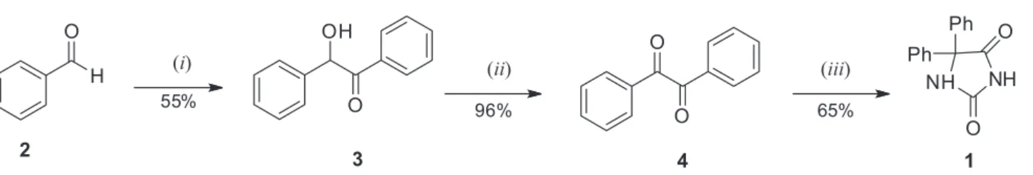
Phenytoin was synthesized using the method described
by Vogel19 (Scheme 1) in which benzaldehyde undergoes
self-condensation followed by oxidation to benzil (4).
Benzil condensation with urea then produces phenytoin (1)
in good overall yield.
Structural modification of pharmacologically active compounds is of interest since the introduction of other pharmacophoric groups in the right position of these compounds can generate a new class of molecules with potential action on living organisms. In this sense, we envisaged the association of the phenytoin nucleus with triazolic rings containing different substituents, since some triazolic compounds have shown a broad spectrum of activities such as antibacterial, anticonvulsant, anti-inflammatory, antiviral, anticancer and antitumor, among others.27
catalyzed by copper(I) salts, a process known as Huisgen
cycloaddition.29 This reaction was studied by Huisgen in
the 1960’s and, due to its versatility, the alkyne and azide groups can be incorporated into various compounds through
many methodologies.30
In this work, several 3-substituted imidazolic-like compounds were prepared from synthetic phenytoin, aiming at potential new drugs presenting inhibitory activity against human metapneumovirus (HMPV). The synthetic methodology is straightforward and is summarized in
Scheme 2. The title compounds (7-9) were obtained in
good overall yields from phenytoin and the formation of the triazolic rings was confirmed by NMR analysis in which the CH protons appear between 7.5-9.0 ppm and the carbons between 130-150 ppm.
Regarding antiviral activity, compounds 8 and 9 showed
a mild potential against HMPV, inhibiting 67.5% and
42.9% of viral replication, respectively, while compound 7
inhibited 99.9%, the same as ribavirin (Table 1).
A dose-response curve was performed in order to determine
the ED50 values for each triazolic compound (Figure 1).
Nevertheless, this was not possible for compound 9, since
at the highest concentration tested it inhibited less than 50%
of viral replication. The antiviral activity for compounds 7
and 8 was dose-dependent, meaning that the lower the
concentration of the compound, the lower the inhibition.
The ED50 values were 85.89 µg mL-1 for compound 8,
3.85 µg mL-1 for compound 7, and 1.68 µg mL-1 for ribavirin.
It was not possible to determine the selectivity index
(SI) of compound 9, since no dose-response curve was
made. But compounds 8, 7 and ribavirin showed SI of
2.74, > 129.87 and > 119.04, respectively.
Four different mechanisms of action were evaluated (Table 2): virucidal, interaction with cellular receptors, inhibition of the viral penetration and the activity in intracellular events (post-penetration). The occurrence of one mechanism does not exclude any other as some researches
have already reported.31-35
O
H
2
( )i
OH
O
( )ii ( )iii
O
O
3 4
NH NH
O
O Ph
Ph
1
55%
96% 65%
Scheme 1. (i) EtOH, NaCN (aq.), reflux, 30 min; (ii) HNO3, reflux, 2 h; (iii) EtOH, KOH, H2NCONH2, reflux, 3 h.18
NH NH
O
O Ph
Ph
1
NH N
O
O Ph
Ph
X
(5) X = Br
(6) X = N
3
NH N
O
O Ph
Ph
N
( )ii, 93%
N N R
(7) R = Ph, 83%
(8) R =
(9) R = OH
O ( )iv,
92%
69%
66%
( )i ( )iii
Scheme 2. (i) BrCH2CH2CH2Br, DBU, CH2Cl2, reflux, 3 h; (ii) NaN3, EtOH/H2O 20%, reflux, 35 h; (iii) PhCCH or CH3CH(OH)CCH, CuSO4.5H2O, Na
ascorbate, reflux, 24 h; (iv) MnO2, CH2Cl2, r.t., 24 h.
Table 1. Cytotoxicity and antiviral activity of the triazolic compounds 7-9
Compound CC20a / (µg mL-1) Inhibition at CC20 / % CC50b / (µg mL-1) ED50c / (µg mL-1) SId / (CC50b/ED50c)
7 500 99.9 > 500 3.85 > 129.87
8 125 67.5 236.05 85.89 2.74
9 500 42.9 > 500 NDe NDe
Ribavirinf 50 99.9 > 200 1.68 > 119.04
Regarding antiviral potential, compound 7 was the most
active, with ED50 value of 3.85 µg mL-1, a value 22 times
smaller than that of the second best compound (ED50 of
85.89 µg mL-1 for compound 8). Furthermore, compound
7 was less toxic for LLC-MK2 cells, which contributed for
its high selectivity index (129.87).
When we studied the mechanisms of action, both
compounds (7 and 8) showed activity in the early stages
of HMPV replication, mainly virucidal (98.3% and 63.9%, respectively), interacting with the cellular receptors, thus preventing HMPV adsorption to cell surface (98.9%
and 67.4%, respectively). Compound 7 also showed
activity during the penetration of HMPV particles into cells, inhibiting 73.8% of viral replication. In this stage,
compound 9 had no activity and compound 8 a mild
one (19.3%). None of the three compounds was capable of inhibiting intracellular events of HMPV replication, unlike ribavirin, probably due to the low solubility of these compounds.
Conclusions
According to the results shown here, we conclude
that compounds 7, 8 and 9 may be used as a prophylactic
treatment for infections caused by HMPV because they act by preventing adsorption and penetration of viral particles in the host cell. Probably, its use in combination with ribavirin may be an alternative to minimize the action of the virus.
Supplementary Information
Supplementary data are available free of charge at http://jbcs.sbq.org.br as PDF file.
Acknowledgments
The authors thank Universidade de Brasília, Universidade Católica Dom Bosco, Universidade Federal do Rio de Janeiro, Conselho Nacional de Desenvolvimento Científico e Tecnológico (CNPq), Coordenação de Aperfeiçoamento de Pessoal de Nível Superior (CAPES) and Fundação Carlos Chagas de Amparo à Pesquisa do Estado do Rio de Janeiro (FAPERJ) for financial support.
References
1. van den Hoogen, B. G.; de Jong, J. C.; Groen, J.; Kuiken, T.; de Groot, R.; Fouchier, R. A.; Osterhaus, A. D.; Nat. Med. 2001,
7, 719.
2. van den Hoogen, B. G.; van Doornum, G, J.; Fockens, J. C.; Cornelisse, J. J.; Beyer, W. E.; de Groot, R. R.; Osterhaus, A. D.; Fouchier, R. A.; J. Infect. Dis.2003, 188, 1571.
3. Landry, M. L.; Cohen, S.; Ferguson, D.; J. Clin. Microbiol. 2008, 46, 1098.
4. Dare, R.; Sanghavi, S.; Bullotta, A.; Keightley, M. C.; St. George, K.; Wadowsky, R. M.; Paterson, D. L.; McCurry, K. R.; Reinhart, T. A.; Husain, S.; Rinaldo, C. R.; J. Clin. Microbiol. 2007, 45, 548.
5. Gerna, G. P.; Vitulo, P.; Rovida, F.; Lilleri, D.; Pellegrini, C.; Oggionni, T.; Campanini, G.; Baldanti, F.; Revello, M. G.;
J. Med. Virol. 2006, 78, 408.
6. Cuevas, L. E.; Nasser, A. M.; Dove, W.; Gurgel, R. Q.; Greensill, J.; Hart, C. A.; Emerg. Infect. Dis. 2003, 9, 1626. 7. da Silva, L. H.; Spilki, F. R.; Riccetto, A. G.; de Almeida, R. S.;
Baracat, E. C.; Arns, C. W.; J. Clin. Virol. 2008, 42, 78. 0 10 20 30 40 50 60 70 80 0 1000 2000 3000 4000 5000 6000
VC 15.6 31.2 62.5 125
Inhibi
on / %
No. copies x 10
3 Inhibi on Inhibi on No. copies No. copies (a) (b) 0 10 20 30 40 50 60 70 80 90 100 0 1000 2000 3000 4000 5000 6000
VC 1.9 3.9 7.8 15.6 31.2 62.5 125 250 500
Inhibi
on / %
No. copies x 10
3
Concentra on / (µg mL Concentra on / (µg mL
-1 1 ) )
Figure 1. Dose-response curves of the triazolic compounds against HMPV
(a) compound 8; (b) compound 7.
Table 2. Mechanisms of action of compounds 7, 8 and 9
Virucidala Receptora Penetrationa Intracellulara
7 98.3 98.9 73.81 0
8 63.9 67.4 19.33 0
9 43.3 46.4 0 0
Ribavirin 0 0 0 99.9
aResults in percentage of inhibition.
0 10 20 30 40 50 60 70 80 0 1000 2000 3000 4000 5000 6000
VC 15.6 31.2 62.5 125
Inhibi
on / %
No. copies x 10
3 Inhibi on Inhibi on No. copies No. copies (a) (b) 0 10 20 30 40 50 60 70 80 90 100 0 1000 2000 3000 4000 5000 6000
VC 1.9 3.9 7.8 15.6 31.2 62.5 125 250 500
Inhibi
on / %
No. copies x 10
3
Concentra on / (µg mL Concentra on / (µg mL
8. Albuquerque, M. C.; Pena, G. P.; Varella, R. B.; Gallucci, G.; Erdman, D.; Santos, N.; Emerg. Infect. Dis. 2009, 15, 806. 9. Debur, M. C.; Vidal, L. R.; Stroparo, E.; Nogueira, M. B.;
Almeida, S. M.; Takahashi, G. A.; Rotta, I.; Pereira, L. A.; Silveira, C. S.; Delfraro, A.; Nakatani, S. M.; Skraba, I.; Raboni, S. M.; Mem. Inst. Oswaldo Cruz 2010, 105, 1010.
10. Pilger, D. A.; Cantarelli, V. V.; Amantea, S. L.; Leistner-Segal, S.;
Mem. Inst. Oswaldo Cruz 2011, 106, 56.
11. Watanabe, A. S.; Carraro, E.; Moreira, L.; Camargo, C.; Sinohara, J.; Puerari, D.; Guatura, S.; Granato, C.; Bellei, N.;
Braz. J. Infect. Dis. 2011, 15, 220.
12. Wyde, P. R.; Chetty, S. N.; Jewell, A. M.; Boivin, G.; Piedra, P. A.; Antiviral Res. 2003, 60, 51.
13. Brunton, L. L.; Chabner, B. A.; Knollman, B. C.; Goodman &
Gilman’s, The Pharmacological Basis of Therapeutics, 12th ed.;
McGraw-Hill: New York, 2011.
14. Oliveira, S. M.; Silva, J. B. P.; Hernandes, M. Z.; Lima, M. C. A.; Galdino, S. L.; Pitta, I. R.; Quim. Nova2008, 31, 614. 15. Botros, S.; Khalil, N. A.; Naguib, B. H.; El-Dash, Y.; Eur. J.
Med. Chem. 2013, 60, 57; Deodhar, M.; Sable, P.; Bhosale, A.;
Juvale, K.; Dumbare, R.; Sakpal, P.; Turk. J. Chem. 2009, 33, 367.
16. Ali, O. M.; Amer, H. H.; Mosaad, A. A.; Abdel-Rahman, A. A.-H.; Chem. Heterocycl. Compd. 2012, 48, 1043; Ali, O. M.; El-Sayed, W. A.; Eid, S. A.; Abdelwahed, N. A. M.; Abdel-Rahman, A. A.-H.; Acta Polon. Pharm. 2012, 69, 657. 17. Ciechanowicz-Rutkowska, M.; Stadnicka, K.;
Kiec-Kononowicz, K.; Byrtus, H.; Filipek, B.; Zygmunt, M.; Maciag, D.; Arch. Pharm. 2000, 333, 357.
18. Ferreira, M. L. G.; Pinheiro, L. C. S.; Santos-Filho, O. A.; Peçanha, M. D. S.; Sacramento, C. Q.; Machado, V.; Ferreira, V. F.; Souza, T. M. L.; Boechat, N.; Med. Chem. Res. 2014, 23, 1501.
19. Vogel, A. I.; Textbook of Practical Organic Chemistry, 5th ed.; John Wiley & Sons: New York, 1989.
20. Borefreund, E.; Puerner, J.; Toxicol. Lett. 1985, 24, 119. 21. Brittain-Long, R.; Nord, S.; Olofsson, S.; Westin, J.;
Lars-Magnus, A.; Lindh, M.; J. Clin. Virol. 2008, 41, 53.
22. Chen, M.; Griffith, B. P.; Lucia, H. L.; Hsiung, G. D.;
Antimicrob. Agents Chemother. 1988, 32, 678.
23. Mendes, G. S.; Soares, A. R.; Martins, F. O.; Albuquerque,
M. C. M.; Costa, S. S.; Yoneshigue-Valentin, Y.; Gestinari, L. M. S.; Santos, N.; Romanos, M. T. V.; Rev. Inst. Med. Trop.
2010, 52, 3
24. Mendes, G.; Soares, A. R.; Sigiliano, L.; Machado, F.; Kaiser, C.; Romeiro, N.; Gestinari, L.; Santos, N.; Romanos, M. T. R.; Molecules2011, 16, 8437.
25. Mendes, G. S.; Duarte, M. E. R.; Colodi, F. G.; Noseda, M. D.; Ferreira, L. G.; Berté, S. D.; Cavalcanti, J. F.; Santos, N.; Romanos, M. T. V.; Carbohydr. Polym.2014, 101, 313. 26. Karimi, A.; Moradi, M. T.; Saeedi, M.; Asgari, S.;
Rafieian-Kopaei, M.; Adv. Biomed. Res. 2013, 2, 36; Astani, A.; Schnitzler, P.; Iran. J. Microbiol. 2014, 6, 149.
27. Siddiquia, N.; Ahsana, W.; Alama, M. S.; Alia, R.; Jainb, S.; Azada, B.; Akhtar, J.; Int. J. Pharm. Sci. Rev. Res.2011, 8, 161. 28. Singhal, N.; Sharma, P. K.; Dudhe, R.; Kumar, N.; J. Chem.
Pharm. Res. 2011, 3, 126; Garzón, R. L.; Valero, D. G.;
Calahorro, C. V.; Pérez, N. C.; Rodriguez, A. G.; Monatsh.
Chem. 1987, 118, 553.
29. Huisgen, R.; Szeimies, G.; Möbius, L.; Chem. Ber. Recl. 1967,
100, 2494.
30. Amblard, F.; Cho, J. H.; Schinazi, R. F.; Chem. Rev. 2009, 109, 4207; Kolb, H. C.; Finn, M. G.; Sharpless, K. B.; Angew. Chem.
Int. Ed. 2001, 40, 2004; Kolb, H. C.; Sharpless, K. B.; Drug
Discov. Today2003, 8, 1128.
31. Carlucci, M. J.; Scolaro, L. A.; Errea, M. I.; Matulewicz, M. C.; Damonte, E. B.; Planta Med. 1997, 63, 429.
32. Barbosa, J. P.; Pereira, R. C.; Abrantes, J. L.; dos Santos, C. C.; Rebello, M. A.; Frugulhetti, I. C.; Texeira, V. L.; Planta Med.
2004, 70, 856.
33. Matsuhiro, B.; Conte, A. F.; Damonte, E. B.; Kolender, A. A.; Matulewicz, M. C.; Mejías, E. G.; Pujol, C. A.; Zúñiga. E. A.;
Carbohydr. Res. 2005, 340, 2392.
34. Cassolato, J. E.; Noseda, M. D.; Pujol, C. A.; Pellizzari, F. M.; Damonte, E. B.; Duarte, M. E.; Carbohydr. Res. 2008, 343, 3085.
35. Bouhlal, R.; Haslin, C.; Chermann, J. C.; Colliec-Jouault, S.; Sinquin, C.; Simon, G.; Cerantola, S.; Riadi, H.; Bourgougnon, N.; Mar. Drugs2011, 9, 1187.
Submitted: June 10, 2015