1 Research paper title:
1
Persistence of wastewater antibiotic resistant bacteria and their genes in human fecal 2 material 3 4 Authors: 5
Nazareno Scaccia, Ivone Vaz-Moreira, Célia M. Manaia 6
7
Universidade Católica Portuguesa, CBQF - Centro de Biotecnologia e Química Fina – 8
Laboratório Associado, Escola Superior de Biotecnologia, Rua Diogo Botelho 1327, 9 4169-005 Porto, Portugal. 10 11 Corresponding author: 12 Célia M. Manaia, Ph.D. 13
Escola Superior de Biotecnologia 14
Universidade Católica Portuguesa 15 4169-005 Porto 16 Portugal 17 Email: [email protected] 18 19 20 21 22 23 24 25 26 27 28 29 30 31 32 33
2 Abstract
34
Domestic wastewater is a recognized source of antibiotic resistant bacteria and antibiotic 35
resistance genes (ARB&ARGs), whose risk of transmission to humans cannot be ignored. 36
The fitness of wastewater ARB in the complex fecal microbiota of a healthy human was 37
investigated in feces-based microcosm assays (FMAs). FMAs were inoculated with two 38
wastewater isolates, Escherichia coli strain A2FCC14 (MLST ST131) and Enterococcus 39
faecium strain H1EV10 (MLST ST78), harboring the ARGs blaTEM, blaCTX, blaOXA-A and 40
vanA, respectively. The FMAs, incubated in the presence or absence of oxygen or in the
41
presence or absence of the antibiotics cefotaxime or vancomycin, were monitored based 42
on cultivation, ARGs quantification and bacterial community analysis. 43
The fecal bacterial community was dominated by members of the phyla Firmicutes, 44
Bacteroidetes, Actinobacteria, Proteobacteria and Verrucomicrobia. The ARGs
45
harbored by the wastewater isolates could be quantified after one week, in FMAs 46
incubated under both aerobic and anaerobic conditions. These observations were not 47
significantly different in FMAs incubated anaerobically, supplemented with sub-48
inhibitory concentrations of cefotaxime or vancomycin. The observation that ARGs of 49
wastewater ARB persisted in presence of the human fecal microbiota for at least one week 50
supports the hypothesis of a potential transmission to humans, a topic that deserves further 51 investigation. 52 53 Keywords: 54
human fecal microbiota, microcosm assays, microcosm effect, antibiotic resistance 55
transmission, antibiotic resistant bacteria, antibiotic resistance genes. 56
57
One sentence summary: 58
3
Antibiotic resistance genes harbored by wastewater bacterial isolates persisted in healthy 59
infant´s stool-based microcosms under different conditions, including the presence of 60
sub-inhibitory concentrations of antibiotics. 61
62
1. Introduction 63
Antibiotic resistance, defined as the capability of bacteria to survive and proliferate in the 64
presence of antibiotics, is a natural bacterial property (Davies and Davies 2010). Bacteria 65
resistant to antibiotics, against which were once susceptible, owe that capability to the 66
acquisition of antibiotic resistance genes (ARGs), most of the times through horizontal 67
gene transfer (Bengtsson-Palme et al., 2018; Summers 2006). Acquired antibiotic 68
resistance emergence and proliferation have been attributed to factors such as the 69
presence of antibiotics and other antimicrobials, metals or conditions still unknown, 70
which through their stressor effects exert what has been designated as selective pressures 71
(Martinez 2008; Rosenblatt-Farrell 2009). Ubiquitous bacteria, harboring acquired 72
antibiotic resistance genes, can thrive in the environment, in particular in wastewater, 73
water, soil and wildlife (Berendonk et al., 2015; Huddleston 2014). The paths of 74
transmission of these bacteria back to humans are not fully understood and the probability 75
of such occurrence hardly can be estimated based on the current knowledge (Manaia 76
2017). A still unanswered question refers to the capability of antibiotic resistant bacteria 77
(ARB) from environmental origin, as well as their genes, to is survive or persist in the 78
human body (Bengtsson-Palme et al., 2018; Larsson et al., 2018; Manaia 2017). 79
Specifically, if it is assumed that the digestive tract is the entry portal, one of the questions 80
would be if these bacteria would be able to survive the competition of complex intestinal 81
microbiome (Manaia 2017; Vaz-Moreira et al., 2014). The role of the digestive tract as a 82
relevant entry portal assumes a particular likeliness in situations of ingestion of raw 83
vegetable-based food products, therefore acting as potential sources of ARB to humans 84
4
(Holzel et al., 2018; Valerio et al., 2006; Zhang et al., 2019). The risks of ARB occurrence 85
in vegetables can be enhanced in scenarios of manure soil amendment or water reuse for 86
irrigation (Becerra-Castro et al., 2015; Heuer et al., 2011; Marti et al., 2013). Indeed, 87
several recent studies have shown the presence of ARB and ARGs in raw-eaten products 88
(e.g. lettuce), raising concerns for consumers, particularly for immunocompromised 89
people (Araujo et al., 2017; Blau et al., 2018; O'Flaherty et al., 2019; Zhang et al., 2017; 90
Zhu et al., 2017). However, even after ingestion, the success of allochthonous bacteria in 91
the intestinal tract is supposedly antagonized by the gut microbiota, a complex and 92
dynamic community of microorganisms colonizing the gastrointestinal tract of humans 93
since birth (Donaldson et al., 2016; Gibson et al., 2014). While indigenous (or 94
autochthonous) microorganisms may have the intestine as a long term or almost 95
permanent niche, allochthonous microorganisms may colonize transiently the human gut, 96
although some, the most fitted, will be able to persist for long time periods (Milani et al., 97
2017; Ventura et al., 2009). Among the most fitted bacterial groups, it can be 98
hypothesized that bacteria of enteric origin, like Escherichia coli or Enterococcus spp., 99
are good candidates to survive the competition of the fecal microbiome of a healthy 100
individual. These can certainly be part of the 106 to 109 bacterial cells that, depending on 101
the different dietary intake, can be ingested daily (Derrien and van Hylckama Vlieg 2015; 102
Lang et al., 2014). If part of these bacteria carry acquired ARGs, such fact can 103
hypothetically increase their fitness in the presence of antibiotics and/or facilitate the 104
interchange of those genes with the native community (Salyers et al., 2004). Therefore, 105
the contamination of the human food chain with environmental ARB might represent a 106
risk for the subsequent transmission to humans. Among the multiple bottlenecks that may 107
hamper the successful human-gut colonization by allochthonous bacteria, are the capacity 108
to survive the complex native microbiome and/or the stability of the respective ARGs. 109
5
These questions boosted this study that used the fecal material of a healthy infant as a 110
model of human gut microbiota to assess the persistence of enteric ARB isolated from 111
wastewater, Escherichia coli strain A2FCC14 and Enterococcus faecium strain H1EV10, 112
harboring the ARGs blaTEM, blaCTX-M, blaOXA-A and vanA, respectively. Specifically, the 113
objectives of this work were to assess: 1) if those wastewater ARB and the respective 114
ARGs were outcompeted in the presence of the human fecal microbiota, 2) if the cell-free 115
ARGs could persist in that environment, and 3) the influence of the presence of oxygen 116
or of antibiotics on the survival and persistence of the ARB and ARGs measured in 1) 117
and 2). Assuming successful colonization, for which it is necessary that the exogenous 118
enteric bacteria can thrive in the presence of fecal material, an additional question is if 119
the acquired ARGs will be lost because represent a fitness cost. 120
121
2. Material and methods 122
The survival of wastewater antibiotic resistant isolates and persistence of the respective 123
ARGs was assessed in the presence of the complex human fecal microbial community. 124
The experiments were conducted in FMAs composed of human stool specimens spiked 125
with two wastewater isolates: E. coli strain A2FCC14 (harboring the ARGs blaTEM, 126
blaCTX and blaOXA-A) and Ent. faecium strain H1EV10 (harboring the ARG vanA), or with 127
the respective DNA extracts. The environmental variables tested in the FMAs were 128
aerobic vs. anaerobic conditions, and the effect of single or multiple doses of sub-129
inhibitory concentrations of cefotaxime or vancomycin, under anaerobic conditions. The 130
FMAs were incubated at 37 ºC, and samples were collected at 0, 1, 3 and 7 days. 131
Monitoring was based on the enumeration of culturable bacteria, quantitative PCR 132
(qPCR) analysis of antibiotic resistance and 16S rRNA genes and bacterial community 133
analyses based on 16S rRNA gene amplicon sequencing. 134
6 135
2.1. Bacterial strains
136
The strains E. coli A2FCC14 (isolated from raw municipal wastewater) and Ent. faecium 137
H1EV10 (isolated from untreated hospital effluent) (Varela et al., 2013) were used to 138
inoculate the fecal microcosm assays (FMAs). The Whole Genome Shotgun projects of 139
strains A2FCC14 and H1EV10 have been deposited at DDBJ/ENA/GenBank under the 140
accession numbers WSZB00000000 and WSZC00000000, respectively. These strains 141
have an Average Nucleotide Identity (ANI) of orthologous gene pairs shared between two 142
microbial genomes with the type strains of the species of 98.5% and 99.5%, respectively. 143
ARGs conferring resistance to beta-lactam (blaTEM, blaCTX-M, and blaOXA-A), harbored by 144
the E. coli strain and to vancomycin (vanA) harbored by the Ent. faecium strain were 145
monitored in FMAs. For inoculum preparation, both strains were handled as follows: E. 146
coli A2FCC14 was cultivated on m-FC agar medium (fecal coliform agar, Difco BD)
147
supplemented with cefotaxime 4 mg L-1 and incubated overnight at 37 °C and Ent. 148
faecium H1EV10 was cultivated on m-Enterococcus agar medium (Difco BD)
149
supplemented with vancomycin 16 mg L-1 and incubated for 48 hours at 37 °C. The 150
biomass collected from each of those bacterial cultures was used to prepare a bacterial 151
suspension in saline solution (0.85% (w/v) NaCl) with a cell density of approximately 152
108 and 107 Colony Forming Units (CFU) mL-1 for E. coli A2FCC14 and Ent. faecium 153
H1EV10, respectively. This cell density was in accordance with the average density of 154
bacteria of these groups in the human microbiome and although higher than can be 155
expected from a possible ingestion and digestion, it overcomes the experimental risk of 156
reaching the values below the limits of quantification. After an initial calibration of CFU 157
versus turbidity at 610 nm, the cell suspensions used to inoculate each FMA were 158
measured by spectrophotometry. 159
7 160
2.2. Feces-based microcosm assays (FMAs)
161
The FMAs prepared with fecal material of a healthy child were used as a model to assess 162
the fate of ARB and ARGs. All assays used fecal material supplied by a single healthy 163
donor, aged 40 to 58 months during this study, and who was never submitted to 164
antibiotherapy. The option for a donor with these characteristics was because although 165
the gut microbiota was certainly affected by diet and lifestyle, it was not rearranged due 166
to previous antibiotic exposure, a control considered relevant for the present experimental 167
design. In total, were collected twelve stool samples, each used to run an independent 168
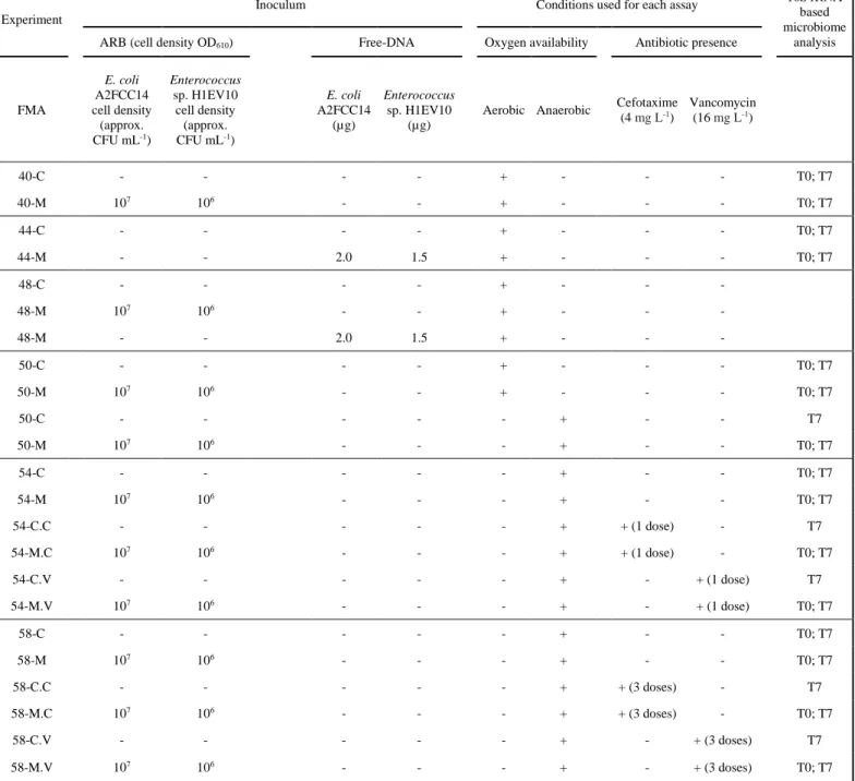
FMA (Table 1). For each FMA experiment, were collected ≥ 80 g of fecal material that 169
was stored at 4 °C for no more than 3 days. Each FMA comprised inoculated assays (M-170
assays) and the respective non-inoculated controls (C-assays). FMAs were tested under 171
aerobic and anaerobic conditions or under anaerobic conditions spiked with antibiotics. 172
The evaluation of the effect of oxygen was considered of interest given the fact that E. 173
coli are facultative anaerobes and enterococci are aerotolerant, supporting some inference
174
about the influence of the fitness of these exogenous bacteria versus the effect of 175
competition by the fecal microbiota. In total, four FMAs were incubated under aerobic 176
conditions [40 (C and M); 44 (C and M); 48 (C and M); 50 (C and M)], three under 177
anaerobic conditions [50 (C and M); 54 (C and M); 58 (C and M)] and four under 178
anaerobic conditions, spiked with subinhibitory concentrations of antibiotics. Antibiotic 179
spiking was done in a single-dose [54-C+cefotaxime C.C), 54-C+vancomycin (54-180
C.V), 54-M+cefotaxime (54-M.C) and 54-M+vancomycin (54-M.V)] or in multiple-181
doses [58-C+cefotaxime C.C), 58-C+vancomycin C.V), 58-M+cefotaxime (58-182
M.C) and 58-M+vancomycin (58-M.V)] (Table 1). Anaerobic FMAs were handled and 183
incubated in an anaerobic chamber (Whitley Workstation A35, containing a gas mixture 184
of 85% carbon dioxide, 10% nitrogen, 5% hydrogen). Each experimental set (FMA) 185
8
comprised 24 vial assays, corresponding to triplicates of spiked and non-spiked assays to 186
be sacrificed for analyses after 0, 1, 3 and 7 days of incubation (3 replicates x spiked/non-187
spiked x 4 incubation periods). Although the transit time of bacteria in the large intestine 188
is estimated to be ~2.5 days, ingested bacteria can be detected in the intestine for one 189
week (Berg 1996; Derrien and van Hylckama Vlieg 2015). Based on this note and 190
preliminary assays, an incubation period of 7 days (extended to 30 days for occasional 191
analysis) was selected. 192
To prepare these experimental sets, stool samples were diluted five times with sterile 193
saline solution (0.85% (w/v) NaCl) and divided in 24 aliquots of 15 mL each. Half of 194
these 24 aliquots were inoculated with 2 mL of a bacterial cocktail, described in the 195
previous section, composed by the mixture of both strains, E. coli A2FCC14 and Ent. 196
faecium H1EV10 (M-assays). The other half of the aliquots, corresponding to
non-197
inoculated controls (C-assays), was spiked with 2 mL of sterile saline solution. FMAs 40, 198
44 and 48 were incubated aerobically. FMA50 comprised two parallel FMAs (24 vials 199
incubated aerobically and 24 anaerobically). FMA54 and FMA58 were incubated 200
anaerobically and were spiked with antibiotics. Briefly, FMA54 comprised 24 vials 201
without antibiotic, 24 spiked with one dose of cefotaxime (4 mg L-1 for each microcosm) 202
and 24 spiked with one dose of vancomycin (16 mg L-1 for each microcosm). FMA58 203
differed from FMA54 on the method of antibiotic spiking which in FMA58 was supplied 204
at three antibiotic moments (3 x 4 mg L-1 for cefotaxime or 3 x 16 mg L-1 for vancomycin) 205
at time 0, 1 day and 3 days, before each sample collection. Cell-free DNA supplemented 206
FMAs, FMA44 and FMA48, were spiked with DNA extracts from E. coli A2FCC14 and 207
Ent. faecium H1EV10. The DNA fragment length of the DNA extract of E. coli A2FCC14
208
was assessed in an agarose gel (1 %) electrophoresis, originating a single band with a 209
molecular weight 10-50 000 bp, with no smearing effect, which would suggest that DNA 210
9
was degraded. The quantity of DNA used was 2.0 and 1.5 µg of DNA extracted from E. 211
coli A2FCC14 and Ent. faecium H1EV10, corresponding to the cell density used in the
212
ARB inoculated FMAs (Table 1). FMA44 was designed aiming at assessing the 213
persistence of the spiked free DNA and FMA48 aimed at assessing the potential 214
occurrence of natural transformants, able to uptake the free DNA. Therefore FMA48 was 215
composed of 3 separated assays (6 vial assays corresponding to triplicates of non-spiked 216
assays, sampled at T0 and T7, 6 vial assays corresponding to triplicates of bacteria-spiked 217
assays, sampled at T0 and T7 and, 6 vial assays corresponding to triplicates of assays 218
spiked with cell-free DNA extracted from the test bacteria, sampled at T0 and T7). 219
Natural transformants were tentatively isolated on the m-FC or m-Enterococcus culture 220
media supplemented with antibiotic (cefotaxime or vancomycin, respectively). Cultivable 221
bacteria counts were processed immediately, and aliquots for dry weight determination 222
and total DNA extraction were stored at -20 and -80 ºC, respectively, until used. All 223
determinations were done in triplicate. The stool dry weight was determined in 1 mL fecal 224
slurry samples by incubation at 60 ºC, until a constant weight was reached, which 225
corresponded to approx. 5 days. This study was approved by the Ethics Committee of the 226
Universidade Católica Portuguesa in Porto. 227
228
2.3. Enumeration of cultivable bacteria in FMAs
229
Luria-Bertani Agar (LA) (Invitrogen), m-FC agar (Difco BD) and m-Enterococcus agar 230
(Difco BD) were used for enumeration of total heterotrophic bacteria (HB), enterobacteria 231
and enterococci, respectively. When necessary, these culture media were supplemented 232
with cefotaxime (4 mg L-1; Sigma-Aldrich, St Louis, USA) or vancomycin (16 mg L-1; 233
Sigma-Aldrich, St Louis, USA), at concentrations corresponding to minimum inhibitory 234
concentration (MIC) for E. coli or enterococci (CLSI 2014). For bacterial enumeration, 235
10
volumes of 1 mL were collected from each FMA, serially diluted in sterile saline solution 236
(0.85% (w/v) NaCl) and plated on the adequate culture medium using the Miles and Misra 237
method (Miles et al., 1938). Cultures were incubated at 37 ºC for 24 h on LA and m-FC, 238
or 48 h on m-Enterococcus agar. All bacterial counts were performed in triplicate. 239
240
2.4. DNA extraction
241
Total DNA was extracted from 1 mL of fecal slurry (corresponding to approximately 240 242
mg of wet sedimented stool) using the NZY Tissue gDNA Isolation kit (Nzytech, 243
Portugal) according to the manufacturer’s instructions. The DNA concentration in the 244
extracts was quantified using Qubit fluorometer (Thermo Fisher Scientific, USA). 245
Extracts were preserved at -20 ºC until qPCR or microbial community analyses were 246 performed. 247 248 2.5. Quantitative PCR 249
The abundance of the genes blaTEM, blaCTX, blaOXA-A and vanA, harbored by the 250
inoculated bacteria, was monitored based on real-time quantitative PCR (qPCR). The 251
total bacterial abundance was assessed based on the housekeeping gene 16S rRNA. The 252
qPCR was conducted in a StepOne™ Real-Time PCR System (Life Technologies, 253
Carlsbad, CA) following the conditions previously described (Narciso-da-Rocha et al., 254
2018) for the ARGs blaTEM, blaCTX and blaOXA-A and the 16S rRNA gene. The qPCR 255
conditions for the detection of the gene vanA were set up in 25 µL volume using the 256
Power SYBR Green mastermix (Thermo Fisher Scientific, Austin, USA) containing 10 257
µM of each primer: VnF ATCGGCAAGACAATATGACAGC-3’ and VnR 5’-258
AGCCTGATTTGGTCCACCTC-3’ (Lata et al., 2009).The PCR program was initiated 259
by a period of 5 min at 95 °C, followed by 40 cycles of 95 °C for 15 sec and 60 °C for 30 260
11
sec. The standard curve for vanA gene was prepared using a clone of the vanA gene of 261
Ent. faecium H1EV10 with an efficiency between 96 and 105%.
262 263
2.6. Bacterial community analysis
264
The bacterial community composition of FMA40, 44, 50, 54 and 58 was analyzed at time 265
zero (T0) and after 7 days of incubation (T7), based on the hypervariable region V3/V4 266
of the 16S rRNA gene, using paired-end Illumina MiSeq® Sequencing (Genoinseq, 267
Portugal) as previously described by Narciso-da-Rocha et al. (2018). The primers used 268
were forward primer Bakt_341F 5’- CCTACGGGNGGCWGCAG -3’ and reverse primer 269
Bakt_805R 5’- GACTACHVGGGTATCTAATCC -3’ according to the manufacturer’s 270
instructions (Illumina, San Diego, CA, USA). Demultiplexed raw reads were extracted 271
from Illumina MiSeq® System in fastq format and the reads were processed and analyzed 272
using Quantitative Insights Into Microbial Ecology (QIIME2) pipeline (version 2017.10; 273
http://qiime2.org/) (Bolyen et al., 2018). Sequences shorter than 200 bp and with average 274
quality scores lower than 25 were eliminated. Sequences with average quality lower than 275
25 in a window of 5 bases were trimmed using the software PRINSEQ (Schmieder and 276
Edwards 2011). Sequences were filtered, merged and, chimeric reads were removed by 277
the DADA2 software package enclosed in QIIME2 (Callahan et al., 2016). Taxonomy 278
was assigned to the amplicon sequence variants (ASVs), sequences with 100% identity, 279
using the ARB SILVA database release 132 (Yilmaz et al., 2014). A total of 4 750 244 280
reads (ranging from 22 176 to 112 134 reads per sample) and 1714 ASVs (ranging from 281
171 to 263 per sample) were obtained from the 105 datasets, corresponding to triplicates 282
of 35 samples of C and M FMAs. 283
284
2.7. Statistical analyses
12
Cultivable bacteria counts were expressed as log values of colony-forming units (CFUs) 286
per g of stool dry weight . Gene abundance was expressed as gene copy number per g of 287
dry weight (abundance) or per 16S rRNA gene copy number (relative abundance or 288
prevalence). One-way analysis of variance (ANOVA), Tukey's and Bonferroni post-hoc 289
tests and t-test (SPSS Statistics for Windows v.24.0; IBM Corp., Armonk, NY, USA) 290
were used for determination of statistically significant differences (p < 0.01) of cultivable 291
bacteria counts, abundance and prevalence of measured genes when comparing different 292
incubation times or conditions. The bacterial community composition was expressed as 293
the relative abundance of reads number of a specific bacterial group per total reads 294
number. Correlations between the relative abundance of bacterial groups at phylum and 295
family level were analyzed using the statistical analysis of taxonomic and functional 296
profiles using the software STAMP v2.1.3 and Canoco 5.01 (Parks et al., 2014; Šmilauer 297
and Lepš 2014). Statistically significant differences between bacterial phyla or family 298
relative abundances were determined using a two-tailed t-test (p <0.01)and the p values 299
were corrected for multiple testing using the Benjamini-Hochberg FDR (Benjamini and 300
Hochberg 1995). 301
302
3. Results and discussion 303
3.1. Microbial community composition of the fecal material
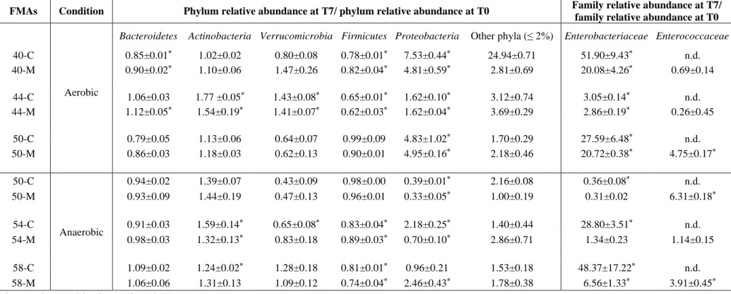
304
Since the fecal microbiota composition and the respective temporal variations could 305
somehow influence the fate of exogenous ARB and ARGs, the analysis of the fecal 306
bacterial community was necessary in this study. This part of the work had two aims, 307
assess the phylogenetic diversity in the fecal material and assess the variations that were 308
due to the microcosm effect. Over the study period, the infant (40-58 months) fecal 309
microbiota was dominated by members of the bacterial phyla Firmicutes (41.6-47.1%) 310
13
and Bacteroidetes (23.5-36.2%), followed by Actinobacteria (7.2-22.5%),
311
Proteobacteria (1.5-7.4%) and Verrucomicrobia (1.2-7.4%) (Table S1). These results are
312
in line with similar studies involving healthy individuals in the same age range (Monira 313
et al., 2011). Statistically significant variations were observed over the study period for
314
all the above mentioned phyla, although none with a clear trend of increase or decrease. 315
These variations might be due to diet and/or the natural dynamic processes observed in 316
infants gut microbiota (Milani et al., 2017; Zmora et al., 2019). Although these variations 317
were smooth and with little expected impacts on the survival of the exogenous bacteria, 318
whenever adequate they will be used to discuss the results. 319
320
3.2 Effect of incubation condition on the microbial community composition
321
The microcosm effect, meaning the bacterial community variations that occurred during 322
the incubation period (7 days), which could allegedly influence the survival or persistence 323
of exogenous bacteria or their ARGs, was assessed in non-inoculated FMAs, under both 324
aerobic and anaerobic conditions. Besides the variation of phyla composition, it was also 325
assessed the variation in the relative abundance of members of the families that include 326
the wastewater ARB surrogates used in the inoculated FMAs, Enterobacteriaceae and 327
Enterococcaceae (Table 2). The relative abundance of Proteobacteria significantly
328
(p<0.01) increased (ratio T7/T0 > 1) during the incubation under aerobic, but not under 329
anaerobic conditions (Table 2). It was also observed that the relative abundance of 330
Enterobacteriaceae in the non-inoculated assays was always higher than in inoculated
331
microcosms (Table 2), which might be due to a steady-state-like for Enterobacteriaceae 332
in the fecal microbial community, in which these bacteria, in equilibrium with the 333
remaining community, are kept at a certain level and eventually exogenous bacteria, as 334
were the wastewater isolates in this case, may have a limited proliferation capacity. 335
14
The relative abundance of the bacterial phylum Firmicutes was fairly stable over the 7 336
days period, under both aerobic and anaerobic conditions, with the ratio T7/T0 varying 337
between 0.65 and 0.99 in non-inoculated FMAs (Table 2). Curiously, members of family 338
Enterococcaceae, of the phylum Firmicutes, were not detected in non-inoculated FMAs.
339
The observation that anaerobic conditions had a lower impact on the microcosm effect 340
than aerobic conditions, mainly in the groups that include the surrogates used, 341
recommended that the effect of antibiotics would be better examined under anaerobiosis. 342
In the FMA54 and FMA58, the addition of cefotaxime or vancomycin only affected 343
significantly (p<0.01) the relative abundance of Proteobacteria (Table S3). Curiously, 344
this effect was different when a single- (FMA54) or multiple-antibiotic-dose (FMA58) 345
was used (Table S3). A single cefotaxime dose (54-C.C and 54-M.C) significantly 346
(p<0.01) reduced the relative abundance of Proteobacteria in both inoculated and non-347
inoculated FMAs (ratio antibiotic/no antibiotic < 1). In contrast, multiple cefotaxime 348
doses led to a significant (p<0.01) reduction on the relative abundance of Proteobacteria 349
only in inoculated FMAs (58-M.C). The use of a single vancomycin dose significantly 350
(p<0.01) reduced the relative abundance of Proteobacteria in the non-inoculated samples 351
(54-C.V; Table S3). In contrast, three doses of vancomycin led to a significant (p<0.01) 352
increase of the Proteobacteria relative abundance, only in non-inoculated FMA (58-C.V). 353
As noted above for oxygen, also the antibiotic effect on members of the 354
Enterobacteriaceae followed the same pattern as for the Proteobacteria phylum (Table
355
S3). In contrast, no noticeable effects of antibiotics were observed on Enterococcaceae 356
in inoculated FMAs. 357
These different behaviors of the bacterial community in presence of a single or multiple 358
doses of antibiotics may suggest the rearrangement of the community or the charity 359
among community members that able to degrade the antibiotics. The Principal 360
15
Component Analysis (PCA) of the non-inoculated assays suggest that irrespective of the 361
antibiotic and spiking method, members of the families Burkholderiaceae (phylum 362
Proteobacteria) and Lachnospiraceae (phylum Firmicutes) were those whose relative
363
abundance decreased during the incubation period. The contribution of these groups to 364
the community rearrangement may have been exacerbated in the presence of antibiotics 365
(Figure S1 A, B, C, and D). In general, the results suggest that antibiotics might have 366
important effects on the fecal bacterial community composition and structure, with effects 367
being noticed in the relative abundance of members of the phyla Firmicutes, 368
Actinobacteria and Bacteroidetes (Table S3). The relative abundance of Actinobacteria
369
was significantly lower in the fecal material used in the FMA where a single antibiotic 370
dose was tested (FMA 54) than in that where three successive doses were assayed (FMA 371
58). The opposite was observed for the relative abundance of Bacteroidetes (Table S1). 372
Since both phyla members were observed to have strong correlation with antibiotic 373
spiking, the distinct behavior observed in response to one or multiple antibiotic doses may 374
be also a result of distinct microbiota composition. The importance of these bacterial 375
groups as potential barriers or facilitators for antibiotic-induced bacterial community 376
rearrangements may deserve further investigation. In general, the PCA results (Figure S1) 377
are in agreement with the literature that suggests that the oral or intravenous antibiotic 378
administration can promote the disturbance of the human gut microbiome (Bhalodi et al., 379
2019; Francino 2016). The PCA analysis (Figure S1) suggests that the significant 380
variations observed in Proteobacteria relative abundance (Table S3), might be due to 381
complex fecal bacterial community rearrangements rather than to the increase or decrease 382
of a specific bacterial lineage. Indeed, it is this type of indirect effect that was 383
demonstrated for vancomycin, a glycopeptide antibiotic used against Gram-positive 384
bacterial infection. In that case, it was associated with the decrease of Firmicutes that led 385
16
to the increase of Proteobacteria in the fecal microbial community (Isaac et al., 2017; 386
Vrieze et al., 2014). Indeed, although there is no evidence specifically for vancomycin 387
and cefotaxime, the ability of sub-inhibitory concentrations of antibiotics to modify the 388
competition between bacterial species within a microbial community has been discussed 389
(Hall and Corno 2014; Martinez 2009). 390
391
3.3. Monitoring of culturable bacteria and antibiotic resistance genes
392
The native and exogenous culturable populations of enterobacteria, enterococci and/or 393
total heterotrophic bacteria were monitored over time and under distinct conditions. 394
Heterotrophic and enterobacteria counts presented similar variation patterns in inoculated 395
and non-inoculated FMAs and, in both cases, it was possible to infer about the beneficial 396
oxygen effect on the first day of incubation, characterized by a slight CFU increase. 397
Therefore, it is suggested that the fitness of enteric bacteria cannot be explained based 398
only on the fecal microbiota competition, it is also explained by the survival capacity, in 399
this case, higher in the presence of oxygen (Figure 1). In contrast, the behavior of the 400
aerotolerant enterococci was identical in the presence or absence of oxygen. Also, 401
enterococci counts presented a similar variation pattern in inoculated and non-inoculated 402
assays (Figure 1). In general, under anaerobic conditions, enterococci, enterobacteria and 403
heterotrophic bacteria counts presented in average reductions of 1.1, 1.0 and 0.1 log units 404
over the 7 incubation days, respectively (Figure 1). However, the stochastic nature of 405
these variations is suggested, when different FMAs are compared (Figure 1). 406
The exogenous bacteria were wastewater isolates, E. coli A2FCC14 and Ent. faecium 407
H1EV10, belonging to the multilocus sequence types ST131 and ST78, respectively, 408
therefore genetically related with widespread pathogens (Khan et al., 2010; Nicolas-409
Chanoine et al., 2014). These exogenous bacteria were traced based on their ARGs 410
17
blaTEM, blaCTX-M, blaOXA-A and vanA (Figure 2). Under aerobic conditions, the 16S rRNA 411
gene abundance per gram of feces dry weight, decreased during the incubation period on 412
average 0.26 log-units in inoculated FMAs (40-M and 50-M) and 0.32 log-units for non-413
inoculated FMAs (40-C and 50-C). These values were comparatively higher under 414
anaerobic conditions, of 0.48 log-units in inoculated FMAs (50-M, 54-M, and 58-M) and 415
of 0.42 log-units in non-inoculated FMAs (50-C, 54-C and 58-C). 416
Among the analyzed ARGs, only blaTEM was detected in non-inoculated microcosms 417
(Figure 2). The blaTEM gene may have been ingested by the donor (e.g. fresh produce)
418
(Blau et al., 2018), and its occurrence in the gut microbiota of healthy individuals, even 419
if never exposed to antibiotics, has been reported (Fouhy et al., 2014; Sommer et al., 420
2009). In general, the vanA gene abundance (per gram of dry weight) did not vary 421
significantly from T0 to T7, with average measurements at T0 and T7 of 5.87 ± 0.11 and 422
5.79 ± 0.15 log-units under aerobic conditions, and 7.34 ± 1.17 and 7.17 ± 1.12 log-units 423
under anaerobic conditions (Figure 2). In contrast, occasionally, although under both 424
aerobic and anaerobic conditions (40-M and 58-M, Figure S2) the prevalence of vanA 425
(per 16S rRNA gene) increased significantly (p<0.01), which may be due to the decrease 426
of the overall bacterial population, herein measured in 16S rRNA gene abundance. 427
Aerobically, on average (FMAs 40-C, 50-C, 40-M and 50-M), the abundance of blaTEM 428
gene had a significant (p<0.01) increase from 7.13 ± 0.55 log-units at T0 to 8.23 ± 0.44 429
log-units at T7 (Figure 2). Anaerobically, blaTEM was not detected in one of the non-430
inoculated assays (58-C) and it decreased significantly (p<0.01) in the FMA50 (C, 50-431
M; Figure 2). In the FMA54, blaTEM gene increased and decreased significantly (p<0.01) 432
in the non-inoculated and inoculated assays, respectively (54-C, 54-M; Figure 2). On the 433
average of the FMAs (50-C, 54-C, 50-M, 54-M, and 58-M), its abundance varied from 434
7.61 ± 1.12 log-units at T0 to 8.18 ± 0.74 log-units at T7 (Figure 2). The prevalence of 435
18
the blaTEM gene (per 16S rRNA gene) followed the same pattern of variation of the blaTEM 436
gene abundance along time, which suggests that the variations of blaTEM gene are mainly 437
due to total bacteria variations (Figure 2, Figure S2). 438
The abundance (per gram of dry weight) of the blaCTX-M gene increased significantly 439
(p<0.01) under aerobic incubation (40-M; Figure 2) ranging from 6.83 ± 0.15 to 7.80 ± 440
0.05 log-units and, the same gene decreased significantly (p<0.01) under anaerobic 441
incubation (50-M; Figure 2) varying from 7.04 ± 0.21 to 5.63 ± 0.46 log-units. Similar 442
results were observed for blaOXA-A gene, which abundance (per gram of dry weight) varied 443
significantly (p<0.01) from 6.37 ± 0.22 to 7.49 ± 0.06 log-units under aerobic conditions 444
(40-M; Figure 2) and from 6.68 ± 0.14 to 5.75 ± 0.42 log-units under anaerobic conditions 445
(50-M; Figure 2). 446
In general, it was observed that the abundance of the beta-lactamase genes increased 447
under aerobic conditions and decrease under anaerobic conditions (Figure 2). This result 448
that is aligned with the enumeration of culturable bacteria, suggests that the fitness of the 449
exogenous bacteria, and not only the competition by the native microbiota, may dictate 450
the fate of enterobacteria in the FMAs. The prevalence (expressed per 16S rRNA gene 451
copy number) of both blaCTX-M and blaOXA-A increased under aerobic (40-M, 50-M) and 452
anaerobic conditions (54-M, 58-M, except 50-M) (Figure S2). Comparing the FMA-40 453
and FMA-50 the abundance and prevalence of the blaCTX-M and blaOXA-A follows the same 454
pattern suggesting that the variations of the beta-lactamase genes are mainly due to 455
bacterial host variations (Figure 2, Figure S2). 456
To test the hypothesis that the fate of the exogenous ARGs is mainly dictated by the 457
survival and integrity of the host cell, cell-free DNA extracts were used for spiking the 458
FMAs (FMA44). The ARGs blaCTX-M, blaOXA-A, and vanA, detected after inoculation at 459
doses of 4-5 log units were below the detection limit after 1 day of incubation (Figure 2), 460
19
suggesting the rapid DNA degradation by the native fecal microbiota (e.g. extracellular 461
enzymes or bacteria feeding in naked DNA). This result supported the hypothesis that 462
viable or at least integer bacterial hosts are necessary to ensure the ARGs persistence. 463
Moreover, any attempts to isolate transformants from free-DNA spiked FMA48 were 464
unfruitful, therefore failing the evidence of antibiotic resistance acquisition by 465
transformation. 466
467
3.3.1 Antibiotics effect on exogenous antibiotic resistant bacteria
468
Culturable enterococci showed a different pattern of variation in inoculated and non-469
inoculated assays spiked with vancomycin, although in both was observed a significant 470
(p<0.01) decrease along time (Figure 1). In the presence of a single-dose of cefotaxime 471
(FMA54; Figure 1), culturable enterobacteria in inoculated and non-inoculated FMAs 472
undergone a significant (p<0.01) decrease of approximately 2 log-units after 7 days. A 473
similar, although less intense effect, was observed after administration of a multiple-dose 474
of cefotaxime, with enterobacteria reductions of approximately 1 log-unit in inoculated 475
and non-inoculated microcosms after a week (Figure 1). Generally, in the presence of low 476
concentrations of cefotaxime or vancomycin, heterotrophic bacteria counts presented a 477
similar trend along the time in both inoculated and non-inoculated microcosms (Figure 478
1). Using a single-dose of cefotaxime (FMA 54-M.C, 54-C.C; Figure 1) or vancomycin 479
(FMA 54-M.V, 54-C.V; Figure 1), heterotrophic bacteria counts of inoculated and non-480
inoculated samples decreased significantly (p<0.01) of approximately 3 or 1 log-units, 481
respectively, after 7 days. A similar, albeit less intense effect, was observed after 482
administration of a multiple-dose of cefotaxime (FMA 58-M.C, 58-C.C; Figure 1) where 483
heterotrophic bacteria counts of inoculated and non-inoculated assays decreased 484
significantly (p<0.01) of approximately 2 log-units after 7 days. 485
20
The abundance of the 16S rRNA gene per gram of feces dry weight, a measure of bacterial 486
abundance, was not significantly different in inoculated and non-inoculated assays, and 487
decreased significantly (p<0.01) over the incubation period, in inoculated (from 11.45 ± 488
0.19 to 10.88 ± 0.28 log-units; FMA 54-M.C, 54-M.V; FMA 58-M.C and 58-M.V) and 489
in non-inoculated microcosms (from 11.38 ± 0.11 to 10.89 ± 0.11 log-units; FMA 54-490
C.C, 54-C.V; FMA 58-C.C and 58-C.V), irrespective of the use of one or three doses of 491
antibiotic (Figure 3). In the presence of one or three doses of cefotaxime or vancomycin, 492
the abundance of vanA gene did not change significantly (FMA 54-M.C, 54-M.V; FMA 493
58-M.C and 58-M.V; Figure 3). However, in the presence of one or three doses of 494
cefotaxime the vanA per 16S rRNA gene copy number (prevalence) increased 495
significantly (p<0.01) on average from -3.66 ± 0.04 to -3.27 ± 0.08 log-units in inoculated 496
microcosms (FMA 54-M.C and FMA 58-M.C; Figure S3). Similarly, in the presence of 497
three doses of vancomycin, the prevalence (per 16S rRNA gene) of vanA increased 498
significantly (p<0.01) from -3.51 ± 0.06 to -2.95 ± 0.01. Since culturable enterococci 499
decreased over this period, the observed increase is probably due to the sharp decrease of 500
bacteria (16S rRNA gene abundance) observed during incubation. In the presence of one-501
dose of cefotaxime, the abundance (per dry weight) of the ARGs blaTEM, blaCTX-M and 502
blaOXA-A decreased significantly (p<0.01) (FMA 54-M.C; Figure 3) while in the presence 503
of three-doses of cefotaxime, the abundance of these genes did not vary significantly 504
(FMA 58-M.C; Figure 3). The prevalence (per 16S rRNA gene) of the ARGs blaTEM, 505
blaCTX-M and blaOXA-A increased significantly (p<0.01), but only when three doses of 506
cefotaxime were supplied (FMA 58-M.C; Figure S3). 507
In the presence of one or three doses of vancomycin, the abundance (per dry weight) of 508
the ARGs blaTEM significantly (p<0.01) decreased and of blaCTX-M significantly (p<0.01) 509
increased, while the gene blaOXA-A no significant variations were observed (FMA 54-M.V 510
21
and FMA 58-M.V; Figure 3). The prevalence of blaTEM, blaCTX-M and blaOXA-A increased 511
significantly (p<0.01) only in the presence of three doses of vancomycin (FMA 58-M.V; 512
Figure S3). The abundance and prevalence of the genes blaTEM and blaCTX-M followed the 513
same pattern of variation when vancomycin was supplemented at three doses suggesting 514
that the variations of these two genes are mainly due to bacterial host variations. 515
Nevertheless, the prevalence increment of the gene blaOXA-A in the presence of three-516
doses of vancomycin suggests that this antibiotic might favor the survival or the 517
persistence of bacteria harboring the blaOXA-A gene along the period of incubation. 518
519
3.4 Relationship between bacterial community and antibiotic resistance genes
520
To unravel the possible relationship between ARGs persistence and the variation of the 521
fecal bacterial community in the presence of a single- or multiple-dose of cefotaxime or 522
vancomycin, a Canonical Correspondence Analysis (CCA) was performed. This analysis 523
confirmed the microbiota rearrangements due to the microcosm effect (Table 2) by the 524
separation between T0 and T7 samples and highlighted the effects of antibiotics (Table 525
S3, Figure S1) (T7 vs T7.C or T7.V, Figure 4). The use of a single- or multiple- antibiotic 526
doses highlighted distinct patterns of correlation between the fecal bacterial community 527
composition and the quantified genes. While with a single-dose, the ARGs variation was 528
co-linear, with all genes showing the same pattern of variation (Figure 4 A and B), for 529
multiple-doses were observed distinct patterns of variation (Figure 4 C and D). With a 530
single cefotaxime or vancomycin dose, the quantified genes were correlated with groups 531
observed to decrease over the incubation period such as Ruminococcaceae and 532
Burkholderiaceae (for cefotaxime), and Lachnospiraceae (for both antibiotics), as could
533
be observed over axis 1, which explains >85% of the variation. The use of multiple doses 534
of cefotaxime or of vancomycin produced distinct correlation patterns. For three 535
22
cefotaxime doses, the genes 16S rRNA, blaCTX-M and blaTEM had a co-linear variation and 536
were correlated with the decrease during incubation of populations such as 537
Lachnospiraceae, as can be seen over axis 1 that explains >70% of the variation. The
538
variation of the genes blaOXA-A and vanA was co-linear and negatively correlated with 539
Firmicutes of the families Christensenellaceae and Enterococcaceae (Figure 4C). For
540
three vancomycin doses, the 16S rRNA gene was positively correlated with the 541
Lachnospiraceae and Burkholderiaceae, as can be observed over axis 1 explaining >88%
542
of the variation. With opposite distributions over axis 2 that explains < 8% of variation 543
were the genes blaOXA-A and vanA, blaCTX-M and blaTEM with a co-linear variation. The 544
three latter were positively correlated with Tannerellaceae (phylum Bacteroidetes) and 545
the Enterococcaceae, Acidaminococcaceae and Christensenellaceae (phylum 546
Firmicutes) (Figure 4 D).
547
This study aimed at assessing if ARGs harbored by exogenous wastewater ARB in fecal 548
material and if aerobiosis or antibiotics could influence their survival. The rationale was 549
that variations of exogenous bacteria could be due to their fitness or due to the influence 550
of the fecal microbiota rearrangements, or both. Previous studies have shown that 551
enterobacteria spiked in animal feces can maintain viability for up to three months in 552
ambient air (Scott et al., 2006; Segura et al., 2018; Sinton et al., 2007; Walters and Field 553
2009). In the present study, it was used a period of 7 days, considering this would be an 554
acceptable time period for transient intestinal colonization with wastewater ARB. The 555
PCA and CCA results suggest that exogenous bacteria survival or proliferation is 556
unpaired by the autochthonous fecal microbiota, mainly Firmicutes and Bacteroidetes 557
families. However, the assayed exogenous bacteria could not be eliminated until 7 days 558
and their genes could be detected in FMAs incubated for 30 days (data not shown). 559
23
Overall these results suggest that: i) the fate of ARGs is mainly determined by the fitness 560
of the host bacteria; ii) in spite of the ARGs host decay, the overall decrease of the fecal 561
bacterial population (e.g. due to adverse conditions or antibiotherapy) may lead to 562
apparent increases of ARGs prevalence, and iii) even if ARGs host cells lose viability 563
they may protect the ARGs, as long as cells integrity is maintained. The role of the 564
competitor native fecal microbiota is unquestionable, and it may be influenced by both 565
diversity and abundance. Although it is difficult to estimate the likelihood of transmission 566
of wastewater-ARB to humans, the fact that these may be able to survive the competition 567
by native microbiota is a topic that should not be neglected. 568
569
4. Funding 570
This work is part of a project that has received funding from the European Union's 571
Horizon 2020, under the Innovative Training Networks (ITN-ETN) programme Marie 572
Skłodowska-Curie grant (ANtibioticS and mobile resistance elements in WastEwater 573
Reuse applications: risks and innovative solutions) agreement No 675530. 574
This work benefited from the scientific collaboration under the FCT project 575
UID/Multi/50016/2013. 576
577
Disclaimer: The content of this publication reflects only the authors’ views and the 578
Research Executive Agency is not responsible for any use that may be made of the 579 information it contains. 580 581 5. References 582
Araujo S, I ATS, Tacao M et al. Characterization of antibiotic resistant and pathogenic 583
Escherichia coli in irrigation water and vegetables in household farms. International 584
journal of food microbiology 2017;257: 192-200.
24
Becerra-Castro C, Lopes AR, Vaz-Moreira I et al. Wastewater reuse in irrigation: a 586
microbiological perspective on implications in soil fertility and human and 587
environmental health. Environment international 2015;75: 117-35. 588
Bengtsson-Palme J, Kristiansson E, Larsson DGJ. Environmental factors influencing the 589
development and spread of antibiotic resistance. FEMS microbiology reviews 2018;42: 590
fux053. 591
Benjamini Y, Hochberg Y. Controlling the False Discovery Rate: A Practical and Powerful 592
Approach to Multiple Testing. Journal of the Royal Statistical Society: Series B 593
(Methodological) 1995;57: 289-300.
594
Berendonk TU, Manaia CM, Merlin C et al. Tackling antibiotic resistance: the environmental 595
framework. Nature reviews Microbiology 2015;13: 310-7. 596
Berg RD. The indigenous gastrointestinal microflora. Trends in microbiology 1996;4: 430-5. 597
Bhalodi AA, van Engelen TSR, Virk HS et al. Impact of antimicrobial therapy on the gut 598
microbiome. Journal of Antimicrobial Chemotherapy 2019;74: i6-i15. 599
Blau K, Bettermann A, Jechalke S et al. The Transferable Resistome of Produce. mBio 2018;9: 600
e01300-18. 601
Bolyen E, Rideout JR, Dillon MR et al. QIIME 2: Reproducible, interactive, scalable, and 602
extensible microbiome data science. PeerJ Preprints 2018;6: e27295v2. 603
Burdet C, Grall N, Linard M et al. Ceftriaxone and Cefotaxime Have Similar Effects on the 604
Intestinal Microbiota in Human Volunteers Treated by Standard-Dose Regimens. 605
Antimicrobial Agents and Chemotherapy 2019;63: e02244-18.
606
Callahan BJ, McMurdie PJ, Rosen MJ et al. DADA2: High-resolution sample inference from 607
Illumina amplicon data. Nature Methods 2016;13: 581. 608
Chu ND, Smith MB, Perrotta AR et al. Profiling Living Bacteria Informs Preparation of Fecal 609
Microbiota Transplantations. PLoS One 2017;12: e0170922. 610
CLSI C. Performance standards for antimicrobial susceptibility testing; twenty-fourth 611
informational supplement. M100-S24 January 2014. 612
Davies J, Davies D. Origins and Evolution of Antibiotic Resistance. Microbiology and Molecular 613
Biology Reviews 2010;74: 417-33.
614
Derrien M, van Hylckama Vlieg JE. Fate, activity, and impact of ingested bacteria within the 615
human gut microbiota. Trends in microbiology 2015;23: 354-66. 616
Donaldson GP, Lee SM, Mazmanian SK. Gut biogeography of the bacterial microbiota. Nature 617
reviews Microbiology 2016;14: 20-32.
618
Fouhy F, Ogilvie LA, Jones BV et al. Identification of aminoglycoside and beta-lactam resistance 619
genes from within an infant gut functional metagenomic library. PLoS One 2014;9: 620
e108016. 621
Francino MP. Antibiotics and the Human Gut Microbiome: Dysbioses and Accumulation of 622
Resistances. Frontiers in Microbiology 2016;6. 623
Gibson MK, Pesesky MW, Dantas G. The yin and yang of bacterial resilience in the human gut 624
microbiota. J Mol Biol 2014;426: 3866-76. 625
Hall AR, Corno G. Tetracycline modifies competitive interactions in experimental microcosms 626
containing bacteria isolated from freshwater. FEMS microbiology ecology 2014;90: 627
168-74. 628
Heuer H, Schmitt H, Smalla K. Antibiotic resistance gene spread due to manure application on 629
agricultural fields. Current opinion in microbiology 2011;14: 236-43. 630
Holzel CS, Tetens JL, Schwaiger K. Unraveling the Role of Vegetables in Spreading 631
Antimicrobial-Resistant Bacteria: A Need for Quantitative Risk Assessment. Foodborne 632
pathogens and disease 2018;15: 671-88.
633
Huddleston JR. Horizontal gene transfer in the human gastrointestinal tract: potential spread 634
of antibiotic resistance genes. Infect Drug Resist 2014;7: 167-76. 635
25
Isaac S, Scher JU, Djukovic A et al. Short- and long-term effects of oral vancomycin on the 636
human intestinal microbiota. The Journal of antimicrobial chemotherapy 2017;72: 128-637
36. 638
Khan MA, Northwood JB, Loor RGJ et al. High prevalence of ST-78 infection-associated 639
vancomycin-resistant <em>Enterococcus faecium</em> from hospitals in 640
Asunción, Paraguay. Clinical Microbiology and Infection 2010;16: 624-7. 641
Lambert-Zechovsky N, Bingen E, Aujard Y et al. Impact of cefotaxime on the fecal flora in 642
children. Infection 1985;13: S140-S4. 643
Lang JM, Eisen JA, Zivkovic AM. The microbes we eat: abundance and taxonomy of microbes 644
consumed in a day's worth of meals for three diet types. PeerJ 2014;2: e659. 645
Larsson DGJ, Andremont A, Bengtsson-Palme J et al. Critical knowledge gaps and research 646
needs related to the environmental dimensions of antibiotic resistance. Environment 647
international 2018;117: 132-8.
648
Lata P, Ram S, Agrawal M et al. Real time PCR for the rapid detection of vanA gene in surface 649
waters and aquatic macrophyte by molecular beacon probe. Environmental science & 650
technology 2009;43: 3343-8.
651
Laursen MF, Andersen LBB, Michaelsen KF et al. Infant Gut Microbiota Development Is Driven 652
by Transition to Family Foods Independent of Maternal Obesity. mSphere 2016;1: 653
e00069-15. 654
Manaia CM. Assessing the Risk of Antibiotic Resistance Transmission from the Environment to 655
Humans: Non-Direct Proportionality between Abundance and Risk. Trends in 656
microbiology 2017;25: 173-81.
657
Marti R, Scott A, Tien Y-C et al. Impact of Manure Fertilization on the Abundance of Antibiotic-658
Resistant Bacteria and Frequency of Detection of Antibiotic Resistance Genes in Soil 659
and on Vegetables at Harvest. Applied and Environmental Microbiology 2013;79: 5701. 660
Martinez JL. Antibiotics and antibiotic resistance genes in natural environments. Science (New 661
York, NY) 2008;321: 365-7.
662
Martinez JL. Environmental pollution by antibiotics and by antibiotic resistance determinants. 663
Environmental pollution (Barking, Essex : 1987) 2009;157: 2893-902.
664
Michea-Hamzehpour M, Auckenthaler R, Kunz J et al. Effect of a single dose of cefotaxime or 665
ceftriaxone on human faecal flora. A double-blind study. Drugs 1988;35 Suppl 2: 6-11. 666
Milani C, Duranti S, Bottacini F et al. The First Microbial Colonizers of the Human Gut: 667
Composition, Activities, and Health Implications of the Infant Gut Microbiota. 668
Microbiology and molecular biology reviews : MMBR 2017;81.
669
Miles AA, Misra SS, Irwin JO. The estimation of the bactericidal power of the blood. J Hyg 670
(Lond) 1938;38: 732-49.
671
Monira S, Nakamura S, Gotoh K et al. Gut Microbiota of Healthy and Malnourished Children in 672
Bangladesh. Frontiers in Microbiology 2011;2. 673
Narciso-da-Rocha C, Rocha J, Vaz-Moreira I et al. Bacterial lineages putatively associated with 674
the dissemination of antibiotic resistance genes in a full-scale urban wastewater 675
treatment plant. Environment international 2018;118: 179-88. 676
Nicolas-Chanoine MH, Bertrand X, Madec JY. Escherichia coli ST131, an intriguing clonal group. 677
Clinical microbiology reviews 2014;27: 543-74.
678
O'Flaherty E, Solimini AG, Pantanella F et al. Human exposure to antibiotic resistant-679
Escherichia coli through irrigated lettuce. Environment international 2019;122: 270-80. 680
Papanicolas LE, Choo JM, Wang Y et al. Bacterial viability in faecal transplants: Which bacteria 681
survive? EBioMedicine 2019;41: 509-16. 682
Parks DH, Tyson GW, Hugenholtz P et al. STAMP: statistical analysis of taxonomic and 683
functional profiles. Bioinformatics (Oxford, England) 2014;30: 3123-4. 684
Rosenblatt-Farrell N. The landscape of antibiotic resistance. Environ Health Perspect 2009;117: 685
A244-A50. 686
26
Salyers AA, Gupta A, Wang Y. Human intestinal bacteria as reservoirs for antibiotic resistance 687
genes. Trends in microbiology 2004;12: 412-6. 688
Schmieder R, Edwards R. Quality control and preprocessing of metagenomic datasets. 689
Bioinformatics (Oxford, England) 2011;27: 863-4.
690
Scott L, McGee P, Sheridan JJ et al. A comparison of the survival in feces and water of 691
Escherichia coli O157:H7 grown under laboratory conditions or obtained from cattle 692
feces. Journal of food protection 2006;69: 6-11. 693
Segura A, Auffret P, Bibbal D et al. Factors Involved in the Persistence of a Shiga Toxin-694
Producing Escherichia coli O157:H7 Strain in Bovine Feces and Gastro-Intestinal 695
Content. Frontiers in Microbiology 2018;9. 696
Sinton LW, Braithwaite RR, Hall CH et al. Survival of indicator and pathogenic bacteria in 697
bovine feces on pasture. Applied and environmental microbiology 2007;73: 7917-25. 698
Šmilauer P, Lepš J. Multivariate analysis of ecological data using CANOCO 5: Cambridge 699
university press, 2014. 700
Sommer MOA, Dantas G, Church GM. Functional characterization of the antibiotic resistance 701
reservoir in the human microflora. Science (New York, NY) 2009;325: 1128-31. 702
Summers AO. Genetic linkage and horizontal gene transfer, the roots of the antibiotic multi-703
resistance problem. Animal biotechnology 2006;17: 125-35. 704
Valerio F, De Bellis P, Lonigro SL et al. In vitro and in vivo survival and transit tolerance of 705
potentially probiotic strains carried by artichokes in the gastrointestinal tract. Applied 706
and environmental microbiology 2006;72: 3042-5.
707
Varela AR, Ferro G, Vredenburg J et al. Vancomycin resistant enterococci: from the hospital 708
effluent to the urban wastewater treatment plant. The Science of the total 709
environment 2013;450-451: 155-61.
710
Vaz-Moreira I, Nunes OC, Manaia CM. Bacterial diversity and antibiotic resistance in water 711
habitats: searching the links with the human microbiome. FEMS microbiology reviews 712
2014;38: 761-78. 713
Ventura M, O'Flaherty S, Claesson MJ et al. Genome-scale analyses of health-promoting 714
bacteria: probiogenomics. Nature reviews Microbiology 2009;7: 61-71. 715
Vrieze A, Out C, Fuentes S et al. Impact of oral vancomycin on gut microbiota, bile acid 716
metabolism, and insulin sensitivity. Journal of hepatology 2014;60: 824-31. 717
Walters SP, Field KG. Survival and persistence of human and ruminant-specific faecal 718
Bacteroidales in freshwater microcosms. Environmental microbiology 2009;11: 1410-719
21. 720
Yilmaz P, Parfrey LW, Yarza P et al. The SILVA and "All-species Living Tree Project (LTP)" 721
taxonomic frameworks. Nucleic acids research 2014;42: D643-8. 722
Zhang H, Li X, Yang Q et al. Plant Growth, Antibiotic Uptake, and Prevalence of Antibiotic 723
Resistance in an Endophytic System of Pakchoi under Antibiotic Exposure. 724
International journal of environmental research and public health 2017;14.
725
Zhang YJ, Hu HW, Chen QL et al. Transfer of antibiotic resistance from manure-amended soils 726
to vegetable microbiomes. Environment international 2019;130: 104912. 727
Zhu B, Chen Q, Chen S et al. Does organically produced lettuce harbor higher abundance of 728
antibiotic resistance genes than conventionally produced? Environment international 729
2017;98: 152-9. 730
Zmora N, Suez J, Elinav E. You are what you eat: diet, health and the gut microbiota. Nature 731
reviews Gastroenterology & hepatology 2019;16: 35-56.
732 733
27
Figure 1. Colony forming units (CFUs) enumeration per gram of dry weight of stool samples, non-inoculated (C, dashed grey lines with empty symbols) or ARB-inoculated (M, solid colorful lines with full symbols) for FMAs incubated under aerobic (column on the left hand side) and anaerobic (column in the middle) conditions, and in the presence of antibiotics (column on the right hand side). FMA54 were performed in the presence of a single-dose of cefotaxime (54-M.C, blue squares) or vancomycin (54-M.V, blue triangles), while FMA58 were conducted in the presence of multiple-doses of cefotaxime (58-M.C, green circles) or vancomycin (58-M.V, green rhombus). CFUs were enumerated on m-Enterococcus, m-FC and LA medium for enterococci, enterobacteria and heterotrophic bacteria (HB) counts, respectively. The CFU values are the average of triplicates with the standard deviation. Note: At T0 of FMA-54 and FMA-58, the log CFUs on antibiotic supplemented culture media per gram of feces dry weight of the ARB-inoculated FMAs was 9.03 ± 0.11 and 9.13 ± 0.14 (54 and 58) on mFC agar with cefotaxime and, 8.55 ± 0.06 and 8.47 ± 0.55 (54 and 58) on m-Enterococcus agar with vancomycin, being below the detection limit in non-inoculated controls. At time 7 days of FMA-54 and FMA-58, the log CFUs on antibiotic supplemented culture media per gram of feces dry weight of the ARB-inoculated FMAs was 6.38 ± 0.07 and 8.06 ± 0.12 (54 and 58) on mFC agar with cefotaxime and, 7.40 ± 0.07 and 6.58 ± 0.08 (54 and 58) on m-Enterococcus agar with vancomycin, being below the detection limit in non-inoculated controls.
28
Figure 2. Variation of 16S rRNA and antibiotic resistance genes over time. The abundance of the genes (vanA, blaOXA-A, blaCTX-M, blaTEM, and 16S rRNA) per g of dry weight of stool, of non-inoculated (C) and ARB-inoculated (M) FMAs under aerobic (column on the left side) and anaerobic (column on the right) conditions is shown. The variation of each gene along time among each FMA is indicated by colored boxplot graphs (blue, red, green and orange corresponding to 0, 1, 3 and 7 days, respectively). The FMAs 40-C, 40-M, 50-C and 50-M were performed aerobically. The FMAs 50-C, 50-M, 54-C, 54-M, 58-C and 58-M were performed in anaerobic condition. With exception of the gene blaTEM, the ARGs blaCTX, blaOXA-A and vanA were not detected in
the non-inoculated microcosms. However, the gene blaTEM was not detected in the
non-inoculated microcosms 58-C. Genes abundance are the average values of all FMAs replicates with the standard deviation. Stars indicate statistically significant variation (p<0.01) of the genes abundance between T0 and T7 for each FMA.
29
In cell-free DNA supplemented FMAs, FMA44 and FMA48, the spiked genes (in log copy number per gram of stool dry weight) were 5.92 ± 0.09 for blaCTX-M, 5.99 ± 0.43 for
blaOXA-A, and 4.02 ± 0.13 for vanA and 9.24 ± 0.30 for blaTEM, a value that might be overestimated, given the natural occurrence of this gene in the non-inoculated microcosms. After one day of incubation the blaCTX-M, blaOXA-A, and vanA genes were below the limit of quantification.
30
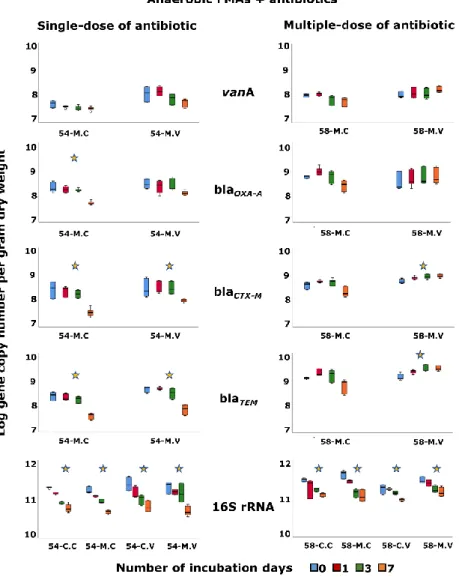
Figure 3. Effect of antibiotic on the variation of 16S rRNA and antibiotic resistance genes along the time. The abundance of the genes (vanA, blaOXA-A, blaCTX-M, blaTEM and 16S rRNA) per g of dry weight of stool, of non-inoculated (C) and ARB-inoculated (M) FMAs under anaerobic conditions in the presence of antibiotics is shown. The variation of each gene along time for each FMA is indicated by colored boxplot graphs (blue, red, green and orange corresponding to 0, 1, 3 and 7 days, respectively). FMAs were performed in the presence of a single-dose of cefotaxime (C.C, M.) or vancomycin (C.V, 54-M.V) or, in the presence of multiple-doses of cefotaxime (58-C.C, 58-M.C) or vancomycin (58-C.V, 58-M.V). The ARGs blaTEM , blaCTX, blaOXA-A and vanA were not
detected in the non-inoculated microcosms. Genes abundance are the average values of all FMAs replicates with the standard deviation. Stars indicated statistically significant variation (p<0.01) of genes abundance between T0 and T7 for each FMA.
31
Figure 4. Canonical Correspondence Analysis (CCA) of bacterial families (with relative abundance > 1%, and with the highest fit values, > 0.90) of the ARB-inoculated assays in the presence of a single-dose of cefotaxime (A) or vancomycin (B) or, in the presence of a multiple-dose of cefotaxime (C) or vancomycin (D). All the CCA represent microcosms at the beginning (T0) and after seven days of incubation (T7) without antibiotic (black circle and light blue circle, respectively), T0 and T7 in the presence of a single-dose of cefotaxime (T0.C, yellow circle; T7.C, green circle) or vancomycin (T0.V, yellow circle; T7.V, green circle) and, T0 and T7 in the presence of a multiple-dose of cefotaxime (T0.C, yellow circle; T7.C, purple circle) or vancomycin (T0.V, yellow circle; T7.V, purple circle). The red arrows show the significant explanatory variables (p<0.05) while grey arrows represent the explanatory variables with no significant correlation.