Prevalence and factors related
to mouth breathing in school
children at the Santo Amaro
project-Recife,2005
Summary
Valdenice Aparecida De Menezes1, Rossana
Barbosa Leal2, Rebecca Souza Pessoa3,
Ruty Mara E. Silva Pontes4
1 PhD, Professor.
2 MS. General Clinician, Dentalpediatrics. 3 Student.
4 Student.
Universidade de Pernambuco - UPE.
Mailing Address: Rossana Barbosa Leal - R. Conselheiro Silveira e Souza 604/103 Coredeiro Recife PE 50721-170.
Paper submitted to the ABORL-CCF SGP (Management Publications System) on February 2nd, 2006 and accepted for publication on April 24th, 2006.
A
im: To determine the prevalence of mouth breathing children at the santo amaro project/ esef/ upe, and study their main facial and behavior alterations. Study design: transversal study. Materials and methods: there were 150 children in the sample, with ages ranging from 8 to 10 years. Data was collected by means of a questionnaire and clinical examinations. As for their breathing assessment, two tests were carried out: test 1- breath steam against a mirror; and test 2 -water remains in the mouth with lips closed for 3 minutes. Results: mouth breathing prevalence was of 53.3%. There was no significant difference between gender, age and type of breathing. Facial alterations were:incomplete lip closure ( 58.8% X 5,7%), fallen eyes ( 40.0% X 1.4%), High palate ( 38.8% X 2.9%), Anterior open bite ( 60.0% Versus 30.0%), Hypotonic lips ( 3.8% X 0.0%), Circles under the eyes (97.5% Versus 77.1%). Conclusion: high mouth breathing prevalence without significant statistical difference between genders,age and type of mouth breathing. There was no association between behavior characteristics and type of breathing. There were significant differences between physical traits and breathing pattern.Keywords: dentistry pediatric, prevalence, mouth breathing. ORIGINAL ARTICLE
INTRODUCTION
Breathing is one of the vital functions of the human
body1. Normal breathing should happen through the nose.
However, it may be detoured to the oral via when there
is some airway obstruction2,3.
According to the literature, it is rare to have ex-clusive oral breathing; commonly, patients have a mixed
respiratory pattern: partially oral and partially nasal2,4-7.
Few are the papers related to the prevalence of oral breathing in the literature, and they present
percent-ages that vary from 5%8 to 75%7. As to gender, there is a
slight predominance of this pathology in females when
compared to their male counterparts9.
Depending on how long it lasts, oral breathing may cause functional, structural, pathological, postural, occlusal
and behavioral alterations10,11. The most common
com-plaints of oral breathers are: breathlessness or respiratory failure, gets tired easily during physical activities, back or neck pain, olfaction and/or taste impairments, halitosis, dry mouth, wake up chocking during the night, bad sleep, day time sleepiness, dark spots underneath the eyes, sneezing,
abundant saliva when speaking, among others12.
As physical consequences, the oral breathing child has many physical traits: long face, dropped eyes, dark spots underneath the eyes, open lips, hypotonic and dry lips, narrow nostrils, hypotonic cheek muscles, high palate, narrowing of the upper arch, and occlusal relation tending
to Angle’s Class II2,3. Oral breathing also alters posture,
morphology and tonicity of phonoarticulatory organs13.
As to behavioral alterations, we stress: restless sleep, irritability, difficulty concentration followed by a reduction in school performance and impaired sports skills, amongst others14,2,5.
Middle and long run alterations, accruing from these alterations, may have harmful consequences for the individual’s quality of life due to its personal, physical,
psychological and social impacts1,2,15,16. Therefore, oral
breathing is considered a syndrome and one of the most
preoccupying public health problems today17.
Knowing the importance of epidemiological re-search today, working to reduce populational health problems, this paper aims at contributing to the study of oral breathing, through research about its prevalence and main facial and behavioral alterations associated to this respiratory pattern in school children. The participants in this study are enrolled in the Projeto Santo Amaro da Escola Superior de Educação Física (ESEF).
MATERIALS AND METHODS
This study was carried out at the Escola Superior de Educação Física (ESEF) of the University of Pernambuco (UPE) from June to August, 2005 and is a transversal and observational type of study.
Our sample included children regularly enrolled in the Santo Amaro Project. Their ages varied between 8 and 10 years, because dental and facial alterations in the oral
breather are already present at this age range18.
The total sample comprised 236 children; through-out a sample calculation, the necessary sample size for this study was 147 children. Thus, the number of subjects per age range was of 50 children, adding up to a total of 150.
The sample was picked by randomly selecting the students in the aforementioned age ranges. Sample inclu-sion criteria were: children between 8 and 10 years of age, from both genders. Sample exclusion criteria were: Children who refused to participate; Children who were not properly authorized by their parents/guardians to par-ticipate and they did not sign the Informed Consent form; children with severe respiratory disorders.
In order to collect data in order to fulfill the objec-tives of the present study, we used an identification form for the children, a questionnaire to identify the behav-ioral alterations of the oral breather, and another form about facial alterations observed at visual inspection, and the results of the tests applied aiming at completing the diagnosis of oral breathing. Clinical exams were carried out by the investigators, using PPE (personal protection equipment) and the information gathered was recorded in the standardized forms.
In order to analyze facial alterations, we checked to see whether or not the children had the following clinical signs: elongated face, dropped eyes, dark circles under-neath the eyes, thin upper lip, dry lips, hypotonic lips, everted lower lip, narrow nostrils, high palate, inadequate lip sealing and anterior open bite. In order to assess some of these criteria, we were careful enough as to observe the children in their natural environment, without letting them see that they were being observed. The children who participated in the study underwent the mirror test and mouth moisture test to aid the diagnosis as follows:
Test 1: the mirror was placed underneath the child’s nostrils and we checked for steam build up in the mir-ror face (upper or lower) due to breathing. Steam on the upper face indicated nasal breathing and on the lower or
lower/upper face indicated oral breathing2.
Test 2: we asked the child to put some water in her/his mouth and kept the lips closed, without swallow-ing the water, for 3 minutes, and we observed through the lips commisure if there was any effort along the time. The children who were unable to keep their lips closed
were considered oral breathers2,18,35,36.
In order to consider a child as an oral breather, she/he should have at least 3 facial alterations, or steam in the lower mirror face and/or in both mirror faces, or spend
less than 3 minutes with water in their mouths12,18,19,35,36.
assigned into two groups: nasal breathers and oral breath-ers, the latter also comprised those children with mixed breathing or exclusive oral breathers.
Calibration was carried out in four phases, in-tra-examiner and inter-examiner, using pictures of the children.
In order to carry out data analysis we obtained absolute and percentage distributions (Descriptive Statis-tics Method), and used the Pearson’s Chi-Squared test (or Fisher’s Exact test when there was no favorable condi-tion to use the Chi-Squared test) and the equality in two proportions in independent group testing (inferential statistics method).
The significance level used in the statistical test was of 5% (0.05) and the intervals were obtained considering a 95.0% confidence interval. Data were plotted in the Excel spreadsheet and the software used to obtain the statisti-cal statisti-calculations was the SAS (Statististatisti-cal Analysis System) version 8.
RESULTS
Assessment of facial alterations by type of breathing in the whole group
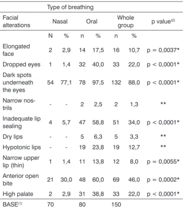
Table 1 shows the list of facial alterations by type of breathing and in the whole group. From this table we stress: in the whole group the highest percentage frequen-cies were recorded for dark circles underneath the eyes (88.0%), anterior open bite (46.0%), inadequate lip sealing (34.0%), dropped eyes (22.0%) and high palate (22.0%).
Among types of breathing patterns, it is possible to see that the percentages of facial alterations were correspondently higher among children who were oral breathers when compared to the nasal breathers. It may be established that the percentage differences presented in a descending order were recorded for: inadequate lip sealing (58.8% versus 5.7%), dropped eyes (40.0% x 1.4%), high palate (38.8% x 2.9%), anterior open bite (60.0% ver-sus 30.0%), hypotonic lips (23.8% x 0.0%) and dark circles underneath the eyes (97.5% versus 77.1%).
Through the statistical analysis we see the signifi-cant difference between the two types of breathing, at the level of 5.0%, for all the items to which we could apply the comparative test (p < 0.05).
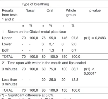
Assessment of the breathing tests results: Glatzel plate and water in the mouth
Table 2 depicts the results of the two breathing tests. In this table we can see that in the Glatzel plate steam test, most of the children (97.3%) were considered (assessed) as having upper steam formation, being 100.0% among the children with nasal breathing and 95.0% among those who were oral breathers and we did not see any significant relation between the two types of breathing (p > 0.05).
As to the test about the time through which the child
Table 1. Assessment of breathing type according to facial alterations seen at visual inspection.
Type of breathing Facial
alterations Nasal Oral Whole group p value(2) N % n % n %
Elongated
face 2 2,9 14 17,5 16 10,7 p = 0,0037* Dropped eyes 1 1,4 32 40,0 33 22,0 p < 0,0001* Dark spots
underneath
the eyes 54 77,1 78 97,5 132 88,0 p < 0,0001* Narrow
nos-trils - - 2 2,5 2 1,3 ** Inadequate lip
sealing 4 5,7 47 58,8 51 34,0 p < 0,0001* Dry lips - - 5 6,3 5 3,3 ** Hypotonic lips - - 19 23,8 19 12,7 ** Narrow upper
lip (thin) 1 1,4 11 13,8 12 8,0 p = 0,0055* Anterior open
bite 21 30,0 48 60,0 69 46,0 p = 0,0002* High palate 2 2,9 31 38,8 33 22,0 p < 0,0001* BASE(1) 70 80 150
(*) – Significant difference at the level of 5.0%.
(**) – It was not possible to apply the test because of a null fre-quence
(1) – Considering that the same child could present more than one facial alteration, only the base is recorded in order to calculate the percentages, and not the whole.
(2) – Through the equality between two proportions test in different groups.
kept the water in the mouth and the lips sealed, we could see that most of them (86.7%) went up to 3 minutes, and this number was 25.0% higher among the nasal breathers investigated (100.0% versus 75.0%), and such difference reveals a significant relation between the two types of breathing, as far as test 2 is concerned (p < 0.05).
DISCUSSION
In diagnosing a respiratory problem, it is of fun-damental importance to obtain information from the par-ents/guardians during the medical interview. Therefore, questions about the child’s sleep patterns, if he/she sleeps with the mouth opened, if there is noisy breathing, if the child lacks concentration at school, if the child feels sleepy during the day, if the pillow is wet in the morning; these questions should all be recorded, because they represent
important elements in the diagnosis of oral breathing20.
the Glatzel metal plate test and the time through which the child keeps water in her/his mouth with the lips sealed and without swallowing it, since we have seen that the results differ and complete each other.
As to the prevalence of oral breathing, despite the few studies that have been carried out with this pur-pose, we have seen disagreements in the literature. In the present study we noticed that most of the children (53.3%) were considered oral breathers. The highest per-centages of this alteration were seen in a study about oral suction habits in a low income population, which was
of 77.78%7 in prevalence. Other epidemiological surveys
have reported on percentages that varied between 4.5% and 34%8,21,22,24,26,28.
These percentage differences may be justified by the diagnostic criteria and the different methodologies used in the studies. In this investigation, we did not record exclusive oral breathers, gathering both the exclusive oral breathers and the mixed breathing children in the same group, as advocated in the literature. However, some stud-ies did not specify the criteria adopted in details.
According to many authors2,4-7,27 it is rare to find
an exclusively oral breathing pattern, and what is more common is for the patient, for some factor that makes it difficult for him/her to breathe freely through the nose (allergies, adenoid, hypertrophied tonsils, tumors, sinusitis, rhinitis, etc) to carry out a mixed type of breathing, partially oral and partially nasal. Within this context, some authors believe that the use of the term oral breather is somewhat improper and it should be replaced by the term: insuf-ficient nasal breather. We also agree as to the inadequate
use of the term “oral breather” since in our study and in the others hereby mentioned, exclusive oral breathing is rare or inexistent.
As to gender, we noticed that despite a higher percentage seen in males (53.75%) when compared to females (46.25%), this difference was not statistically sig-nificant among oral breathers, and such data corroborate other studies in which there was a slight difference in the variable being analyzed8. On the other hand, other investigations reported that there is a slight predominance
of this pathology in females when compared to males9.
However, these data do not seem to be relevant, since this gender predominance is mild, seen both in our study as in others mentioned above.
As to facial alterations that affect oral breathers, the highest percentages seen in the present study were: anterior open bite (60%), inadequate lip sealing (58.8%) and high palate (38.8%). These results are in agreement with those from a study in which the main craniofacial alterations seen in oral breathers were: open bite, high
palate, malocclusion28. In a retrospective study, it was
also possible to notice that most oral breathers also had malocclusion, and the anterior open bite was the most
frequent type found29.
Other alterations such as dark spots underneath the eyes (97.5%) and dropped eyes (40%) which repre-sented high percentages among the population studied were also mentioned by other authors as being facial alterations commonly found in patients with oral
breath-ing syndrome2,11.
Among oral breathers and nasal breathers, respec-tively, the percentage differences seen in descending order were: inadequate lip sealing (58.8% versus 5.7%), dropped eyes (40.0% x 1.4%), high palate (38.8% x 2.9%), anterior open bite (60.0% versus 30.0%), hypotonic lips (23.8% x 0.0%) and dark spots underneath the eyes (97.5% versus 77.1%).
In the long run, individuals who have trouble breath-ing may develop a number of disorders, such as: craniofa-cial alterations (long and narrow face), malocclusion, high palate, hypotonic lips and tongue, dry lips, sleepy face, deep dark spots underneath the eyes, greater likelihood of developing dental cavities, speech disorders, postural and gait alterations, all which interfere in school and work
performance, and also in social relationships2,5,30-33. Oral
breathers are more prone to having repeated flue episodes, spasmodic cough and hoarseness. Moreover, they develop
facial deformations called “facies adenoid”20.
Analyzing the data from the present study, it was also possible to see that of the facial alterations seen, percentages were correspondingly higher in oral breath-ing children than in nasal breathers, with a significant association, in agreement with the results found by afore-mentioned authors, despite the fact that these data support the statement that not all individuals with oral breathing Table 2. Evaluation of the breathing type according to test 1 (steam
on the Glatzel plate) and test 2 (time span with water in the mouth and lips sealed) results.
Type of breathing Results
from tests 1 and 2
Nasal Oral Whole
group p value
n % n % n %
1 – Steam on the Glatzel metal plate face
Upper 70 100,0 76 95,0 146 97,3 p(1) = 0,2483 Lower - - 3 3,7 3 2,0
Both - - 1 1,3 1 0,7 TOTAL 70 100,0 80 100,0 150 100,0 2 – Time span with water in the mouth and lips sealed 3 minutes 70 100,0 60 75,0 130 86,7 p(1) <
0,0001* Less than
3 minutes - - 20 25,0 20 13,3 TOTAL 70 100,0 80 100,0 150 100,0 (*) – Significant difference at 5.0%.
patterns present these characteristics34.
Notwithstanding, today, oral breathing is one of the
most concerning problems for public heath12,17. Depending
on its duration, it may cause a number of alteratins1,10,11,16.
These alterations may bring about harmful consequences to the individual’s quality of life due to its personal, physical, psychological and social impact. Therefore, its treatment should be multidisciplinary, involving early prevention and treatment strategies in order to avoid
symptomatic treatments2,3,11.
Considering the high prevalence of oral breathing in the population studied, we submit that health policies should be implemented in order to improve the life qual-ity of oral breathing children, and we stress the need for new studies with a larger number of children.
CONCLUSIONS
1. The prevalence of oral breathing was high, with-out significant difference between genders.
2. There were no relations between breathing pat-tern and behavioral alterations.
3. Physical characteristics related to oral breathing were: elongated face, dropped eyes, dark spots underneath the eyes, narrow nostrils, inadequate lip sealing, dry and hypotonic lips, narrow upper lip (thin), anterior open bite and high palate.
REFERENCES
1. Jorge TM et al. Hábitos bucais - Interação entre Odontopediatria e Fonoaudiologia. JBP 2002;5(26):342-50.
2. Paiva JB. Identificando o respirador bucal. (entrevista). Revista da APCD 1999;53(4):265-74.
3. Parizotto SPCAL, Nardão GT, Rodrigues CRMD. Atuação multidisci-plinar frente ao paciente portador da síndrome da respiração bucal. JBC 2002;6(36):445-9.
4. Fourniol Filho A. Pacientes Especiais e a Odontologia. 1a ed. São Paulo: Santos; 1998. p. 455-9.
5. Queluz DP, Gimenez CMM. A síndrome do respirador bucal. Revista do CROMG 2000;6(1).
6. Lusvarghi L. Identificando o respirador bucal. Revista da APCD 1999;53(4):265-74.
7. Cavassani VGS et al. Hábitos orais de sucção: estudo piloto em população de baixa renda. Revista Brasileira de Otorrinolaringologia 2003;69(1):106-10.
8. Kharbanda OP et al. Oral habits in school going children of Delhi:a prevalence study. J Indian Soc Pedod Prev Dent 2003;21(3):120-4. 9. Polanco CMS et al. Respiración bucal. Ortodoncia, edição
especial;9:5-11.
10. Mocellin M. Respirador Bucal. In: Petrelli E. et al. Ortodontia para Fonoaudiologia. Curitiba: Lovise; 1992.
11. Spinelli MLM, Casanova PC. Respiração Bucal. [serial online] 2002 Feb [cited 2005 Jan 12]. Available from: URL::http://www.odontologia. com.br/artigos.asp?id=224&idesp+14&ler=s
12. Marchesan IQ, Krakauer LH. A Importância do Trabalho Respiratório na Terapia Miofuncional. In: Marchesan IQ, Bolaffi C, Gomes ICD, 20 RZI, JL. Tratado de Fonoaudiologia. São Paulo: Lovise; 1995. p. 155-60.
13. Carvalho MP, Brandão G, Vinha PP. Os respiradores bucais e as desordens bucodentais. In: Cardoso RJA, Gonçalves EAN (Org.).
Odontopediatria: prevenção. São Paulo: Artes Médicas; 2002;4(11). 14. Carvalho GD. Alterações Comportamentais Comuns na S.R.B [serial
online] 2000 jan [cited 2005 Feb 10]. Available from: URL: http://www. ceaodontofono.com.br/artigos/art/2000/jan00.htm
15. Martinez JE et al. Análise crítica dos parâmetros de qualidade de vida de pacientes com fibromialgia. Acta Fisiátrica 1998;5(2):116-20. 16. Leal RB. Elaboração e validação de instrumento para avaliar a
quali-dade de vida do respirador oral. [dissertação]. Recife (BR):FOP/UPE Univ.;2004.
17. Carvalho GD. S.O.S Respirador Bucal - Obstáculos nas Diferentes Estruturas Dificultando ou Impedindo o Livre Processo Respiratório [serial online] 1999 Oct-Nov-Dec [cited 2005 Feb 10]. Available from: URL:http://www.ceaodontofono.com.br/artigos/art/1999/out99.htm 18. Ponte STD. Respiração Bucal [monograph], CEFAC (Centro De
Especialização Em Fonoaudiologia Clínica Motricidade Oral). Londrina(BR);2000.
19. Di Francesco RC. Respirador Bucal: a visão do otorrinolaringologista. JBO - Jornal Brasileiro de Ortondotia & Ortopedia Facial 1999; Ano 4:21.
20. Miranda PPC et al. Enfoque Multidisciplinar na Síndrome do Respira-dor Bucal. Revista Paulista de Odontologia 2002; Ano XXIV: 03. 21. Rojas V et al. Prevalencia de malos hábitos orales y respiración bucal
en niños de 5 a 17 años del área de Santiago Centro / Prevalence of bad oral habits and mouth breathing in children of 5 to 17 years old from the center area of Santiago Rev Fac Odontol Univ Chile 2001;19(1):9-19.
22. Wendel A et al. Relação Causal Entre a Respiração Oral e Dificul-dades na Aprendizagem. Revista CEFAC - Atualização Científica em Fonoaudiologia 2002;4(2).
23. Montiel J, María Elena. Frecuencia de maloclusiones y su asociación con hábitos perniciosos en una población de niños mexicanos de 6 a 12 años de edad / Frequency of malocclusions and its associa-tion with pernicious habits in a 6- to 12- year-old Mexican children population. Rev ADM 2004;61(6):209-14.
24. Chelotti VL, Vieira MM. Relação entre oclusão dental, respiração, hábito oral deletério e alimentação. Saúde 1997;23 (1-2):178-87. 25. Shetty SR, Munshi AK. Oral habits in children - a prevalence study.
J Indian Soc Pedod Prev Dent 1998;16(2):61-6.
26. Löfstrand-Tideström B et al. Department of Otorhinolaryngology, University of Uppsala, Sweden Breathing obstruction in relation to craniofacial and dental arch morphology in 4-year-old children. Eur J Orthod 1999;21(4):323-32.
27. Ellingsen R et al. Temporal variation in nasal and oral breathing in children. American Journal of Orthodontics and Dentofacial Ortho-pedics 1995 April.
28. Motonaga SM, Berti LC, Anselmo-Lima WT. Respiração Bucal: Causas e Alterações no Sistema Estomatognático. Revista Brasileira de Otor-rinolaringologia 2000;66(4).
29. Ribeiro F et al. Respiração Oral: Alterações Oclusais e Hábitos Orais. Rev CEFAC 2002;4:187-90.
30. Aragão W. Respirador Bucal (RB). OM 1986;XIII(7):39-41.
31. Prates NS, Magnani MBA, Valdrigh HC. Respiração bucal e problemas ortodônticos. Relação causa-efeito. Revista Paulista de Odontologia 1997; Ano XIX(4).
32. Pinto CSCD, Bommarito S. Avaliação dos tipos de respiração e sua correlação com as más oclusões Classe I e II de Angle. Revista Odonto 2003; Ano 11(22).
33. Amaral CSF, Martins ER, Rios JBM. A Respiração Bucal e o Desen-volvimento do Complexo Dentofacial. Rev Bras Alerg Imunopatol 2002;25(4).
34. Seixas CAO, Almeida EF, Fattori L. Diagnóstico, Prevenção e Trata-mento Precoce para Hábitos Bucais Deletérios. JBP - Jornal Brasileiro de Odontopediatria & Odontologia do Bebê 1998;1(1).
35. Padovan BAE. Deglutição atípica. Separata Reeducação mioterápica nas pressões atípicas da língua. Ortodontia 1976;9(1-2):5-59. 36. Rakoski T, Schimith GPF: Análise cional. In: Graber TM, Newmann B.