DOI: 10.1590/0004-282X20150175
ARTICLE
Longer epilepsy duration and multiple lobe
involvement predict worse seizure outcomes
for patients with refractory temporal lobe
epilepsy associated with neurocysticercosis
Longa duração de epilepsia e envolvimento de múltiplos lobos são fatores preditivos
de pior controle das crises convulsivas em pacientes com epilepsia refratária do lobo
temporal associada a neurocisticercose
Lucas Crociati Meguins1, Rodrigo Antônio Rocha da Cruz Adry1, Sebastião Carlos da Silva Júnior1, Carlos
Umberto Pereira2, Jean Gonçalves de Oliveira3,4, Dionei Freitas de Morais1, Gerardo Maria de Araújo Filho5,
Lúcia Helena Neves Marques6
Neurocysticercosis (NCC) is an infection of the central nervous system in which the meninges are infected with
the larval stage of the pork tapeworm Taenia solium1,2,3.
his tapeworm is endemic in the majority of low-income
countries in which pigs are raised and continues to be one of the most important causes of seizures in the world4,5,6. he
World Health Organization (WHO) lists NCC as a neglected tropical disease and estimates that approximately 50 million
1Faculdade de Medicina de São José do Rio Preto, Hospital Base, Departamento de Ciências Neurológicas, Divisão de Neurocirurgia, São José do Rio Preto SP, Brazil;
2Universidade Federal de Sergipe, Departamento de Medicina, Aracaju SE, Brazil;
3University Nove de Julho, Faculdade de Medicina, Departamento de Ciências Médicas, Divisão de Neurocirurgia, São Paulo SP, Brazil;
4Hospital Beneicência Portuguesa de São Paulo, Centro de Neurologia e Neurocirurgia Associados, Departamento de Cirurgia Cerebrovascular e Base do Crânio, São Paulo SP, Brazil;
5Faculdade de Medicina de São José do Rio Preto, Departamento de Psiquiatria e Psicologia Médica, São Paulo SP, Brazil;
6Faculdade de Medicina de São José do Rio Preto, Hospital Base, Departamento de Ciências Neurológicas, Divisão de Neurologia, São José do Rio Preto SP, Brazil.
Correspondence: Lucas Crociati Meguins; Rua Pedro Palotta, 101/31B; 15092-205 São José do Rio Preto SP, Brasil; E-mail: [email protected]
Conflict of interest: There are no conlicts of interest to declare.
Received 12 April 2015; Received in inal form 01 August 2015; Accepted 21 August 2015.
ABSTRACT
Objective: To investigate the surgical outcomes of temporal lobe epilepsy associated with hippocampal sclerosis (TLE-HS) and neurocysticercosis (NCC). Methods: A retrospective investigation of patients with TLE-HS was conducted in a tertiary center. Results:
Seventy-nine (62.2%), 37 (29.1%), 6 (4.7%), and 5 (3.9%) patients were Engel class I, II, III, and IV, respectively. Fifty-two (71.2%) patients with epilepsy durations ≤ 10 years prior to surgery were seizure-free 1 year after the operation compared to 27 (50.0%) patients with epilepsy durations > 10 years (p = 0.0121). Forty-three (72.9%) patients with three or fewer lobes affected by NCC were seizure-free one year after the operation, and 36 (52.9%) patients with more than three involved lobes were seizure-free after surgery (p = 0.0163). Conclusions:
Longer epilepsy durations and multiple lobe involvement predicted worse seizure outcomes in TLE-HS plus NCC patients.
Keywords: temporal lobe epilepsy, hippocampal sclerosis, neurocysticercosis.
RESUMO
Objetivo: Investigar o resultado cirúrgico da epilepsia do lobo temporal associada à esclerose hipocampal (TLE-HS) e neurocisticercose (NCC). Métodos: Estudo retrospectivo realizado em um centro de epilepsia. Resultados: Cinqüenta e dois pacientes (71,2%) com 10 anos ou menos de epilepsia antes da cirurgia tornaram-se livres de crises após um ano da operação, enquanto que 27 (50,0%) com mais de dez anos tornaram-se livres de crises após a cirurgia (p = 0,0121). Quarenta e três pacientes (72,9%), com três ou menos lobos afetados pela NCC tornaram-se livres de crises após um ano de operação, enquanto que 36 pacientes (52,9%) com mais de três lobos envolvidos estavam livres de crises após a cirurgia (p = 0,0163). Conclusão: A duração mais longa da epilepsia e o envolvimento de múltiplos lobos prevê pior resultado após a cirurgia para TLE-HS mais NCC.
people worldwide have NCC and that it causes approximate -ly 50,000 deaths each year7. Recent Brazilian investigations
have reported that NCC seems to contribute or even cause refractory epileptic seizures associated with hippocampal sclerosis8,9. According to these investigations, inlammatory
and/or electrogenic mechanisms promoted by NCC may in -duce epileptogenic discharges9.
In the present study, we investigated the surgical out
-comes of patients with temporal lobe epilepsy associated with hippocampal sclerosis and NCC.
METHODS
Study delineation
A retrospective observational investigation was conduct
-ed with data collect-ed from all patients treat-ed in the epilepsy clinic of the Faculdade de Medicina de Sao Jose do Rio Preto (FAMERP, a Brazilian tertiary referral epilepsy center) with di
-agnoses of temporal lobe epilepsy associated with hippocam -pal sclerosis (TLE-HS) from January 2000 to March 2013. he
clinical data were retrospectively obtained from the patient
records and iles. For all patients with a diagnosis of TLE-HS
based on magnetic resonance imaging (MRI), the following data were collected: sex, age at surgery, handedness, type and number of antiepileptic drugs (AEDs) used, and formal neu -ropsychological evaluation results. NCC was evaluated with
brain computed tomography (CT). he present study was
ap-proved by the ethical committee of our institution.
Pre-surgical evaluation
he patients were submitted to
video-electroencephalog-raphy (EEG) monitoring using Neuro Workbench software and Nihon Kohden hardware to record all epileptic events for later evaluation. Patient data were analyzed by an ex
-perienced epileptologist as an integral part of the inpatient assessment.
All patients submitted to pre- and post-surgical (at 12
months) neuropsychological assessments. Verbal memory
was assessed with a list-learning task, and igural
memo-ry was assessed with a learning test involving independent
items. Memory deicits were deined by performances that
were one standard deviation below the normal performance
of age-matched controls.
Brain MRI was performed obtained according to a spe -ciic epilepsy protocol using a Philips 1.5-Tesla scanner at
the Department of Neuroradiology of our institution. All MRI data were analyzed by an experienced neuroradiologist who
conirmed the visual radiological diagnoses of TLE-HS. NCC
was evaluated with brain CT, and the number of involved lobes was documented. All patients underwent MRI within
30 days of surgery and at each year of follow-up.
Biopsy specimens were collected from all patients with
chronic drug-resistant TLE-HS with radiological evidence
who underwent surgery. Standardized neuropathological
analyses were performed for all studied patients. he surgical
specimens submitted for neuropathological evaluation were
microscopically analyzed using hematoxylin-eosin staining. he pathologists reported their indings blind to the clinical
and imaging data.
Surgical technique
he surgical approaches were similar for all patients, and
a single neurosurgeon who was experienced with epilepsy
surgery (SCS Jr.) performed all of the procedures. he patient
positioning included the placement of a shoulder roll to el -evate the trunk followed by turning of the head 15-20 degrees
from the midline so that the operative side was facing up.
he head was slightly extended to bring the sylvian issure
to a plane that was perpendicular to the operating approach.
Finally, the vertex was dropped down toward the loor to
im-prove the surgeon’s access to the mesial structures and allow for less temporal lobe retraction. A reverse question mark in
-cision was made from immediately above the zygoma and extending back into the temporal region. An anterior tem
-poral craniotomy was performed respecting the anatomical landmarks of the temporal lobe from the root of the zygoma
to the anatomic keyhole. he remaining anterior and lateral
bone was removed by drilling down to the limits of the me
-dial fossa loor. At the end of the craniotomy, all of the bone
edges were waxed as necessary, any exposed air cells were
sealed, and take-up sutures were performed prior to opening
the dura mater to prevent epidural bleeding. A maximum of 4.0 to 5.0 cm of the anterior lateral temporal lobe was resect
-ed. he mesial resection included removal of the amygdala
and the anterior 2.0 to 3.0 cm of the hippocampus.
Outcome assessments and follow-up
Follow-up investigations were performed on the operat-ed patients. At the 12-month follow-up, all patients
under-went a neurological examination that included observations of behavior disorders, explorations of seizure outcomes and
1.5-Tesla cerebral MRI. Seizure outcomes were classiied ac-cording to the Engel classiication.
Ethical statement
he ethical committee of our institution analyzed the project and approved the investigation. his study complied
with the Declaration of Helsinki. Informed consent was ac
-quired from all patients and/or guardians.
Statistical analysis
he data collected from all patients were organized in ta-bles. he data are expressed as the means ± the SDs for para
-metric variables and the median values for nonpara-metric vari
-ables. Statistical analyses were performed with Fisher’s exact
RESULTS
A total of 136 patients with medically intractable TLE-HS
plus NCC were diagnosed and treated at our institution. However, nine patients were excluded from this sample be
-cause they did not achieve a minimum of 1 year of follow up.
Table 1 summarizes the clinical indings of 127 patients with TLE-HS plus NCC who underwent operations. here
were more males than females, and the mean age at surgery was 34.7 ± 11.9 (21-68 years). he mean age at seizure onset
was 14.4 ± 18.6 (8-65 years). he seizure frequency was 12.3 ± 21.2 (1-90) per month. he mean time from seizure onset to
surgery was 11.8 ± 22.9 (1-33 years). Complex partial seizures
were the most common type and were present in 116 (91.3%)
patients, followed by generalized tonic-clonic seizures in
9 (7.1%) patients and multiple seizure types in 2 (1.6%).
Interictal EEG indings revealed that 97 (76.4%) patients
pre-sented with unilateral epileptic discharges, and 30 (23.6%) exhibited bilateral epileptic discharges. Based on neuropsy -chological testing, the right hemisphere was the dominant side for memory in 5 (3.9%) patients, and the left hemisphere
was dominant in 122 (96.1%). he mean long-term follow-up
was 8.2 ± 5.8 (1-13 years).
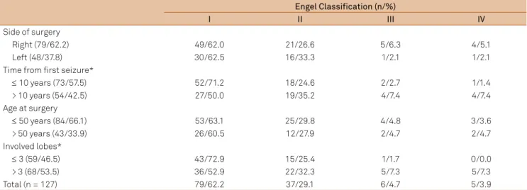
Table 2 summarizes the surgical outcomes of the 127 pa
-tients with TLE-HS plus NCC who underwent surgery. After
1 year of follow up, 79 (62.2%) patients were Engel class I, 37 (29.1%) were Engel class II, 6 (4.7%) were Engel class III,
and 5 (3.9%) were Engel class IV. Signiicant diferences in
the achievement of seizure freedom (Engel class I) were ob -served according to the time from irst seizure to surgery and the number of lobes afected by NCC. First, we found
that 52 (71.2%) patients with epilepsy durations ≤ 10 years
prior to surgery were seizure-free (Engel I) 1 year after the
operation, whereas 27 patients (50.0%) with epilepsy dura -tions > 10 years were seizure-free (Engel class I) following
surgery (Fisher’s exact test, p = 0.0121). Second, we observed
that 43 patients (72.9%) with three or fewer lobes afected by NCC were seizure-free (Engel class I) 1 year after the
opera-tion, and 36 patients (52.9%) with more than three involved
lobes were seizure-free (Engel class I) (Fisher’s exact test,
p = 0.0163). Neither the age at the time of surgery nor the side
of the operation had a signiicant efect. No relationship
be-tween the side of the NCC and the side of the hippocampal sclerosis was observed in the present study.
Table 3 summarizes the complications that occurred in the 127 surgical patients. A total of 18 patients (14.2%) ex -perienced post-operative complications. Infections were
ob-served in 14 (11.0%) patients, and 1 of these required bone removal. Two (1.6%) patients exhibited transitory contralat
-eral hemiparesis, and two (1.6%) had clinical complications Table 1. Clinical indings from 127 patients with TLE-HS plus
NCC who underwent operations.
Number of cases/%
Sex
Female 59/46.5
Male 68/53.5
Mean age at surgery (years) 34.7 ± 11.9 (21-68 years) Mean age at seizure onset (years) 14.4 ± 18.6 (8-65 years) Seizure frequency/month 12.3 ± 21.2 (1-90) Time from seizure onset to surgery (years) 11.8 ± 22.9 (1-33 years) Seizure type
Partial complex 116/91.3
Tonic-clonic generalized 9/7.1
Multiple 2/1.6
EEG indings
Unilateral 97/76.4
Bilateral 30/23.6
Hemispheric dominance
Right 5/3.9
Left 122/96.1
Mean follow-up (years) 8.2 ± 5.8 (1-13 years)
EEG: electroencephalography; NCC: neurocysticercosis; TLE-HS: temporal lobe epilepsy associated with hippocampal sclerosis
Table 2. Seizure outcomes of 127 patients with TLE-HS plus NCC who underwent operations.
Engel Classification (n/%)
I II III IV
Side of surgery
Right (79/62.2) 49/62.0 21/26.6 5/6.3 4/5.1
Left (48/37.8) 30/62.5 16/33.3 1/2.1 1/2.1
Time from irst seizure*
≤ 10 years (73/57.5) 52/71.2 18/24.6 2/2.7 1/1.4
> 10 years (54/42.5) 27/50.0 19/35.2 4/7.4 4/7.4
Age at surgery
≤ 50 years (84/66.1) 53/63.1 25/29.8 4/4.8 3/3.6
> 50 years (43/33.9) 26/60.5 12/27.9 2/4.7 2/4.7
Involved lobes*
≤ 3 (59/46.5) 43/72.9 15/25.4 1/1.7 0/0.0
> 3 (68/53.5) 36/52.9 22/32.3 5/7.3 5/7.3
Total (n = 127) 79/62.2 37/29.1 6/4.7 5/3.9
that consisted of mild renal insuiciency in one patient and a
pulmonary embolus that was treated with anticoagulation in another. Both of these complications were resolved without
further problems. here were no operative deaths.
DISCUSSION
NCC, an infection caused by the encysted larval stage of the tapeworm T. solium, constitutes one of the most com
-mon parasitic diseases of the nervous system in humans and
is a major public health problem for most of the developing
world10,11. he clinical manifestations of NCC are variable and
strongly depend on the number, type, size, location, and stage of development of the cysticerci, as well as the immune re
-sponse of the host against the parasite12,13,14,15,16.
Seizures are the most frequent manifestations of NCC
(70-90%) followed by headache (38%), focal deicits (16%),
and signs of intracranial hypertension (ICH, 12%). Other manifestations occur in fewer than 10% of symptomatic pa -tients13. Recent Brazilian investigations have reported that
NCC seems to contribute to or even cause refractory epi
-leptic seizures associated with hippocampal sclerosis8,9.
According to these investigations, the inlammatory and/or electrogenic efects elicited by NCC may induce
epileptogen-ic discharges9. In the present study, we presented our clini
-cal experience with refractory TLE-HS and NCC. Because the majority of our patients did not undergo a radiological
inves-tigation of the central nervous system to determine the pres -ence of NCC prior to seizure onset, we cannot infer whether NCC precipitated the hippocampal scleroses; however, we believe that the presence of secondary epileptogenic zones
induced by NCC may have accelerated degenerative process
-es already afecting the m-esial structur-es.
Based on the increased prevalence of inactive NCC in pa -tients with mesial temporal sclerosis (MTS) and refractory epi
-lepsy, a potential causal relationship between NCC and MTS has been proposed17. he authors of this proposal speculated that
in-lammatory lesions or repetitive seizures might play a role in
dis-ease pathogenesis17,18. Additionally, although refractory epilepsy
associated with TLE-HS plus NCC was previously believed not to inluence surgical outcomes19, these patients achieved post
-operative Engel IA statuses less frequently20. In the present study,
we observed that a longer duration of epilepsy and the involve
-ment of multiple lobes predicted worse seizure outcomes in pa
-tients with HS plus NCC. herefore, we propose that pa-tients
with HS plus NCC should be treated earlier in the natural course of the disease because once multiple sites of NCC are present and widely spread throughout the central nervous system, the seizure outcomes following surgery are worse. Bianchin et al.9
also noted that single NCC lesions are more commonly identi
-ied ipsilateral to HS, and this inding is suggestive of an anatom-ical relationship between TLE-HS and NCC.
here are several methodological aspects relevant to the present indings that should be interpreted in the con-text of a number of limitations. First, this study was a
non-randomized retrospective investigation that was performed in a highly selected population of a tertiary epilepsy center.
Secondly, these indings cannot be generalized to all patients with TLE-HS plus NCC because the results represent the
surgical experience of a single institution. However, we de -scribed the surgical outcomes of a relatively large number of patients who underwent surgery due to this pathology over
an extended follow-up duration.
CONCLUSIONS
he present study revealed that TLE-HS plus NCC is
high-ly prevalent in patients with refractory epilepsy and that lon
-ger epilepsy duration and the involvement of multiple lobes may predict worst seizure outcomes in this group of patients. Early diagnosis and treatment may improve the prognoses of
patients with TLE-HS plus NCC.
References
1. Del Brutto OH. Neurocysticercosis. Handb Clin Neurol. 2014;121:1445-59. doi:10.1016/B978-0-7020-4088-7.00097-3
2. Del Brutto OH. Neurocysticercosis: a review. Scientiic World J. 2012;2012:159821. doi:10.1100/2012/159821
3. Mewara A, Goyal K, Sehgal R. Neurocysticercosis: a disease of neglect. Trop Parasitol. 2013;3(2):106-13. doi:10.4103/2229-5070.122111
4. Bruno E, Bartoloni A, Zammarchi L, Strohmeyer M, Bartalesi F, Bustos JA et al. Epilepsy and neurocysticercosis in Latin America:
a systematic review and meta-analysis. PLoS Negl Trop Dis. 2013;7(10):e2480. doi:10.1371/journal.pntd.0002480
5. Winkler AS. Neurocysticercosis in sub-Saharan Africa: a review of prevalence, clinical characteristics, diagnosis, and management. Pathog Glob Health. 2012;106(5):261-74. doi:10.1179/2047773212Y.0000000047
6. Winkler AS, Willingham AL 3rd, Sikasunge CS, Schmutzhard E. Epilepsy and neurocysticercosis in sub-Saharan Africa. Wien Klin Wochenschr. 2009;121(3):3-12. doi:10.1007/s00508-009-1242-3
Table 3. Complications of 127 patients with TLE-HS plus NCC who underwent operations.
Number of cases/%
Infection 14/11.0
Contralateral hemiparesis 2/1.6
Clinical complications 2/1.6
Total 18/14.2
7. Bouteille B. [Epidemiology of cysticercosis and neurocysticercosis]. Med Sante Trop. 2014;24(4):367-74. French. doi:10.1684/mst.2014.0378
8. Bianchin MM, Velasco TR, Santos AC, Sakamoto AC. On the relationship between neurocysticercosis and mesial temporal lobe epilepsyassociated with hippocampal sclerosis: coincidence or a pathogenic relationship? Pathog Glob Health. 2012;106(5):280-5. doi:10.1179/2047773212Y.0000000027
9. Bianchin MM, Velasco TR, Wichert-Ana L, Alexandre Junior V, Araujo Junior D, Santos AC et al. Characteristics of mesial temporal lobe epilepsy associated with hippocampal sclerosis plus neurocysticercosis. Epilepsy Res. 2014;108(10):1889-95. doi:10.1016/j.eplepsyres.2014.09.018
10. Del Brutto OH, Garcia HH. Neurocysticercosis. Handb Clin Neurol. 2013;114:313-25. doi:10.1016/B978-0-444-53490-3.00025-X
11. Newton CR, Preux PM, Singhi P. Parasitic disorders. Handb Clin Neurol. 2013;112:1139-52. doi:10.1016/B978-0-444-52910-7.00034-9
12. Ito A, Takayanagui OM, Sako Y, Sato MO, Odashima NS, Yamasaki H et al. Neurocysticercosis: clinical manifestation, neuroimaging, serology and molecular conirmation of histopathologic specimens. Southeast Asian J Trop Med Public Health. 2006;37 Suppl 3:74-81.
13. Carabin H, Ndimubanzi PC, Budke CM, Nguyen H, Qian Y, Cowan LD et al. Clinical manifestations associated with neurocysticercosis: a systematic review. PLoS Negl Trop Dis. 2011;5(5):e1152. doi:10.1371/journal.pntd.0001152
14. Takayanagui OM, Jardim E. [Clinical aspects of neurocysticercosis: analysis of 500 cases]. Arq
Neuropsiquiatr. 1983;41(1):50-63. Portuguese. doi:10.1590/S0004-282X1983000100004
15. Takayanagui OM, Odashima NS. Clinical aspects of neurocysticercosis. Parasitol Int. 2006;55 Suppl:S111-5. doi:10.1016/j.parint.2005.11.016
16. Pal DK, Carpio A, Sander JW. Neurocysticercosis and epilepsy in developing countries. J Neurol Neurosurg Psychiatry. 2000;68(2):137-43. doi:10.1136/jnnp.68.2.137
17. Bianchin MM, Velasco TR, Takayanagui OM, Sakamoto AC.
Neurocysticercosis, mesial temporal lobe epilepsy, and hippocampal sclerosis: an association largely ignored. Lancet Neurol.
2006;5(1):20-1. doi:10.1016/S1474-4422(05)70269-6
18. Bianchin MM, Velasco TR, Wichert-Ana L, Takayanagui OM, Leite JP, Sakamoto AC. How frequent is the association of neurocysticercosis and mesial temporal lobe epilepsy with hippocampalsclerosis? Epilepsia. 2010;51(11):2359-60. doi:10.1111/j.1528-1167.2010.02735.x
19. Leite JP, Terra-Bustamante VC, Fernandes RM, Santos AC, Chimelli L, Sakamoto AC et al. Calciied neurocysticercotic lesions and postsurgery seizure control in temporal lobe epilepsy. Neurology. 2000;55(10):1485-91. doi:10.1212/WNL.55.10.1485