v. 11 n. 4, 2007ISSN 1809-9246 Histological/functional analysis of knee following ACL reconstruction 221 Rev. bras. fisioter., São Carlos, v. 11, n. 4, p. 221-227, July/Aug. 2007
©Revista Brasileira de Fisioterapia
CORRELATIONS BETWEEN HISTOLOGICAL ANALYSES AND
FUNCTIONAL EVALUATION OF THE KNEES OF PATIENTS UNDERGOING
RECONSTRUCTION OF THE ACL
V
EIGAPHA
1,2, A
LBUQUERQUERFM
1, T
EODOROWPR
1, M
ARTINSJVG
3& A
LVESRLBR
21 Orthopedics and Traumatology Institute, Medicine College, São Paulo University, São Paulo, SP - Brazil
2 Physical Therapy Course, Cathotic University of Pernambuco, Recife, PE - Brazil
3 Orthopedics and Traumatology Institute of Rio Grande do Norte, Natal, RN - Brazil
Correpondence to: Paulo Henrique Altran Veiga, Rua José de Holanda, 510, Bloco B, Apto 602, Bairro Torre, CEP 50710-140, Recife, PE – Brasil, e-mail: [email protected]
Received: 15/05/2006 - Revised: 22/12/2006 - Accepted: 22/06/2007
ABSTRACT
Objective: This study investigated the relationship between range of motion and the histological analysis of the synovium of the knee joint following reconstruction of the anterior cruciate ligament. Method: Histological, pain and range-of-motion (ROM) evaluations were performed on the knees of 26 patients. The Shapiro-Wilk test, Friedman test and Spearman correlation coefficient were utilized for verifying the existence of significant correlations between the variables, using the SPSS for Windows 12.0 statistical software with a significance level of 5%. Results: Comparing before and after the operation, statistical differences were found in relation to knee extension ROM (p= 0.042), flexion ROM (p= 0.001) and hyperextension (p= 0.001). There were also significant correlations between the subsynovial tissue and the flexion ROM of the operated knees (r= 0.53; p= 0.008), between the subsynovial tissue and the sensation of pain (r= 0.46; p= 0.024), and between the extension ROM after the operation and type I collagen (r= 0.52; p= 0.016). Conclusion: The greater the quantity of type I collagen observed, the thicker was the subsynovial tissue, the greater the reported pain, and the less was the ROM of the operated knees.
Key words: knee; anterior cruciate ligament; histological analysis; functional assessment; physical therapy.
INTRODUCTION
Complications after surgery for reconstruction of the ACL (ACL) have been extensively studied in the late decades1.
Formation and installation of knee arthrofibrosis is one of these complications and is part of a process of exacerbated scarring after reconstruction of the ACL that can lead to important functional limitations2-4. Functional limitations of
the knee in such cases are multifactorial and the commonly described causes include anterior pain and adherences in the affected joint, decreased intercondylar space, infrapatellar retraction, extra-articular surgery, inadequate graft placement (away from the isometry point), post-surgery immobilization, infection and reflex sympathetic dystrophy2-4. The surgical
trauma or the inflammatory processes in the knee have been described as the main causes for development of arthrofibrosis, characterized as an abnormal fibroblastic response with intra-articular fibrotic connective tissue that progressively obliterates the intra-articular space and causes limitations in the range of movement of knee5. The deposition of
connective tissue that involves the anterior aspect of the
synovia and articular capsule is secondary to the scarring process that results from the surgical lesion. If the healing response is exacerbated, arthrofibrosis can develop with an incidence of 4% in patients submitted to isolated ACL reconstruction to 23% in patients that undergo reconstruction of the ACL and the medial collateral ligament. Eventually, the incidence of arthrofibrosis can reach 35% in patients who undergo reconstruction of the ACL during the acute inflammatory period6-8.
Physical therapy plays a fundamental role in the prevention of restrictions in the range of movement after surgery. Nowadays, there is practically a consensus among physical therapists working with knee rehabilitation that an accelerated rehabilitation protocol aimed at improving terminal knee extension brings better results9. However, even when
surgery. The objectives of the present study was to make a histological analysis of the synovial membrane of knees submitted to reconstruction of the ACL, and relate histological findings to the functional evolution after surgery.
MATERIALS AND METHODS
The sample was comprised of a random sample of 26 patients with ACL injury who submitted to open surgical reconstruction. Inclusion criteria were: be a male or female, have unilateral injury of the ACL, have no more than one injured structure in addition to the ACL, have a maximum time of 6 months after injury, be submitted to open intra-articular reconstruction with patellar tendon graft and receive physical therapy three times a week after surgery according the Shelbourne accelerated rehabilitation protocol. Individuals had to sign the informed consent form to take part in the study according to the resolution 196/196 of the National Health Council.
Exclusion criteria were: patients with two or more injured structures in addition to the ACL injury, reconstruction by arthroscopy, patients who were not evaluated between the 85th and 95th days after surgery or who had any kind of
complications between the day of surgery and 90 days later, and patients who did not undergo physical therapy treatment. This study was approved by the Clinical Board of the Ethics and Research Committee for Analyses of Research Projects (CAPPesq) under the protocol number 906/06.
Measurement Instruments and Processing
For the purposes of this study, the following instruments were used: Informed Consent Form, functional assessment form adapted from the International Knee Documentation Committee10 and from the Shelbourne classification for
arthrofibrosis2, a universal goniometer (CARCI, Brazil) to
assess knee joint range of movement; a Sony personal computer to store functional data collected with the assessment forms, a HP printer, a CCD Sony DXC-10 photographic camera, a Zeiss Axiplan microscope, a Sony Trinitron monitor, a digitalizing system (Oculus TCX, Coreco Inc., St. Laurent, Quebec, Canada) to capture images on a computer (Pentium 133Mhz), and the Bioscan-Optimas Software version 5.2 (Bioscan Inc., Edmonds, WA) to process all data.
Procedures
The free and informed consent form (FICF) was presented to the subject. After the subject signed the FICF, the researcher performed a pre-surgery functional assessment. Assessment procedures were always performed in the same sequence. First the knee joint range of movement (ROM) was determined with the patient wearing shorts and laying in the supine position. The goniometer was placed with one
arm over the lateral longitudinal length of the thigh, the axis on the joint line and the other arm on the longitudinal length of the tibia. Both the affected and non-affected knees were assessed. After that, the individual functional assessment form was completed. Results of the functional assessments were classified according to an adaptation of the IKDC protocol (normal, near normal, altered, severely altered). Arthrofibrosis was classified according to standards developed by Shelbourne et al.11. This classification is based on the
ROM of the knee submitted to surgery compared to the non-affected knee, including assessment of hyperextension. Subjects with deficits of extension ROM ≤10 were classified as having Type I arthrofibrosis. Patients with extension ROM deficits >10º compared to the non-affected knee, but with normal flexion, were classified as having Type II arthrofibrosis. Patients with extension ROM deficits >10º and flexion ROM deficits >25º compared to the normal knee were classified as having Type III arthrofibrosis. Finally, patients with clinical presentations similar to those classified as Shelbourne Type III but with radiological findings indicating a low patella were classified as having Type IV arthrofibrosis. Results of the Shelbourne classification were analyzed in the form “aº/bº/cº”, with “a” representing the degrees of hyperextension, “b” representing the degrees needed to achieve full extension and “c” representing the degrees of flexion available in the knee. After the ROM assessment, the perceived pain in the injured knee was registered. The patient was asked to indicate the perceived level of pain in the visual analog scale12 that varies from
0 (no pain) to 100 (extreme pain). All responses related to pain were also classified according to the adapted IKDC standards (normal, near normal, altered, severely altered). The graft donation area was also assessed in regard to pain in response to palpation, and the knee as assessed for the presence of joint effusion, increased temperature and paresthesia. Besides the examination by palpation, the presence of hyperemia, hematomas and deformities caused by increased liquid volume inside the joint were also assessed by visual inspection. Results were classified according to the IKDC (normal= absent, near normal= mild, altered= moderate, severely altered= severe).
After the initial evaluation, the patients were submitted to surgery according to the usual routines of the Orthopedics Group. During surgery, the advisor and co-advisor researchers, who were both orthopedists specialized in the procedure, performed a biopsy of a small fragment of the infrapatellar region, since this region is the most commonly affected by fibrosis5,4,11,13,14. Five synovia samples collected from cadaver
v. 11 n. 4, 2007 Histological/functional analysis of knee following ACL reconstruction 223
a. b.
of a car accident. The control samples were used as a normal standard for results of histological analyses, as shown in Figure 1. Collected materials were immersed in a 10% solution of formol and sent to the Laboratory for Studies of Extra-cellular Matrix of the University of São Paulo Medical College. Where the samples were prepared and analyzed in the laboratory.
After ACL reconstruction surgery, the patients were referred to the physical therapy services of the collaborating institutions. Patients underwent rehabilitation according to
the Shelbourne and Nitz9 accelerated protocol. All participants
were submitted to a second assessment 85 to 95 days after surgery, always with the same procedures used in the assessment performed before surgery.
Morphological Study
After fixation, the material was embedded in paraffin. Sections of three to four micrometers were obtained and stained with hematoxiline and eosine, masson trichrome and picrosirius in order to assess the sub-synovial lattice quantified by morphometric analyses (Figure 2).
Immunofluorescence
Cross sectional slices of the infrapatellar synovia of the knees of patients were prepared in slides treated with 3-aminopropiltriethoxy Silano (Sigma Co.). They were immersed in xylol at 60ºC for 20 minutes. After that, the slices were embedded in xylol at room temperature and successively washed with ethylic alcohol in decreasing concentrations (100%, 95% and 75%), fresh water, distilled water, and phosphate-buffered saline (PBS) for re-hydration. To expose and identify antigenic sites (mapping of epitopes), the material was digested with 2mg of pig pepsin (10000 U/ml) (Sigma Co.) mixed with acetic acid 50 mM for 30 minutes at 37°C. The slices were washed with PBS three times for 10 minutes each time. Non-specific antigenic sites were blocked with Molico milk at 5% for 30 minutes and the slices were then
incubated with human polyclonal type I anti-collagen produced in rabbits and monoclonal (oncogene) type III anti-collagen at concentrations of 1:200 and 1:100, respectively. They were maintained at 4°C for one night. The slides were washed with PBS with Tween20 at 0.05% three times for 10 minutes each time and incubated with the secondary rabbit antibody anti-IgG and conjugated with fluorescence (Sigma) diluted in the proportion of 1:50, in a PBS solution with Evans Blue at 0.004% for 1 hour and 30 minutes. The slides were then washed again with PBS with Tween20 at 0.05% six times for five minutes each time. After these procedures, the slides were mounted with glycerin and the reaction was analyzed with a fluorescence microscope (Nikon).
Morphometric Analysis
Ten fields in each case were randomly selected at a magnification of 400 times. The density of synovial surface was determined by optical density in a system of image analysis consisting of a Sony CCD DXC-10 camera attached to a Zeiss Axiplan microscope and a monitor (Sony Trinitron). By means of a digitalizing system, the images were captured on a computer (Pentium 133 Mhz) and processed with the software Bioscan Optimas version 5.1 (Bioscan ; Edmonds, WA). Focus was directed to each field of collagen fibers. Contrast was increased to facilitate identification of blue fibers (collagen fibers), black fibers (elastic fibers) or birefringent bands (collagen fibers). Fiber areas were determined by digital colored densitometry adjusted to the density expressed by
the tone of collagen fibers. Values were expressed in μm2
(Figure 2).
DATA ANALYSIS
Results of microscopic tests were explored for associations with the results of the physical examination. According to the typology of the obtained data and study
Table 1. Comparison between values mean of range of motion (ROM) of extension nomal, pré and pos surgical time.
ROM EXTENSION Mean SD ℵ2 p
ROM Normal 0.38 1.96
ROM pre 1.54 6.12
ROM pos 1.92 3.76
6.32 0.042*
* significantly estatistical diference (p< 0.05).
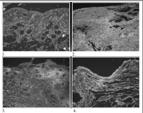
1. 2.
3. 4.
Figure 2. Immunofluorescence of infra patelar subsinovial screen show: 1. normal collagen I; 2. Increase collagen III (without ROM decrease); 3. Increase collagen I (note surround vessel and subsinovial screen compromise ROM extension) and 4. Normal collagen III. 400x.
objectives, results were presented in tables and figures. Initially, exploratory analyses were undertaken to verify the necessary assumptions of statistical testing. Thus, the Shapiro-Wilk test, as well as analyses of symmetry and kurtosis, were used to test for data normality. Since the ROM values had an asymmetric distribution the Friedman test was, therefore, used to verify the presence of statistically significant differences between the central tendency measures of ROM (normal, before and after). To test for correlations between variables of the present study (ROM after surgery, collagen type, pain level after surgery, total collagen and sub-synovial lattice) the Spearman correlation coefficient was used. All data were analyzed with the SPSS statistical software version 12.0 for Windows with a level of significance set to 5%.
RESULTS
Significant differences were found for knee extension ROM between the three assessments (ℵ2= 6.32; p= 0.04), as demonstrated in Table 1. Significant differences were also found in the comparison of flexion range of movement between the normal knee and the affected knee before and after surgery (ℵ2
= 13.26; p= 0.01) (Table 2). Hyperextension ROM was significantly different between the three assessments (ℵ2
= 14.27; p= 0.01) (Table 3). The Spearman
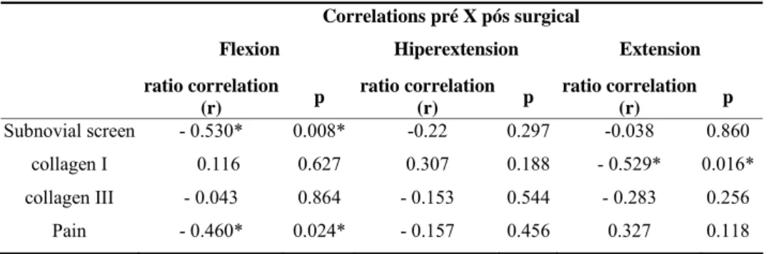
test was used to verify the presence of correlations between the ROM and sub-synovial lattice, type of collagen, and pain after surgery. The following results were found: hyperextension ROM after surgery compared to ROM before surgery was not significantly correlated with the sub-synovial lattice, type of collagen or pain after surgery. Flexion ROM after surgery compared to ROM before surgery was significantly correlated with sub-synovial lattice (r= -0.53;
p= 0.008) and pain perception (r= -0.46; p= 0.024). Extension
ROM after surgery compared to ROM before surgery was significantly correlated with type I collagen (r= -0.529;
p= 0.016) (Table 4). An increased concentration of type I
v. 11 n. 4, 2007 Histological/functional analysis of knee following ACL reconstruction 225
FIELDS N Minimal Full Mean SD
Out points 12.20 37.70 18.55 6.00
Sub sinovial screen 26 23.40 86.70 59.10 19.23
Fat 0 58.70 22.36 19.38
Table 5. Relate values of morfometric analisys results in μm (micrômeters). Increase of 400x.
DISCUSSION
Despite the great number of studies regarding ACL reconstruction and its consequences for functional recovery, there is no consensus in relation to the best rehabilitation protocol for this surgical procedure. It is acknowledged, however, that surgery success depends upon the experience of the surgeon, type of fixation and type of graft used, as well as the fixation in the optimal isometry point and an early rehabilitation program with a focus on promoting gains in
knee extension range15.
In this context, physical therapy intervention is of fundamental importance to prevent the loss of range of movement. Rehabilitation protocols that restrict exercises in terminal extension are associated to the persistence of several symptoms after ACL reconstruction, such as anterior knee pain, mobility blocks and inability to return to previous
activity levels11. It has been demonstrated that earlier
rehabilitation is associated with better results for range of
movement16,17. Shelbourne et al.11 described the difficulties
to reestablish normal mobility in a knee with arthrofibrosis.
Table 2. Comparison between values mean of range of motion (ROM) of flexion nomal, pré and pos surgical time.
* significantly estatistical diference (p< 0.05). * significantly estatistical diference (p< 0.05).
Table 3. Comparison between values mean of range of motion (ROM) of hiperextension nomal, pre and pos surgical time.
ROM flexion Mean SD ℵ2 p
ROM Normal 130.38 7.47
ROM pre 127.5 9.72
ROM pos 121.54 15.12
13.26 0.001*
ROM HIPEREXTENSION Mean SD ℵ2 p
ROM normal 5.0 4.24
ROM pre 4.81 3.87
ROM pos 1.69 3.02
14.27 0.001*
Correlations pré X pós surgical
Flexion Hiperextension Extension
ratio correlation
(r) p
ratio correlation (r) p
ratio correlation (r) p
Subnovial screen - 0.530* 0.008* -0.22 0.297 -0.038 0.860
collagen I 0.116 0.627 0.307 0.188 - 0.529* 0.016*
collagen III - 0.043 0.864 - 0.153 0.544 - 0.283 0.256
Pain - 0.460* 0.024* - 0.157 0.456 0.327 0.118
* significantly estatistical diference (p< 0.05).
Table 4. Correlation between range of motion (ROM), pain and collagen I and III.
Seventy-two patients with arthrofibrosis were assessed and submitted to an efficacious rehabilitation protocol. However, for patients with Type IV arthrofibrosis, that is, extension deficits greater than 10º and flexion deficits equal or greater than 30º, 21% of results were unsatisfactory. One of the most important precautions to be taken after surgery is to prevent the occurrence of fibrotic processes, which is the most
efficacious treatment for arthrofibrosis2. From a practical
viewpoint, the evolution of ACL rehabilitation techniques can be characterized by two very distinct major phases that seem to have determined the main procedures adopted in physical therapy. The first phase was initiated with the study
by Paulos et al.18 who published conservative principles for
ACL rehabilitation, with an emphasis on preserving graft integrity with less aggressive techniques and approaches, and according to them, time is essential for adequate graft healing. Therefore, the rehabilitation of a patient submitted to ACL reconstruction should respect maturation of the “new ligament” and should place minimal loads on the operated limb, as well as restrictions of movements of flexion and extension immediately after surgery.
After using this “conventional” protocol, professionals treating and rehabilitating patients submitted to knee surgery observed several complications such as quadriceps strength deficits and atrophy, anterior pain and, especially, terminal
knee extension blocks19,4,17. With the evolution of surgical
treatment of ACL reconstructions can be reported to have begun after the publication of a study by Shelbourne et al.9.
These authors developed the idea of the so-called “accelerated protocol” with an emphasis on early complete knee extension as its main characteristic. The authors demonstrated that knee joint movement angles, strength and function can be trained early without affecting stability or risking damage to the graft19. Basic research and clinical investigations revealed
that knee immobilization and activities that limited mobility produced unsatisfactory functional results15. It was also
demonstrated that the early return to sports practice and activities of daily living did not increase the incidence of new injuries in the operated knees20. In the present study,
the Shelbourne physical therapy accelerated protocol was used in rehabilitation, since this protocol emphasized gains in terminal knee extension and the prevention of arthrofibrosis of the ifrapatellar area9.
Significant differences were observed for terminal extension of the knee between the three assessments (normal knee and affected knee before and after surgery) (ℵ2= 6.32;
p= 0.042). It could be observed that better classifications
corresponded to greater range of movement. The correlation coefficients between the extension range of movement and the variables: sub-synovial lattice, type I collagen, type III collagen and pain perception are presented in Table 4. According to these results, knee extension range, after ACL injury is significantly associated with type I collagen (r= -0.529; p= 0.016). This association suggests that greater amounts of type I collagen are related to decreased extension range of movement after injury.
The control samples obtained from normal knees demonstrated a larger type III collagen area according to the morphometric analysis of the immunofluorescence. This demonstrates that the presence of type III collagen is more frequent, that is, the presence of immature collagen is normal in healthy knees.
Bradykinin, histamine and serotonin are known to be associated with the inflammatory process21. Douillet et al.22
analyzed the influences of bradykinin on the inflammatory process and tissue fibrosis and found that the levels of bradykinin receptors were induced by hyperglycemia in the vascularization of affected cells. Hyperglycemia stimulated production and accumulation of the extra-cellular matrix and growth factors that served as a substrate for fibrosis. The authors observed that bradykinin significantly stimulates a decrease in the levels of messenger RNA (RNAm) of type I collagen, facilitating its transference to plasma.
Zeichen et al.23 report that the mechanism of stimulation
of type I collagen, in the context of an exacerbated scarring and inflammatory process, can be taken into consideration in the mechanism of chronic tissue repair of knees submitted to ACL reconstruction, as a result of deficient regulation of cellular and vascular synthesis. A factor that contributes
to the complex mechanism of protein deposition in the periarticular extra-cellular matrix in chronic lesions, such as vascular neogenesis, causes the sub-synovial lattice to increase its thickness. Gelse et al.24 performed a detailed
study of the structure, function and biosynthesis of type I collagen and reported that this type of collagen is the most prevalent and most studied to date. Type I collagen (mature collagen) forms more than 90% of the organic osseous mass and is the most common type of collagen in tendons, skin, ligaments, cornea and several interstitial tissues, with the exception of few tissues such as hyaline cartilage, cerebral tissue and aqueous humor. In several organs, especially in tendons and muscular fascia, type I collagen fibers provide great tensile resistance. In bones, collagen has a considerable role in defining biomechanical characteristics and is influenced by weight bearing, tensile strength and angular force. These particular characteristics of type I collagen suggest that excessive accumulation can lead to deficient knee terminal extension and flexion after ACL reconstruction. Type III collagen (immature collagen) is formed by three
α1 (III) chains and is largely distributed in tissues rich in
v. 11 n. 4, 2007 Histological/functional analysis of knee following ACL reconstruction 227
rehabilitation programs specially focused on terminal knee extension. Additionally, subjects with thickening of the sub-synovial lattice had a higher probability of developing deficits in knee flexion range of movement after ACL reconstruction surgery.
FINAL CONSIDERATIONS
Increased thickness of the sub-synovial lattice is a risk indicator for decreased knee flexion range of movement after ACL reconstruction surgery. While, increased amounts of type I collagen is an indication of risk for decreased knee extension range of movement after ACL reconstruction surgery.
The observed correlations between the sub-synovial lattice and increased deposition of type I collagen and the functional results after ACL reconstruction suggest that rehabilitation protocols should prioritize flexion and extension to prevent deficits in the range of movement.
REFERENCES
1. Torry MR, Decker MJ, Jockel JR, Viola R, Sterett WI, Steadman JR. Comparison of tibial rotation strength in patients’ status after anterior cruciate ligament reconstruction with hamstring versus pattelar tendon autografts. Clin J Sport Med. 2004;14(6):325-31.
2. Shelbourne KD, Patel DV. Treatment of limited motion after anterior cruciate ligament reconstruction. Knee Surg Sports Traumatol Arthrosc. 1999;7(2):85-92.
3. Murakami S. Quantitative analysis of synovial fibrosis in the infrapatellar fat pad before and after anterior cruciate ligament reconstruction. Am J Sports Med. 1997;25(1):29-34.
4. Paulos LE, Rosemberg TD, Drawbert J, Manning J, Abbot P. Infrapatellar contracture syndrome: an unrecognized cause of knee stiffnes with patella entrapment and patella infera. Am J Sports Med. 1987;15(4):331-41.
5. Mariani PP, Santori N, Rovere P, Rocca CD, Adriani E. Histo-logical and structural study of the adhesive in knee fibroarthrosis: a clinical-pathological correlation. Arthroscopy. 1997;13(3):313-8.
6. Cosgarea AJ, Sebastianelli WJ, DeHaven KE. Prevention of arthrofibrosis after anterior cruciate ligament reconstruction using the central third patellar tendon-autograft. Am J Sports Med. 1995;23(1):87-92.
7. Noyes FR, Mangine RE, Barber SD. The early treatment of motion complications after reconstruction of the anterior cruciate ligament. Clin Orthop Relat Res. 1992;277:217-28.
8. Strum GM, Friedman MJ, Fox JM, Ferkel RD, Dorey FH, Del Pizzo W, et al. Acute anterior cruciate ligament reconstruction. Analysis of complications. Clin Orthop Relat Res. 1990;253:184-9.
9. Shelbourne KD, Nitz P. Accelerated rehabilitation after ACL reconstruction. Am J Sports Med. 1990;(18):292-9.
10. Hefti F, Muller W. Evaluation of knee ligament injuries with the IKDC form, in knee. Surg Sports Traumatol Arthrosc. 1993;1:226-34.
11. Shelbourne KD, Patel DV, Martini DJ. Classification and mana-gement of arthrofibrosis of the knee after anterior cruciate ligament reconstruction. Am J Sports Med. 1996;24(6):857-62.
12. Jacox A. Management of cancer pain. clinical pratice guideline. 1994; 9. AHCPR Publication, nº 94-0592. Rockville md agency for health care polcy and research, u.s. departament of health and human services, public health service.
13. Sprague N, O‘Conner R, Fox J. Arthroscopy treatment of postoperative knee fibroarthrosis. Clin Orthop. 1982;166: 165-72.
14. Payr E. Zur operativen behandlung der kniegelenksteife. Zentralbl Chir. 1917;44:809.
15. Beyond BD, Johnson RJ, Fleming BC. The science of anterior cruciate ligament rehabilitation. Clin Orthop and Relat Research. 2002;402:9-20.
16. Berbig R, Rillmann P. Timing of the surgery of rupture of the anterior cruciate ligament: Effects of acute or delayed surgery on arthrofibrosis rate and work disability. Unfallchirurg. 2000;103(9):726-30.
17. Noyes FR, Torres SB, Westin SDB, Heckmann TP. Prevention of permanent arthrofibrosis after anterior cruciate ligament reconstruction alone or combined with associated procedures: a prospective, study in 443 knees. Knee Surg Sports Traumatol Arthrosc. 2000;8(4):196-206.
18. Paulos L, Noyes FR, Grood E, Butler DL. Knee rehabilitation after anterior cruciate ligament reconstruction and repair. Am J Sports Med. 1981;9(3):140-9.