Tamoxifen decreases the myofibroblast count in the
healing bile duct tissue of pigs
Orlando Hiroshi Kiono Siqueira,IBenedito Herani Filho,IIRafael Erthal de Paula,IIIFa´bio Otero A´scoli,IV Antonio Cla´udio Lucas da No´brega,IVAngela Cristina Gouveˆa Carvalho,VAndre´a Rodrigues Cordovil Pires,V Nicolle Cavalcante Gaglionone,VIKarin Soares Gonc¸alves Cunha,VIIJose´ Mauro GranjeiroVIII
IFluminense Federal University, Department of General and Specialized Surgery, Nitero´i/RJ, Brazil. IISa˜o Paulo Federal University, Surgical
Gastroenterology, Sa˜o Paulo/SP, Brazil. IIIFluminense Federal University, School of Medicine, Nitero´i/RJ, Brazil. IVFluminense Federal University, Biomedical Institute, Nitero´i/RJ, Brazil.VFluminense Federal University, Pathology, Nitero´i/RJ, Brazil.VIFluminense Federal University, Clinical Research Unit, Nitero´i/RJ, Brazil.VIIFederal Fluminense University, Postgraduate Program in Pathology, Nitero´i, RJ/Brazil.VIIIFederal Fluminense University - Clinical Research Unit, Nitero´i/RJ, Brazil.
OBJECTIVE:The aim of this study was to evaluate the effect of oral tamoxifen treatment on the number of myofibroblasts present during the healing process after experimental bile duct injury.
METHODS:The sample consisted of 16 pigs that were divided into two groups (the control and study groups). Incisions and suturing of the bile ducts were performed in the two groups. Tamoxifen (20 mg/day) was administered only to the study group. The animals were sacrificed after 30 days. Quantification of myofibroblasts in the biliary ducts was made through immunohistochemistry analysis using anti-alpha smooth muscle actin of the smooth muscle antibody. Immunohistochemical quantification was performed using a digital image system.
RESULTS: In the animals treated with tamoxifen (20 mg/day), there was a significant reduction in
immunostaining for alpha smooth muscle actin compared with the control group (0.1155 vs. 0.2021,p= 0.046).
CONCLUSION:Tamoxifen reduced the expression of alpha smooth muscle actin in the healing tissue after bile duct injury, suggesting a decrease in myofibroblasts in the scarred area of the pig biliary tract. These data suggest that tamoxifen could be used in the prevention of biliary tract stenosis after bile duct surgeries.
KEYWORDS: Tamoxifen; Myofibroblasts; Biliary Wound Healing; Bile Duct Stricture; Bile Duct Injury.
Siqueira OH, Herani Filho B, Paula RE, A´ scoli FO, No´brega AC, Carvalho AC, et al. Tamoxifen decreases the myofibroblast count in the healing bile duct tissue of pigs. Clinics. 2013;68(1):101-106.
Received for publication onMay 19, 2012;First review completedJuly 4, 2012;Received for publication onSeptember 21, 2012
E-mail: [email protected]
Tel.: 55 21 2629-9025
& INTRODUCTION
Cholecystectomy is one of the most commonly performed general surgery procedures in the world (1). With the advent of video laparoscopy, nearly 90% of all cholecystectomies are currently performed laparoscopically, which has resulted in an increased incidence of bile duct injuries (BDIs). The incidence has increased from 0.1-0.2% (open cholecystectomy) to 0.4-0.6% (video laparoscopy) (2,3). Despite their low prevalence, iatrogenic BDIs are significant with regard to their absolute numbers (4) and are important in terms of health care costs (5,6); furthermore, they are among the leading causes of negligence claims against surgeons (7-10).
When the bile duct has lost continuity after injury from cholecystectomy or other biliary operations, surgical recon-struction is the only feasible treatment option (11,12). Nevertheless, the management of major BDIs is a surgical challenge (13), even for experienced hepatobiliary surgeons. Due to the small caliber of the main bile duct, anastomosis is difficult to perform and favors the occurrence of stenosis, which is usually secondary to the inflammatory process and fibrosis (14). The prevalence of bile duct stenosis varies from 0.2-0.5% (4) and can potentially progress to cholangitis, biliary cirrhosis, portal hypertension, end-stage liver disease and death (15,16).
Myofibroblast differentiation and activation are critical events in the pathogenesis of human fibrotic diseases (17), as is post-operative biliary stenosis. Myofibroblasts are present in large numbers and represent the main cause of scar contracture and the occurrence of fibrosis (18,19). These cells exhibit features that are intermediate between fibro-blasts and smooth muscle cells; specifically, they produce collagen, they express alpha smooth muscle actin (a-SMA) and their differentiation and activation are induced by Copyrightß2013CLINICS– This is an Open Access article distributed under
the terms of the Creative Commons Attribution Non-Commercial License (http:// creativecommons.org/licenses/by-nc/3.0/) which permits unrestricted non-commercial use, distribution, and reproduction in any medium, provided the original work is properly cited.
No potential conflict of interest was reported.
transforming growth factor-beta 1 (TGF-b1) (17). Studies have indicated that the expression of TGF-b1 in stenotic bile ducts is significantly higher than that in normal bile ducts, suggesting that TGF-b1 is a key factor in the prolonged healing process of the bile duct and in the proliferation of cicatrix (20,21).
Tamoxifen is a synthetic nonsteroidal antiestrogen agent that exhibits antifibrotic properties and has been shown to successfully treat many fibrotic diseases (e.g., hypertrophic scars (22), keloids (22), encapsulating peritoneal sclerosis (23), retroperitoneal fibrosis (24), fibrosing mediastinitis (25), sclerosing cervicitis (25) and recurrent desmoid tumors (26,27)). It is believed that the antifibrotic property of tamoxifen is mainly due to its downregulation of TGF-b1 (22,28).
Considering that tamoxifen may inhibit the increase in myofibroblasts during wound healing, the aim of this study was to experimentally investigate the effect of oral tamox-ifen treatment on the number of myofibroblasts in the healing tissue after BDI.
& MATERIALS AND METHODS
This study was conducted with approval from the Ethics Committee in Animal Experimentation of Fluminense Federal University, Rio de Janeiro, Brazil.
Animals
Female pigs (Sus scrofa domesticus) of the Large White breed that weighed between 20 and 32 kg were used in the experiments. The animals were kept under standard condi-tions (12 h/12 h day/night cycle), were fed a standard diet and received waterad libitum. For the sample size calculation, a pilot study was carried out in which tamoxifen was administered to three of the six animals. Considering an effect size of 0.65, a significance level of 5% (a) and a statistical test power of 80% (1-b), it was estimated that a minimum sample size of nine animals would be needed for each experimental group.
Eighteen pigs were included in this study and divided into two groups: a control group without treatment (Group A) and an experimental group treated with oral tamoxifen (Group B).
Anesthesia and analgesia
The pigs were intramuscularly (IM) pre-medicated with ketamine (5 mg/kg), midazolam (0.5 mg/kg) and aceproma-zine (0.05 mg/kg). Anesthesia was induced with propofol (4 mg/kg). After orotracheal intubation, an epidural block was performed with bupivacaine 0.125% (10 mL) and morphine (0.1 mg/kg). The isoflurane concentration was maintained at 1.5%. Tramadol was administered at a dose of 2 mg/kg to the animals that exhibited pain during the post-operative period and required rescue analgesic. Oxytetracycline (15 mg/kg IM) was administered before and 48 hours after each surgery.
Surgical procedures
The peritoneal cavity was accessed using a right subcostal incision. Dissection, ligation and sectioning of the cystic artery, together with the cystic duct, were performed. The gallbladder was dissected and removed. The hepatoduode-nal ligament was opened and the bile duct was measured using digital calipers from a location that was 1.5 cm away from the superior border of the first portion of the
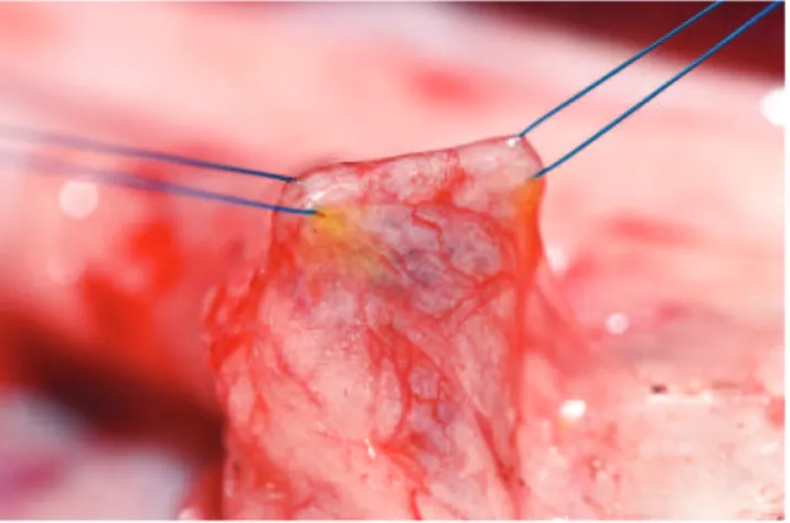
duodenum. In all of the animals, two repair stitches were placed (on each side of the future incision) using 6.0 polypropylene sutures (Figure 1). A longitudinal 5-mm incision was made in the anterior wall of the bile duct. This incision was measured using digital calipers, starting from a distance of 1 cm from the superior border of the first portion of the duodenum and extending in the cranial direction (Figure 2). These wounds were closed using four non-continuous stitches that were distributed in an equidistant manner and placed using 6.0 polydioxanone sutures (Figure 3). To close the abdominal cavity, the musculoapo-neurotic layer was sutured using simple running stitches of 2.0 vicryl sutures. The skin was closed using continuous, simple running 3.0 nylon sutures.
Tamoxifen administration
Following the surgery, the group B animals received tamoxifen at a dose of 20 mg/day orally for 30 days.
Data sampling
The animals were euthanized 30 days after the surgery. After withholding food overnight, the animals were sedated intramuscularly with ketamine (5.0 mg/kg), xylazine (1.0 mg/kg) and acepromazine (0.1 mg/kg). Thiopental sodium (5%) was administered intravenously to induce anesthesia. When an animal was deeply anesthetized, Figure 1 -Repair stiches in the choledoco duct.
potassium chloride (10%) was administered intravenously to stop the heartbeat.
The bile duct from each animal was resected through a median abdominal incision. The resected bile ducts were opened longitudinally along the posterior wall and immersed in 10% buffered formalin. The tissues were routinely processed. Five-micron-thick sections were cut from paraffin blocks, stained with hematoxylin and eosin and submitted for immunohistochemistry analysis.
Immunohistochemical staining
After dewaxing, the presence of a-SMA protein was assessed using immunohistochemistry by employing the polymer method (Mach 4, Biocare Medical, Concord, CA, USA). Endogenous peroxidase activity was blocked by incubation in 1.3% H2O2 in methanol at room temperature for 15 min. Antigen retrieval was performed by incubating samples in Trilogy Antigen Retrieval Solution (Cell Mark, Hot Springs, CA, USA) at 96
˚
C for 40 min. Non-specific protein binding was blocked by incubation with Dako Blocking Solution (Dako, Carpinteria, CA, USA) for 15 min at 37˚
C. The sections were incubated for 1 hour at room temperature with a 1:200 dilution of the primary monoclonal antibody anti-a-SMA (clone 1A4, Cell Mark, Hot Springs, CA, USA). Visualization was performed by incubation in diami-nobenzidine for 5 min. The sections were counterstained with Harris hematoxylin for 30 seconds. Between each step, the sections were washed in phosphate-buffered saline (PBS buffer). All of the incubations were carried out in humidified chambers to prevent evaporation. Histological sections from the uterus were used as a positive control. The negative control was performed by omitting the primary antibody.Image analysis
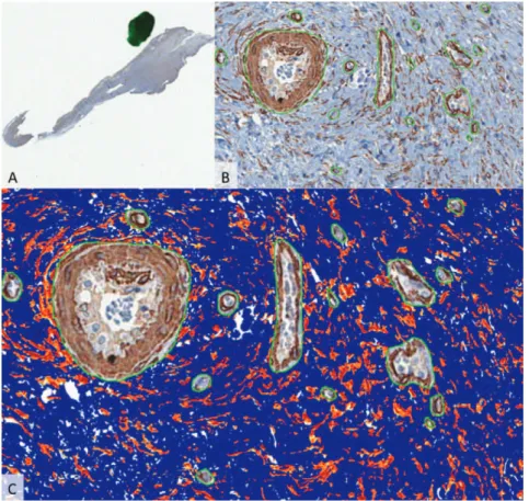
The immunohistochemical quantification of alpha smooth muscle actin was performed using a digital image system (Aperio Technologies, Vista, CA, USA). All of the glass slides were scanned at an objective magnification of 40x using a ScanScopeTM CS scanning system (Aperio Technologies, Vista, CA, USA). The positivity index (PI; positive area/total area) was calculated for each image within the scar area using the positive pixel count algorithm
in the ImageScopeTM software (Aperio Technologies, Vista, CA, USA). The pen tool was used to select the scar areas and to exclude the immunopositive vessels.
Statistics
Statistical analysis was performed using the software SASH
System, version 6.11 (SAS Institute, USA). The results were expressed as the median and interquartile intervals (Q1 and Q3). The Shapiro-Wilk test was employed to examine the normality of the data distribution. Comparisons of the PI and surgical data between the two groups (control and study) were performed using the Mann-Whitney test. The level of significance was set at 5%.
& RESULTS
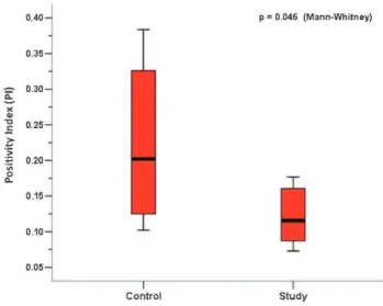
One animal from the control group died on the sixth day after surgery from a biliary fistula and was excluded from the study. Another pig was excluded from the experimental group due to its characterization as an outlier, it once had considerably higher PI value than did the other pigs: in the interval between Q3+1.5*AIQ and Q3+3*AIQ; Q1 and Q3 are the 1st and 3rd quartiles and AIQ corresponds to the interquartilic amplitude (Q3-Q1). Therefore, the results were analyzed and presented based on a total of 16 animals (eight in each group). One representative immunohistochemical image for alpha smooth muscle actin is shown in Figure 4. Table 1 provides the surgical data of the control and study groups. There were no statistically significant differences in any of the parameters evaluated (p.0.05), demonstrating the homogeneity of the groups according to the analyzed parameters. Table 2 shows the descriptive data for the PI values of the control and study groups and the correspond-ing p values (Mann-Whitney test). The data are expressed as the median and interquartile intervals (Q1-Q3). The study group exhibited a significantly lower PI than the control group (p= 0.046), as illustrated in Figure 5.
& DISCUSSION
This study investigated the effect of oral tamoxifen on the myofibroblast count in the healing tissue after BDI in an experimental pig model. To our knowledge, this is the first study to investigate the possible use of oral tamoxifen as a therapeutic option for reducing stenosis after bile duct reconstruction. We observed that the oral treatment of pigs with tamoxifen reduced thea-SMA expression in the healing tissue after BDI, suggesting a reduction of the number of myofibroblasts through the inhibition of TGF-b1 production. Because the biliary tracts of pigs are anatomically similar to those of humans, pigs have been used in many studies to evaluate biliary tract processes (29-32). Therefore, the results obtained in this study approximate the results that can be expected in humans (9) because the greater the physiologi-cal and anatomiphysiologi-cal similarity, the more applicable the conclusions (33). The arterial supply of the common hepatic duct and the bile duct have a longitudinal anastomotic chain arranged in a ladder form in humans (34). Therefore, we performed a longitudinal incision to maintain the best blood supply in the scarring area.
activation of myofibroblasts (19). This process may lead to biliary strictures due to wound contraction and fibrosis (19). In addition toa-SMA, myofibroblasts also express smooth muscle myosin isoforms, which are responsible for the contraction and/or motility of smooth muscle (36). Myofibroblasts also produce matrix proteins and additional growth factors in response to proinflammatory cytokines (36). After repair or scar formation, myofibroblasts are eliminated by apoptosis (36). In dogs, myofibroblasts appear one week after BDI, peak after three weeks and remain for a long period (35). Therefore, we chose to sacrifice the animals 30 days after the surgery to ensure that the peak number of myofibroblasts had been achieved.
In this study, myofibroblasts were identified using immunohistochemistry with an anti-a-SMA antibody. It is important to note that we used computerized digital image analysis procedures to calculate the immunopositive areas. Many studies use conventional pathologist-based manual scoring of the immunohistochemical results (37-39), but this approach suffers from a greater risk of interobserver and intraobserver variability (40).
In a model of nephropathy in rats, tamoxifen exhibited an antifibrotic effect that, according to the authors, could be explained by its ability to reduce myofibroblast prolifera-tion, as there was also a reduction ofa-SMA expression (41). In another study, tamoxifen inhibited keloid fibroblast
Table 1 -Surgical data for the control and study groups.
Variable Group Median Q1 Q3 p-valuea
Weight at surgery (kg) control 26.5 25.0 28.7 0.83
study 27.0 24.8 30.3
Weight at necropsy (kg) control 33.3 31.0 36.8 0.71
study 33.3 27.8 35.0
Caliber of the bile duct at surgery (mm) control 7.31 6.07 10.43 0.79
study 7.53 6.99 8.15
Caliber of the bile duct at necropsy (mm) control 5.59 5.30 6.65 0.17
study 5.01 3.67 5.96
Duration of the surgery (min) control 55.0 46.0 71.0 0.95
study 58.5 54.5 65.0
Q1: 1st quartile; Q3: 3rd quartile,aMann-Whitney test.
proliferation and decreased collagen production by decreas-ing the expression of TGF-b1 (42). It is known that biliary epithelial cells and periductal myofibroblasts are closed linked because damage to the former leads to activation and proliferation of the latter (19). Various experimental models in rats have previously shown that estrogen plays a key role in the biology of cholangiocytes (43,44) because the administration of tamoxifen or ovariectomy reduces biliary tree growth and induces cholangiocytes apoptosis (45,46). This concept is consistent with the findings in humans that estrogen receptors are over-expressed in the cholangiocytes of primary biliary cirrhosis patients (47).
The present results should be interpreted in light of the potential limitations of the study. First, it is not possible to know whether the modulatory effect of tamoxifen would be present in male animals because we only examined females. However, this deliberate choice provided us with a more homogeneous sample and enhanced the translational impact of the results, considering that there is a known higher prevalence of biliary gallstone disease in women. Second, because we addressed the biliary wound healing process after mechanical injury at a single one-month follow-up time point, it is not known whether tamoxifen exerts a long-lasting action. Further studies are needed to describe the time course of this modulatory action of tamoxifen. The one-month time point was chosen because myofibroblast proliferation reaches its peak after three weeks (35). Third, because our aim was specifically to investigate the number of myofibroblasts present during the biliary wound healing process, we did not obtain an overview of the entire wound healing process, including the changes in collagen. Although a more comprehensive approach would be desirable, the present results are
straightforward and provide novel evidence for the mod-ulatory role of tamoxifen in myofibroblast proliferation in biliary wound healing.
Biliary duct reconstruction is complex and although a large variety of suggested reconstruction techniques and materials exist in the literature, the BDI treatment results are still not satisfactory [49]. Therefore, other options must be investigated to improve reconstruction outcomes. In con-clusion, our results demonstrate that oral tamoxifen is a promising therapeutic option for reducing stenosis after bile duct reconstruction because it reduces the number of myofibroblasts present in the healing tissue. More studies must be undertaken to validate the inclusion of oral tamoxifen in clinical practice.
& AUTHOR CONTRIBUTIONS
Siqueira OH, Herani Filho B, de Paula RE and No´brega AC designed the study. Siqueira OH, A´ scoli FO, Carvalho AC, Cunha KS, Pires AR and Gaglionone NC performed the research. Siqueira OH and Cunha KSG analyzed the data. Siqueira OH, No´brega AC, Cunha KS and Granjeiro JM wrote the paper.
& REFERENCES
1. Pofahl WE, Pories WJ. Current status and future directions of geriatric general surgery. Journal of the American Geriatrics Society. 2003;51(7 Suppl):S351-4, http://dx.doi.org/10.1046/j.1365-2389.2003.51347.x. 2. Linhares BL, Magalha˜es AG, Cardoso PM, Filho JPP. Lesa˜o iatrogeˆnica
de via biliar po´s-colecistectomia. Rev. Col. Bras. Cir. 2011;38:95-9, http:// dx.doi.org/10.1590/S0100-69912011000200005.
3. de Santibanes E, Palavecino M, Ardiles V, Pekolj J. Bile duct injuries: management of late complications. Surg Endosc. 2006;20(11):1648-53, http://dx.doi.org/10.1007/s00464-006-0491-8.
4. Sikora SS, Srikanth G, Agrawal V, Gupta RK, Kumar A, Saxena R, et al. Liver histology in benign biliary stricture: fibrosis to cirrhosis . . . and reversal? J Gastroenterol Hepatol. 2008;23(12):1879-84, http://dx.doi. org/10.1111/j.1440-1746.2007.04901.x.
5. Pottakkat B, Sikora SS, Kumar A, Saxena R, Kapoor VK. Recurrent bile duct stricture: causes and long-term results of surgical management. J Hepatobiliary Pancreat Surg. 2007;14(2):171-6, http://dx.doi.org/10. 1007/s00534-006-1126-0.
6. Van de Sande S, Bossens M, Parmentier Y, Gigot JF. National survey on cholecystectomy related bile duct injury--public health and financial aspects in Belgian hospitals--1997. Acta Chir Belg. 2003;103(2):168-80. 7. Berney CR. Major common bile duct injury and risk of litigation: a
surgeon’s perspective. Am J Surg. 2011 Aug 25. [Epub ahead of print]. 8. de Reuver PR, Rauws EA, Bruno MJ, Lameris JS, Busch OR, van Gulik
TM, et al. Survival in bile duct injury patients after laparoscopic cholecystectomy: a multidisciplinary approach of gastroenterologists, radiologists, and surgeons. Surgery. 2007;142(1):1-9, http://dx.doi.org/ 10.1016/j.surg.2007.03.004.
9. de Reuver PR, Dijkgraaf MGW, Gevers SKM, Gouma DJ. Poor agreement among expert witnesses in bile duct injury malpractice litigation: an expert panel survey. Ann Surg. 2008;248(5):815-20, http://dx.doi.org/10. 1097/SLA.0b013e318186de35.
10. Scurr JRH, Brigstocke JR, Shields DA, Scurr JH. Medicolegal claims following laparoscopic cholecystectomy in the UK and Ireland. Ann R Coll Surg Engl. 2010;92(4):286-91, http://dx.doi.org/10.1308/ 003588410X12664192076214.
11. Helmy AA, Hamad MA, Aly AM, Sherif T, Hashem M, El-Sers DA, et al. Novel technique for biliary reconstruction using an isolated gastric tube with a vascularized pedicle: a live animal experimental study and the first clinical case. Ann Surg Innov Res. 2011;5:8, http://dx.doi.org/10. 1186/1750-1164-5-8.
12. Mercado-Dı´az MA´ , Ramı´rez-Morales R, Medinilla-Cruz MA, Poucel-Sa´nchez Medal F. Fe´rula transhepa´tica-transanastomo´tica en lesiones de las vı´as biliares: evolucio´n a largo plazo; Transhepatic transanastomotic stents for bile duct injuries: long-term evolution. Cir Cir. 2008;76(3):219-23.
13. Sicklick JK, Camp MS, Lillemoe KD, Melton GB, Yeo CJ, Campbell KA, et al. Surgical management of bile duct injuries sustained during laparoscopic cholecystectomy: perioperative results in 200 patients. Ann Surg. 2005;241(5):786-92; discussion 793-5, http://dx.doi.org/10.1097/ 01.sla.0000161029.27410.71.
14. Starling SV, Abrantes WL. Common bile duct injury: ligation and cholecystojejunostomy as surgical option: [case report]. Rev Col Bras Cir. 2003;3:244-6.
Table 2 -Positivity indices of the control and study groups.
Group Median Q1 Q3 p-valuea
Control 0.2021 0.1249 0.3259 0.046
Study 0.1155 0.0873 0.1607
Q1: 1s t quartile; Q3: 3rd quartile.aMann-Whitney test.
15. Sampaio JA, Kruse CK, Passarin TL, Waechter FL, Nectoux M, Fontes PRO, et al. Benign biliary strictures: repair and outcome with the use of silastic transhepatic transanastomotic stents. ABCD, arq. bras. cir. dig. 2010;23:(4)259-265, http://dx.doi.org/10.1590/S0102-67202010000400011. 16. Lillemoe KD. Evaluation of suspected bile duct injuries. Surg Endosc.
2006;20:1638-43, http://dx.doi.org/10.1007/s00464-006-0489-2. 17. Thannickal VJ, Lee DY, White ES, Cui Z, Larios JM, Chacon R, et al.
Myofibroblast differentiation by transforming growth factor-beta1 is dependent on cell adhesion and integrin signaling via focal adhesion kinase. J Biol Chem. 2003;278(14):12384-9, http://dx.doi.org/10.1074/ jbc.M208544200.
18. Geng Z-M, Yao Y-M, Liu Q-G, Niu X-J, Liu X-G. Mechanism of benign biliary stricture: a morphological and immunohistochemical study. World J Gastroenterol. 2005;11(2):293-5.
19. Demetris A-J, Lunz J-G 3rd, Specht S, Nozaki I. Biliary wound healing, ductular reactions, and IL-6/gp130 signaling in the development of liver disease. World J Gastroenterol. 2006;12(22):3512-22.
20. Geng ZM, Zheng JB, Zhang XX, Tao J, Wang L. Role of transforming growth factor-beta signaling pathway in pathogenesis of benign biliary stricture. World J Gastroenterol. 2008;14(31):4949-54.
21. Geng ZM, Xiang GA, Han Q, Liu XG, Liu QG, Pan CE. The expression and significance of macrophage and TGF-b1 on healing process of bile duct. Zhonghua Shiyan Waike Zazhi. 2000;17:522-3.
22. Gragnani A, Warde M, Furtado F, Ferreira LM. Topical tamoxifen therapy in hypertrophic scars or keloids in burns. Arch Dermatol Res. 2010;302(1):1-4, http://dx.doi.org/10.1007/s00403-009-0983-1. 23. Guest S. Tamoxifen therapy for encapsulating peritoneal sclerosis:
mechanism of action and update on clinical experiences. Perit Dial Int. 2009;29(3):252-5.
24. Costanzi S, Zoli A, Ferraro PM, Danza FM, Ferraccioli GF. A paraneoplastic retroperitoneal fibrosis resistant to corticosteroids treated with tamoxifen. Clin Nephrol. 2008;70(2):172-5.
25. Savelli BA, Parshley M, Morganroth ML. Successful treatment of sclerosing cervicitis and fibrosing mediastinitis with tamoxifen. Chest. 1997;111(4):1137-40, http://dx.doi.org/10.1378/chest.111.4.1137. 26. Ohashi T, Shigematsu N, Kameyama K, Kubo A. Tamoxifen for recurrent
desmoid tumor of the chest wall. Int J Clin Oncol. 2006;11(2):150-2. 27. Gwynne-Jones DP, Theis JC, Jeffery AK, Hung NA. Long-term follow-up
of a recurrent multifocal desmoid tumour treated with tamoxifen: a case report. J Orthop Surg (Hong Kong). 2005;13(2):174-7.
28. Delleˆ H, Rocha JRC, Cavaglieri RC, Vieira JM Jr, Malheiros DMAC, Noronha IL. Antifibrotic effect of tamoxifen in a model of progressive renal disease. J Am Soc Nephrol. 2012;23(1):37-48, http://dx.doi.org/10. 1681/ASN.2011010046.
29. Miyazawa M, Torii T, Toshimitsu Y, Okada K, Koyama I, Ikada Y. A tissue-engineered artificial bile duct grown to resemble the native bile duct. Am J Transplant 2005;5(6):1541-7, http://dx.doi.org/10.1111/j. 1600-6143.2005.00845.x.
30. Aikawa M, Miyazawa M, Okamoto K, Toshimitsu Y, Torii T, Okada K, et al. A novel treatment for bile duct injury with a tissue-engineered bioabsorbable polymer patch. Surgery. 2010;147(4):575-80, http://dx.doi. org/10.1016/j.surg.2009.10.049.
31. Leppa¨niemi A, Wherry D, Pikoulis E, Hufnagel H, Waasdorp C, Fishback N, et al. Common bile duct repair with titanium staples. Comparison with suture closure. Surg Endosc. 1997;11(7):714-7. 32. Li Q, Tao L, Chen B, Ren H, Hou X, Zhou S, et al. Extrahepatic bile duct
regeneration in pigs using collagen scaffolds loaded with human collagen-binding bFGF. Biomaterials. 2012;33(17):4298-308, http://dx. doi.org/10.1016/j.biomaterials.2012.03.003.
33. Calasans-Maia MD, Monteiro ML, Ascoli FO, Granjeiro JM. The rabbit as an animal model for experimental surgery. Acta Cir Bras. 2009; 24(4):325-8.
34. Gadzijev EM. Surgical anatomy of hepatoduodenal ligament and hepatic hilus. J Hepatobiliary Pancreat Surg. 2002;9(5):531-3, http://dx.doi.org/ 10.1007/s005340200068.
35. Xu J, Geng ZM, Ma QY. Microstructural and ultrastructural changes in the healing process of bile duct trauma. Hepatobiliary Pancreat Dis Int. 2003;2(2):295-9.
36. Kosmidis C, Efthimiadis C, Anthimidis G, Basdanis G, Apostolidis S, Hytiroglou P, et al. Myofibroblasts and colonic anastomosis healing in Wistar rats. BMC Surg. 2011;11:6, http://dx.doi.org/10.1186/1471-2482-11-6.
37. Juniantito V, Izawa T, Yuasa T, Ichikawa C, Yamamoto E, Kuwamura M, et al. Immunophenotypical analyses of myofibroblasts in rat excisional wound healing: possible transdifferentiation of blood vessel pericytes and perifollicular dermal sheath cells into myofibroblasts. Histol Histopathol. 2012;27(4):515-27.
38. Angadi PV, Kale AD, Hallikerimath S. Evaluation of myofibroblasts in oral submucous fibrosis: correlation with disease severity. J Oral Pathol Med. 2011;40(3):208-13, http://dx.doi.org/10.1111/j.1600-0714.2010.00995.x. 39. Arai H, Furusu A, Nishino T, Obata Y, Nakazawa Y, Nakazawa M, et al.
Thalidomide prevents the progression of peritoneal fibrosis in mice. Acta Histochem Cytochem. 2011;44(2):51-60, http://dx.doi.org/10.1267/ahc. 10030.
40. Cunha KSG, Caruso AC, Gonc¸alves AS, Bernardo VG, Pires ARC, da Fonseca EC, et al. Validation of tissue microarray technology in malignant peripheral nerve sheath tumours. J Clin Pathol. 2009; 62(7):629-33, http://dx.doi.org/10.1136/jcp.2008.063081.
41. Marinotto DBE, Delleˆ H, da Rocha JRC, Becker LE, Malheiros DMAC, Vieira-Jr JM, et al. Efeito do Tamoxifeno como Droga Anti-Fibro´tica em Modelo de Nefrotoxicidade Croˆnica por Ciclosporina. J Bras Nefrol. 2008;30(1):32-9.
42. Ruffy MB, Kunnavatana SS, Koch RJ. Effects of tamoxifen on normal human dermal fibroblasts. Arch Facial Plast Surg. 2006;8(5):329-32, http://dx.doi.org/10.1001/archfaci.8.5.329.
43. Alvaro D, Onori P, Metalli VM, Svegliati-Baroni G, Folli F, Franchitto A, et al. Intracellular Pathways Mediating Estrogen-Induced Cholangiocyte Proliferation in the Rat. Hepatology. 2002;36(2):297-304, http://dx.doi. org/10.1053/jhep.2002.34741.
44. Alvaro D, Alpini G, Onori P, Perego L, Baroni GS, Franchitto A, et al. Estrogens stimulate proliferation of intrahepatic biliary epithelium in Rats. Gastroenterology. 2000;119(6):1681-91, http://dx.doi.org/10.1053/ gast.2000.20184.
45. Alvaro D, Alpini G, Onori P, Franchitto A, Glaser S, Le Sage G, et al. Effect of ovariectomy on the proliferative capacity of intrahepatic rat cholangiocytes. Gastroenterology. 2002;123(1):336-44, http://dx.doi.org/ 10.1053/gast.2002.34169.
46. Glaser SS, Gaudio E, Miller T, Alvaro D, and Alpini G. Cholangiocyte proliferation and liver fibrosis. Expert Rev Mol Med. 2009;11:e7, http:// dx.doi.org/10.1017/S1462399409000994.
47. Alvaro D, Invernizzi P, Onori P, Franchitto A, Pinto G, De Santis A, et al. Expression of estrogen receptors in cholangiocytes of patients with primary biliary cirrhosis (PBC) and relationship with immunohisto-chemical markers of cell proliferation and death. J Hepatol. 2003; 8(Suppl. 2):184-5, http://dx.doi.org/10.1016/S0168-8278(03)80013-2. 48. Schanaider A, Pannain VLN, Mu¨ller LCCM, Maya MCA. Expanded