einstein. 2017;15(3):366-8
CASE REPORT
This content is licensed under a Creative Commons Attribution 4.0 International License.
1 Hospital do Servidor Público Estadual “Francisco Morato de Oliveira”, São Paulo, SP, Brazil. 2 Hospital Universitário, Universidade Federal de Sergipe, Aracaju, SE, Brazil.
Corresponding author: Marcelo Protásio dos Santos – Rua Cedro, 213 – São José – Zip code: 49020-170 – Aracaju, SE, Brazil – Phone: (55 79) 3211-9119 – E-mail: [email protected]
Received on: Dec 1, 2016 – Accepted on: Feb 10, 2017
DOI: 10.1590/S1679-45082017RC3942
ABSTRACT
An asymptomatic 79-year-old woman, with incidental finding on abdominal ultrasound of a solid nodule in the tail of the pancreas. Magnetic resonance imaging showed a 12mm solid tumor. The suggested diagnosis was pancreatic neuroendocrine tumor. The pathological examination showed an intrapancreatic splenic tissue. This is a rare ectopic location of spleen tissue and it should be considered in the differential diagnosis of pancreatic solid tumors.
Keywords: Spleen; Pancreas; Neuroendocrine tumors/diagnosis; Diagnosis, differential; Case reports
RESUMO
Mulher de 79 anos, com achado incidental em ultrassonografia de abdome de lesão sólida em cauda de pâncreas. Em ressonância magnética, foi confirmada lesão de 12mm. Foi aventada a hipótese diagnóstica de tumor neuroendócrino não secretor, sendo realizada pancreatectomia distal laparoscópica. Em estudo anatomopatológico, diagnosticou-se tecido esplênico, configurando baço acessório intrapancreático. Este tipo de manifestação topográfica de baço é rara, mas deve fazer parte dos diagnósticos diferenciais em tumores sólidos do pâncreas.
Descritores: Baço; Pâncreas; Tumores neuroendócrinos/diagnóstico; Diagnóstico diferencial; Relatos de casos
INTRODUCTION
The accessory spleen is a congenital anomaly caused by a failure in embryologic development of spleen tissue with occurring in 10% of general population. In 16% of cases, this tissue is found in tail of the pancreas.(1)
Intrapancreatic accessory spleen is a benign affection and rarely symptomatic. This affection great importance
relays on the fact that of being a differential diagnosis to pancreatic neuroendocrine, but with therapies and
completely different prognosis.(2)
We report a case of an incidental finding in radiology exam that mimicked a neuroendocrine pancreatic tumor.
CASE REPORT
This was an asymptomatic 79-year-old woman followed-up for chronic C hepatitis and who after an abdominal ultrasound had an incidental finding of a hypoechoic nodule measuring 10mm in the tail of the pancreas. Her physical exam did not show changes and laboratorial tests, including the cancer antigen 19.9, were normal.
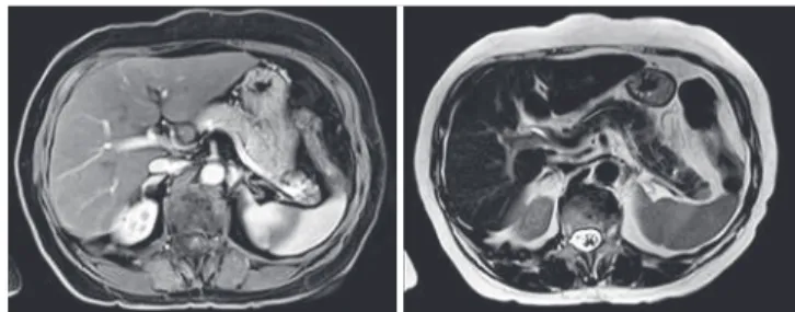
Abdominal magnetic resonance confirmed the finding of nodular injury measuring 12mm in the tail of the pancreas, hypointense in T1, hyperintense in T2, and highlight after infusion of contrast agent (Figure 1).
Based on these findings, we suspected of non-functioning neuroendocrine pancreatic tumor. A Figure 1. Abdominal magnetic resonance imaging (T1 and T2)
Intrapancreatic accessory spleen
Baço acessório intrapancreático
Marcelo Protásio dos Santos1, Aline Pacheco de Rezende1, Paulo Vicente dos Santos Filho2,
367
Intrapancreatic accessory spleen
einstein. 2017;15(3):366-8
distal pancreatectomy was carried out with spleen preservation (Figure 2). The surgical procedure was done without intercurrences by videolaparoscopic access, and pancreatic resection with yellow reload linear stampler
(EndoGIA®) with 60mm. The patient evolved with
pancreatic fistula in the fifth day after surgery without need of surgery. She was discharged 16 days after the surgery.
ectopic spleens (around 80%) are placed closed to splenic hilum, and 16% are located in the tail of the pancreas.(³)
In general, intrapancreatic accessory spleen is an asymptomatic injury and without need of surgical therapy. However, in the majority of described cases, diagnosis was made after surgical resection because of the difficult in the pre-operative differential diagnosis
with pancreatic neuroendocrine tumors.(4)
Pancreatic neuroendocrine tumors are rare neuroendocrine neoplasias with an annual incidence estimation of <1/100,000 in population studies and
correspond for less than 2% of all pancreatic tumors.(5,6)
These tumors are classified as functioning and non-functioning according to hormonal secretion and symptoms observed, but, unfortunately, there are no global consensus to such definition. Most of pancreatic neuroendocrine tumors is no-functioning and the
majority of them are malignant.(6) Primary surgical
resection is an associated factor with increase in long-term survival in these tumors, mainly in injuries greater than 20mm. The conservative therapy is preferred in
cases of tumors smaller than 10mm.(7)
The significant importance to differentiate intrapancreatic accessory spleen from pancreatic neuroendocrine tumors before the surgery relays on the fact that the latter needs surgical intervention whereas the first should be approached conservatively. The difficulty is to perform differential diagnosis, mainly because, so far, there are no laboratorial or radiologic exams to confirm or exclude the diagnosis of
intrapancreatic accessory spleen.(4)
Imaging exams can be useful to differentiate two injuries. However, computed tomography with contrast agent and conventional magnetic resonance are limited
in this evaluation mainly in injuries smaller than 10mm.(8)
Magnetic resonance combined with diffusion-weighted phase revealed to be a high accurate method in diagnosis and differentiation of intrapancreatic ectopic spleen and small solid pancreatic tumors. The intrapancreatic ectopic spleen normally is seen in magnetic resonance of hyperintense diffusion-weighted in T2 and hypointense
weighted in T1 compared to normal pancreatic tissue.(8)
The endoscopic ultrasound with aspiration biopsy, in addition to images, provides a definitive diagnosis by the use of pathological examination, but it constitutes an exam that depends on the researcher and injury site, in
addition to be an invasive method.(9)
The 68Ga-DOTA-TOC PET/CT is a high specificity
method for diagnosis of pancreatic neuroendocrine tumors because there is an important expression of Figure 2. Intraoperative with repaired pancreas and isolated splenic vessels
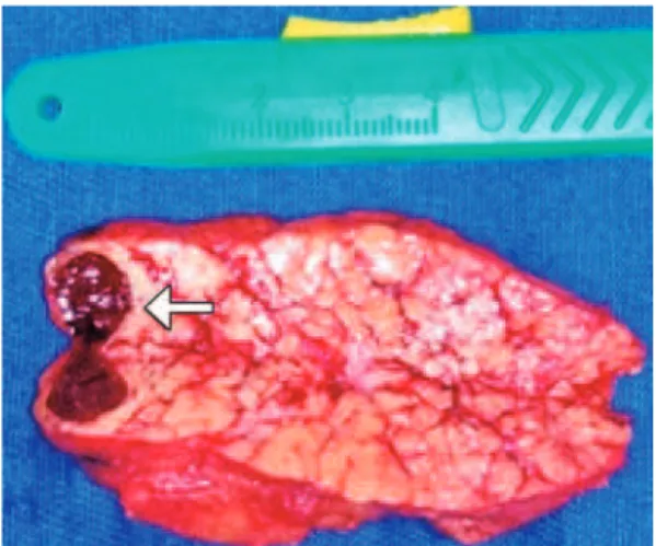
Figure 3. Surgical sample of distal pancreatectomy with intrapancreatic ectopic spleen
Mascroscopic cuts of surgical specimen showed nodular area of smooth aspect and brown color with 12x7x7mm surrounded by pancreatic tissue. The pathological examination confirmed the diagnosis of accessory intrapancreatic spleen (Figure 3).
DISCUSSION
Accessory spleen is a congenital abnormality with
incidence approximately 10% in necropsy studies.(¹) Its
development results in a change during differentiation of mesenchymal cells in formation of splenic tissue
einstein. 2017;15(3):366-8
368 Santos MP, Rezende AP, Santos Filho PV, Gonçalves JE, Beraldo FB, Sampaio AP
receptors of somatostatin on lymphocytes. Therefore, a
physiological accumulation of 68Ga-DOTA-TOC is always
seen in splenic tissue.(10) Scintigraphy with marked
erythrocytes with 99 technetium is one of the most specific methods to diagnose intrapancreatic ectopic spleen, because when marked erythrocytes are injected with radiopharmacy, more than 90% of the material is uptake by the splenic tissue, therefore contributing significantly to detect intrapancreatic splenic tissue and differentiate neuroendocrine pancreatic tumors and,
mainly, avoid unnecessary surgical procedure.(11)
CONCLUSION
Intrapancreatic ectopic spleen is rare. Surgery is not indicated in asymptomatic patients. This affection must be considered as a differential diagnosis before surgery of solid pancreatic injuries suggestive of neuroendocrine neoplasias to avoid unnecessary pancreatic resections.
REFERENCES
1. Halpert B, Gyorkey F. Lesions observed in accessory spleens of 311 patients. Am J Clin Pathol. 1959;32(2):165-8.
2. Octavio A, Castillo C, Pizzi Pl. Aberrant spleen simulating an adrenal mass. Rev Chil Cir. 2013;65(2):162-5.
3. Halpert B, Alden ZA. Accessory spleens in or at the tail of the pancreas. A survey of 2,700 additional necropsies. Arch Pathol. 1964;77:652-4. 4. Zhu HX, Lou WH, Kuang TT, Wang DS. Post-splenectomy intrapancreatic
accessory spleen mimicking endocrine tumor of the pancreas. Int J Surg Case Rep. 2014;5(12):1151-3.
5. Lam KY, Lo CY. Pancreatic endocrine tumour: a 22-year clinico-pathological experience with morphological, immunohistochemical observation and a review of literature. Eur J Surg Oncol. 1997;23(1):36-42. Review.
6. Halfdanarson TR, Rabe KG, Rubin J, Petersen GM. Pancreatic neuroendocrine tumors (PNETs): incidence, prognosis and recent trend toward improved survival. Ann Oncol. 2008;19(10):1727-33.
7. Chung JC, Choi DW, Jo SH, Heo JS, Choi SH, Kim YI. Malignant nonfunctioning endocrine tumors of the pancreas: predictive factors for survival after surgical treatment. World J Surg. 2007;31(3):579-85.
8. Jang KM, Kim SH, Lee SJ, Park MJ, Lee MH, Choi D. Differenciation of an intrapancreatic acessory spleen from a small (<3-cm) solid pancreatic tumor: value of diffusion-wighted MR imaging. Radiology. 2013;266(1):159-67.
9. Bastidas AB, Holloman D, Lankarani A, Nieto JM. Endoscopic ultrasound-guided needle-based probe confocal laser endomicroscopy (nCLE) of intrapancreatic ectopic spleen. ACG Case Rep J. 2016;3(3):196-8.
10. Fülber I, Wurster C, Librizzi D, Röbler M, Gallmeier E, Bartsch DK. Intrapancreatic acessory spleen: a diferencial diagnosis to neuroendocrine tumors of the pancreas on Ga-68- DOTATOC PET/TC. JOP J Pancreas. 2016;17(4):427-30. 11. Kim SH, Lee JM, Han JK, Lee JY, Kim KW, Cho KC, et al. Intrapancreatic