The use of diffusion-weighted magnetic resonance
imaging in the differentiation between benign
and malignant breast lesions*
O uso da difusão por ressonância magnética na diferenciação das lesões mamárias benignas e malignas
Fernanda Philadelpho Arantes Pereira1, Gabriela Martins2, Eduardo Figueiredo3, Marisa Nassar Aidar Domingues2, Romeu Côrtes Domingues4, Lea Mirian Barbosa da Fonseca5
OBJECTIVE: To study the utility of diffusion-weighted magnetic resonance imaging in the differentiation between benign and malignant breast lesions. MATERIALS AND METHODS: Forty-five women (mean age, 46.1 years) with 52 focal breast lesions underwent diffusion-weighted magnetic resonance imaging. The calculation of apparent diffusion coefficient (ADC) was based on the ADC map reflecting five b values (0,
250, 500, 750, and 1000 s/mm2
). The mean ADC value of each lesion was correlated with imaging findings and histopathologic results. Cutoff ADC, sensitivity and specificity of diffusion-weighted imaging in the differentiation between benign and malignant lesions were calculated. P < 0.05 was considered as statistically
significant. RESULTS: The mean ADC was significantly lower for malignant lesions (0.92 ± 0.26 × 10⁻³ mm2
/s) as compared with benign lesions (1.50 ± 0.34 × 10⁻³ mm2
/s) (P < 0.0001). Diffusion-weighted
imaging showed high sensitivity and specificity (both, 92.3%) in the differentiation between benign and malignant lesions. CONCLUSION: Diffusion-weighted imaging is a potential resource as an adjuvant to breast magnetic resonance imaging to differentiate benign from malignant lesions. Such sequence can be easily added to the standard breast magnetic resonance imaging protocol, without implying any significant increase in examination time.
Keywords: Breast cancer; Diffusion-weighted imaging; Magnetic resonance imaging.
OBJETIVO: Estudar a utilidade da sequência pesada em difusão na diferenciação das lesões mamárias benig-nas e maligbenig-nas. MATERIAIS E MÉTODOS: Quarenta e cinco mulheres (idade média de 46,1 anos) com 52 nódulos de mama foram submetidas a ressonância magnética acrescida da sequência difusão. O coeficiente de difusão aparente (ADC) foi calculado através do mapa de ADC obtido pelo uso de cinco valores de b (0,
250, 500, 750 e 1.000 s/mm2
). O valor de ADC médio de cada lesão foi correlacionado com achados de imagem e resultados histopatológicos. Valores de ADC de corte, sensibilidade e especificidade da sequência difusão na diferenciação das lesões benignas e malignas foram calculados. P < 0,05 foi considerado
esta-tisticamente significativo. RESULTADOS: O valor de ADC médio foi significativamente menor para as lesões malignas (0,92 ± 0,26 × 10⁻³
mm2
/s) comparado com as lesões benignas (1,50 ± 0,34 × 10⁻³ mm2
/s) (P < 0,0001). A sequência difusão mostrou altas sensibilidade e especificidade (ambas 92,3%) na
diferen-ciação entre lesões benignas e malignas. CONCLUSÃO: A sequência pesada em difusão representa um re-curso potencial como coadjuvante da ressonância magnética das mamas na diferenciação das lesões benig-nas e maligbenig-nas. Tal sequência pode ser facilmente inserida no protocolo padrão da ressonância magnética das mamas, sem aumento significativo no tempo de exame.
Unitermos: Câncer de mama; Difusão; Imagem por ressonância magnética. Abstract
Resumo
* Study developed at Clínica de Diagnóstico Por Imagem (CDPI), Rio de Janeiro, RJ, Brazil.
1. Medical Residency, Fellow Master degree, Universidade Federal do Rio de Janeiro (UFRJ), MD, Radiologist at Clínica de Diagnóstico Por Imagem (CDPI), Rio de Janeiro, RJ, Brazil.
2. Medical Residency, MDs, Radiologists at Clínicas de Diag-nóstico Por Imagem (CDPI) and Multi-Imagem, Rio de Janeiro, RJ, Brazil.
3. Application GE Healthcare, São Paulo, SP, Brazil.
4. Medical Residency, Medical Director at Clínicas de
Diag-INTRODUCTION
Breast cancer is the second most fre-quent type of cancer in the world and the most common in women. The number of expected new cases of breast cancer in Bra-zil for the year 2008 was 49,400, with an estimated risk of 51 cases for each 100 thousand women(1).
Arantes Pereira FP, Martins G, Figueiredo E, Domingues MNA, Domingues RC, Fonseca LMB. The use of diffusion-weighted magnetic resonance imaging in the differentiation between benign and malignant breast lesions. Radiol Bras. 2009;42(5):283– 288.
nóstico Por Imagem (CDPI) and Multi-Imagem, Rio de Janeiro, RJ, Brazil.
5. PhD, Titular Professor, Universidade Federal do Rio de Ja-neiro (UFRJ), Nuclear Physician at Clínica de Diagnóstico Por Imagem (CDPI), Rio de Janeiro, RJ, Brazil.
Mailing address: Dra. Fernanda Philadelpho Arantes Pereira. Rua Ataulfo de Paiva, 669, 2º andar, Leblon. Rio de Janeiro, RJ, Brazil, 22649-900. E-mail: [email protected]
This neoplasm is a source of anxiety and worry for women, besides affecting their self-image and life expectancy. Despite being considered as a cancer with a rela-tively good prognosis provided the disease is early diagnosed and timely managed, the mortality rates for breast cancer still remain high in Brazil, most probably because the diagnosis is still achieved at advanced stages of the disease. In the world popula-tion, the mean five-year survival rate is 61%(1).
Currently, clinical examination and mammography are recommended for women from 40 years of age, as a method for early detection of the disease. Screen-ing mammography benefits have already been established. Several randomized clini-cal studies have proved that screening mammography can reduce mortality rates(1–4). However, the limitation of this two-dimensional method, particularly for evaluating dense breasts, results in a false-negative rate between 4% and 34% for the diagnosis of cancer(5). And, it is exactly in the youngest women that the cancer inci-dence has increased, generally with a more aggressive presentation(1).
Breast magnetic resonance imaging (MRI) has found a wide clinical application as an adjunctive to mammography and ul-trasonography not only for providing infor-mation regarding the lesion morphology, but also functional aspects such as contrast enhancement kinetics(6). Main indications of the method are associated with cases of proved diagnosis requiring evaluation of the disease extent, residual disease, tumor recidivation, occult primary site in the pres-ence of axillary carcinoma, and response to neoadjuvant chemotherapy(2). Because of the high sensitivity and effectiveness of the method in the evaluation of dense breasts, MRI can be a valuable complementary screening method in women at high genetic risk for breast cancer and in the diagnostic investigation of patients with inconclusive clinical and imaging findings.
In the last years, the availability of high-field units and coils specific for breast tis-sue in association with the development of a system for standardization of the descrip-tion of imaging findings (Breast Imaging Reporting and Data System – BI-RADS®)(4) and the development of the learning curve
of the method have resulted in an increased utilization of MRI with higher safety and efficacy.
MRI has a high sensitivity (89–100%) in the characterization of breast tumors(6–11). However, an overlap between benign and malignant findings still persists, resulting in a variable specificity (50–90%)(8,11–13) due to false-positive results related to the menstrual cycle, hormone replacement therapy, proliferative alterations, fibroad-enomas and papillomas. Thus, sometimes a differential diagnosis between malignant and benign lesions cannot be achieved based only on conventional MRI find-ings(14,15). Some studies have investigated the role played by functional MRI tech-niques, such as diffusion-weighted imag-ing to improve the method specificity in the evaluation of breast lesions(14,16–19).
For two decades, diffusion-weighted se-quences have been utilized for assessing intracranial diseases such as cerebrovascu-lar accidents. In the nineties, technological developments allowed the utilization of diffusion technique in extracranial sites(20,21). The diffusion-weighted sequence de-rives images from the difference of water molecules motion (Brownian motion) in tissues, resulting in quantitative and quali-tative data reflecting changes at cellular level and, consequently, unique informa-tion on the tumor cellularity and cell mem-branes integrity. This sequence seems to be a useful tool in the detection and charac-terization of tumors(17), as well as for moni-toring the response to neoadjuvant therapy(20).
By utilizing diffusion-weighted se-quences one can calculate the apparent dif-fusion coefficient (ADC), a quantitative measurement that is directly proportional to the water molecules diffusion(22). The high cellular proliferation in malignant tu-mors causes an increase in the cellular den-sity, creating additional barriers against the extracellular water molecules, reducing the ADC and resulting in decreased signal in-tensity.
The evaluation of breast lesions can be favored by the development of functional techniques including the diffusion tech-nique in the determination of the differen-tial diagnosis between malignant and be-nign of suspicious lesions detected at
con-ventional MRI. The primary objective of the present study is to evaluate the effec-tiveness of the diffusion technique as an adjunctive to conventional MRI in the dif-ferentiation between benign and malignant breast lesions. This technique would allow the increase in breast MRI specificity, with the consequential reduction of false-posi-tive results and unnecessary biopsies.
MATERIALS AND METHODS
Study population
From August/2007 to June/2008, a pro-spective study on diffusion-weighted se-quence was developed with 50 female pa-tients with 57 breast nodules submitted to MRI in the authors’ institution. Exclusion criteria were the following: non-nodular contrast enhancement of a more “dissemi-nated” tumor with possibility of a partial volume effect(18,23); benign cysts, since they do not pose diagnostic difficulty and the high ADC would artificially increase the benign values mean and variation(17); pa-tient motion that could lead to dubious ADC values; lesions undetected by diffu-sion-weighted sequence mainly due to their small size; and previous neoadjuvant therapy that could determine an increase in the ADC values(22,24). Based on these cri-teria, five lesions of five patients were ex-cluded. As a result, the present study in-cluded 45 patients (22 to 80 years of age; mean, 46.1 years) with 52 breast lesions.
At the histopathological study, 26 ma-lignant lesions were found as follows: in-filtrating ductal carcinoma (n = 19), duc-tal carcinoma in situ (n = 2), tubular carci-noma (n = 2), cystic adenoid carcinoma (n
= 1), mucinous (colloid) carcinoma (n = 1) and malignant phyllodes tumor (n = 1). The mean size of the malignant lesions was 3.09 cm, varying from 1.0 to 11.2 cm.
be-tween two radiologists specialized in breast images (with eleven- and eight-year expe-rience). According to the literature(26,27), the following criteria were considered as pre-dictive of benign disease: lobular shape, regular margins, non-enhancing internal septations or internal septations with lesser enhancement as compared with the adja-cent breast tissue. The presence of non-enhancing internal septations in a lobulated and regular nodule is highly specific for the diagnosis of fibroadenoma (93–97% speci-ficity)(28,29). The mean size of the lesions was 1.68 cm, ranging from 0.8 to 4.7 cm. Additionally, after one-year follow-up with mammography and/or ultrasonography, a there was a significant change in the image pattern of these benign lesions.
All the patients signed a term of free and informed consent.
Images acquisition
All the MRI studies were performed in a 1.5 T unit (Signa Excite HD; GE Health-care, Milwaukee, USA) with a dedicated bilateral eight-channel breast coil. Previ-ously to the diffusion-weighted sequence, conventional sequences were acquired, including axial, T1-weighted spin-echo sequence (TR/TE, 370/15 ms; matrix, 512 × 256; FOV, 340 mm; NEX, 1; slice thick-ness, 5 mm; interval, 1 mm), sagittal, T2-weighted fast spin-echo sequence with fat-suppression (TR/TE, 4200/85 ms; matrix, 320 × 224; FOV, 220 mm; NEX, 2; slice thickness, 5 mm; interval, 0 mm), axial STIR sequence (TR/TE, 4100/85 ms; TI, 150 ms; matrix, 512 × 256; FOV, 340 mm; NEX, 2; slice thickness, 5 mm; interval, 1 mm), and axial, 3D gradient, T1-weighted sequence with fat-suppression (flip angle, 15°; matrix, 352 × 352; FOV, 350 mm; slice thickness, 1 mm; interval, 0 mm) once before and four times after rapid injection in an infusion pump of 0.1 mmol/l de gadoterate meglumine (Dotarem; Guerbet, Roissy, France) per kilogram of body weight, followed by 20 ml saline solution. After examination, the precontrast images were subtracted from the early and delayed postcontrast images.
The diffusion was performed with single-shot echo-planar imaging (EPI) se-quence in the axial plane, centered on the lesions (b = 0, 250, 500, 750, and 1000
s/mm2; TR/TE, 1800/93.8 ms; matrix, 160 × 192; FOV, 360 mm; NEX, 16; number of sections, 10; slice thickness, 5 mm; inter-val, 0 mm; acquisition time, 3:44 minutes).
Analysis of images and data collection
All the images were transferred to a workstation (Advantage Windows version 4.2-07; GE Healthcare, Milwaukee, USA) and the diffusion-weighted sequence was postprocessed with a commercial software (Functool; GE Healthcare, Milwaukee, USA), with the objective of obtaining ADC maps (black/white and color, the latter with a Puh-thallium color scheme ranging from black [restricted diffusion] to red [without restricted diffusion]). The ADC maps for each lesion were calculated with five b
values (0, 250, 500, 750, and 1000 s/mm2). In order to achieve standardized condi-tions for results analysis and avoiding data contamination by adjacent structures, two regions of interest (ROI), with mean area of 61 mm2 (ranging from 40 to 94 mm2), were individually placed on the ADC map at the site of the target lesion and the mean ADC was calculated. Necrotic or cystic components were avoided utilizing the conventional MRI sequences as a refer-ence.
Statistical analysis
The data collected in the present study included patients’ age, size of the lesions, BI-RADS category, histopathological re-sults, ADC values and ROI size.
The Kolmogorov-Smirnov test was uti-lized for evaluating the normality of data such as age, tumor size and ADC value. At the level of 5%, the normality hypothesis was not rejected for none of the variables. The Students’ t-test for independent samples evaluated the difference between the means of the variables age, tumor size and ADC value according to the benign or malignant histopathological result at a 5%
significance level. All the ADC variables were assessed by the Levene’s test for equality of variance at the level of 5%; that is to say that P < 0.05 would indicate sta-tistically significant differences between the benign and malignant groups of lesions. Subsequently, a ROC (receiver operat-ing characteristic) curve of the ADC values according to the histopathological result was analyzed for determining the best cut-off value. The non-parametric distribution was the hypothesis utilized for determining the ROC curve. The measurement utilized for determining the ADC cutoff value, con-sidering the balance between sensitivity/ specificity was the Youden statistics (Y = sensitivity – [1 – specificity]). A higher value for the Youden statistics indicates a better cutoff value and, consequently, bet-ter sensitivity and specificity values.
Data processing and analysis were per-formed with the software SPSS 16.0 (Sta-tistical Software for Social Sciences; Chi-cago, USA).
RESULTS
The mean ADC value corresponding to malignant breast lesions (0.92 ± 0.26 × 10⁻³ mm2/s) was significantly lower than that observed in benign lesions (1.50 ± 0.34 × 10⁻³ mm2/s) (P < 0.0001) (Table 1; Fig-ures 1 and 2).
Considering a cutoff ADC value of 1.21 × 10⁻³ mm2/s, 2/26 benign lesions (papil-loma and epidermoid cyst), and 2/26 ma-lignant lesions (mucinous [colloid] carci-noma and malignant phyllodes tumor) would be erroneously diagnosed. As a re-sult, the diffusion-weighted sequence pre-sented high sensitivity and specificity (92.3% for both) in the differentiation be-tween benign and malignant lesions. The ROC curve demonstrated the value of 0.912 corresponding to the area under the curve.
Table 1 Apparent diffusion coefficient in benign and malignant lesions.
ADC values (× 10–3 mm2/s)
Mean
Standard deviation
Median
Interquartile interval
Benign lesions (n = 26)
1.50
0.34
1.48
1.31–1.68
Malignant lesions (n = 26)
DISCUSSION
The present study evaluated the role played by the diffusion-weighted sequence in the differentiation between benign and
malignant lesions. The mean ADC value of the benign lesions was significantly lower than the value of the malignant lesions.
The diffusion reflects the changes in the water molecules mobility caused by tissue
alterations associated with pathological processes. Thus, the measurement of the water molecules motion provides addi-tional information which may determine an increase in the MRI specificity in the
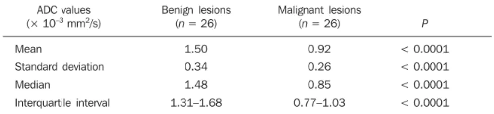
clas-Figure 1. Female, 43-year-old patient presenting fibroadenomas in the left breast. Delayed phase contrast-enhanced 3D gradient, T1-weighted sequence with fat-suppression in the axial plane (A), diffusion-weighted sequence (b 500 s/mm2) in the axial plane (B), and apparent diffusion coefficient (ADC) black/white
map in the axial plane (C) show two nodules with morphology and contrast-enhancement with benign appearance. Note that the nodules present high signal intensity on the diffusion (arrows) and on the ADC map (arrows) suggesting absence of water molecules diffusion restriction.
A B C
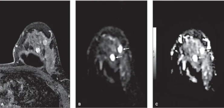
Figure 2. Female, 48-year-old patient presenting infiltrating ductal carcinoma in the left breast. Axial, 3D gradient T1-weighted sequence with fat-suppression at early postcontrast phase (A), diffusion-weighted sequence (b 500 s/mm2) in the axial plane (B), and apparent diffusion coefficient (ADC) black/white map
in the axial plane (C) show microlobulated nodule with suspicious contrast-enhancement. Note that the nodule presents high signal intensity on the diffusion (arrow) and signal loss on the ADC map (arrow), suggesting restricted diffusion of water molecules.
A B C
sification of breast lesions. Previous stud-ies with diffusion-weighted MRI have shown promising results in the differentia-tion between benign and malignant lesions with sensitivity ranging from 81% to 93% and specificity ranging from 80% to 88.5%(12,16–19,30). The results of the present study are in agreement with these previous studies, demonstrating statistical differ-ences between benign and malignant le-sions with high sensitivity and specificity (92.3% for both).
According to the diagnostic criteria adopted in the present study, all the fibroad-enomas and invasive ductal carcinomas were appropriately classified by the ADC, including two fibroadenomas erroneously classified as suspicious by conventional MRI. Such results indicate that the ADC would be effective in the differentiation between fibroadenomas and invasive duc-tal carcinomas, which would be extremely useful in the characterization of the tumor, considering that fibroadenomas may present points of similarity with malignant lesions, both at ultrasonography and MRI(31).
The results of the present study confirm that the mean ADC value of breast tumors is strongly correlated with its cellularity, even in the analysis of false-positive and false-negative results. Malignant breast lesions present higher cellularity and lower ADC than benign breast lesions. Thus, a malignant tumor with low cellularity due to the presence of cystic areas inside, like the malignant phyllodes tumor observed in the present study, demonstrated a high ADC and was erroneously classified as benign lesion. A carcinoma with high sig-nal intensity on a T2-weighted sequence, like the mucinous (colloid) carcinoma, pre-sented high ADC because of the low cel-lular density and the high water component in the extracellular space(32,33). By contrast, benign tumors with high cellularity like papilloma and epidermoid cyst also present in this study, demonstrated reduced ADC e led to an erroneous diagnosis of malig-nancy.
There are some limitations in the present study. Firstly, the patient motion during the acquisition of the diffusion-weighted sequence, leading to the obten-tion of equivocal ADC values. Addiobten-tion- Addition-ally, even in optimum circumstances,
dif-fusion-weighted sequences may fail in the categorization of breast lesions because of the limited capacity to recognize small le-sions (< 1 cm) on the ADC map. In cases where a lesion cannot be visualized on dif-fusion-weighted sequences, it is difficult to determine the exact localization of the ROI on the ADC map. Finally, like in other stud-ies, the sample of the present study is rela-tively small, and future studies with greater populations should be considered, and this is one of the next steps of the authors.
In spite of the limitations, the diffusion-weighted sequence provides additional in-formation for a rapid and easy characteriza-tion of breast nodules. Considering that conventional MRI is known for its good sensitivity and variable specificity in the characterization of breast lesions, a combi-nation of ADC measurement with the in-terpretation of contrast-enhancement pat-terns at conventional MRI can lead to an increase in the MRI accuracy, reducing the number of false-positive results and unnec-essary invasive procedures.
The diffusion-weighted sequence may be useful in the differentiation between malignant and benign breast lesions, in-creasing the specificity of breast MRI. This sequence is performed with no significant increase in the acquisition time and can be easily added to the standard breast MRI protocol.
REFERENCES
1. Instituto Nacional de Câncer, Ministério da Saúde. Estimativa 2008: incidência de câncer no Brasil. Rio de Janeiro: INCA; 2007.
2. Chala LF, Barros N. Avaliação das mamas com métodos de imagem. Radiol Bras. 2007;40(1): iv–vi.
3. Kestelman FP, Souza GA, Thuler LC, et al. Breast Imaging Reporting and Data System – BI-RADS®: valor preditivo positivo das categorias 3, 4 e 5. Revisão sistemática da literatura. Radiol Bras. 2007;40:173–7.
4. Roveda Jr D, Piato S, Oliveira VM, et al. Valores preditivos das categorias 3, 4 e 5 do sistema BI-RADS em lesões mamárias nodulares não-palpá-veis avaliadas por mamografia, ultra-sonografia e ressonância magnética. Radiol Bras. 2007;40: 93–8.
5. Huynh PT, Jarolimek AM, Daye S. The false-negative mammogram. Radiographics. 1998;18: 1137–54.
6. Kuhl C. The current status of breast MR imaging. Part I. Choice of technique, image interpretation, diagnostic accuracy, and transfer to clinical prac-tice. Radiology. 2007;244:356–78.
7. Schnall MD, Blume J, Bluemke DA, et al.
Diag-nostic architectural and dynamic features at breast MR imaging: multicenter study. Radiology. 2006;238:42–53.
8. Macura KJ, Ouwerkerk R, Jacobs MA, et al. Pat-terns of enhancement on breast MR images: in-terpretation and imaging pitfalls. Radiographics. 2006;26:1719–34.
9. Wiener JI, Schilling KJ, Adami C, et al. Assess-ment of suspected breast cancer by MRI: a pro-spective clinical trial using a combined kinetic and morphologic analysis. AJR Am J Roentgenol. 2005;184:878–86.
10. Bedrosian I, Mick R, Orel SG, et al. Changes in the surgical management of patients with breast carcinoma based on preoperative magnetic reso-nance imaging. Cancer. 2003;98:468–73. 11. Pereira FPA, Martins G, Calas MJG, et al.
Resso-nância magnética das mamas: o exame e suas indicações. Femina. 2008;36:565–70. 12. Marini C, Iacconi C, Giannelli M, et al.
Quanti-tative diffusion-weighted MR imaging in the dif-ferential diagnosis of breast lesion. Eur Radiol. 2007;17:2646–55.
13. Kuhl CK, Mielcareck P, Klaschik S, et al. Dynamic breast MR imaging: are signal intensity time course data useful for differential diagnosis of enhancing lesions? Radiology. 1999;211:101–10. 14. Wenkel E, Geppert C, Schulz-Wendtland R, et al. Diffusion weighted imaging in breast MRI: com-parison of two different pulse sequences. Acad Radiol. 2007;14:1077–83.
15. Sinha S, Sinha U. Functional magnetic resonance of human breast tumors: diffusion and perfusion imaging. Ann N Y Acad Sci. 2002;980:95–115. 16. Guo Y, Cai YQ, Cai ZL, et al. Differentiation of clinically benign and malignant breast lesions using diffusion-weighted imaging. J Magn Reson Imaging. 2002;16:172–8.
17. Rubesova E, Grell AS, De Maertelaer V, et al. Quantitative diffusion imaging in breast cancer: a clinical prospective study. J Magn Reson Im-aging. 2006;24:319–24.
18. Woodhams R, Matsunaga K, Iwabuchi K, et al. Diffusion-weighted imaging of malignant breast tumors: the usefulness of apparent diffusion co-efficient (ADC) value and ADC map for the de-tection of malignant breast tumors and evaluation of cancer extension. J Comput Assist Tomogr. 2005;29:644–9.
19. Kuroki Y, Nasu K, Kuroki S, et al. Diffusion-weighted imaging of breast cancer with the sen-sitivity encoding technique: analysis of the appar-ent diffusion coefficiappar-ent value. Magn Reson Med Sci. 2004;3:79–85.
20. Koh DM, Collins DJ. Diffusion-weighted MRI in the body: applications and challenges in oncol-ogy. AJR Am J Roentgenol. 2007;188:1622–35. 21. Woodhams R, Matsunaga K, Kan S, et al. ADC mapping of benign and malignant breast tumors. Magn Reson Med Sci. 2005;4:35–42. 22. Paran Y, Bendel P, Margalit R, et al. Water
diffu-sion in the different microenvironments of breast cancer. NMR Biomed. 2004;17:170–80. 23. Kuroki-Suzuki S, Kuroki Y, Nasu K, et al.
Detect-ing breast cancer with non-contrast MR imagDetect-ing: combining diffusion-weighted and STIR imag-ing. Magn Reson Med Sci. 2007;6:21–7. 24. Pickles MD, Gibbs P, Lowry M, et al. Diffusion
treatment of breast cancer. Magn Reson Imaging. 2006;24:843–7.
25. American College of Radiology (ACR). ACR BI-RADS – magnetic resonance imaging. In: ACR Breast Imaging Reporting and Data System, Breast Imaging Atlas. Reston: American College of Radiology; 2003.
26. Kuhl CK. Concepts for differential diagnosis in breast MR imaging. Magn Reson Imaging Clin N Am. 2006;14:305–28, v.
27. Morris EA. Breast MR imaging lexicon updated. Magn Reson Imaging Clin N Am. 2006;14:293– 303, v.
28. Nunes LW, Schnall MD, Siegelman ES, et al. Di-agnostic performance characteristics of architec-tural features revealed by high spatial-resolution MR imaging of the breast. AJR Am J Roentgenol. 1997;169:409–15.
29. Nunes LW, Schnall MD, Orel SG, et al. Correla-tion of lesion appearance and histologic findings for the nodes of a breast MR imaging interpreta-tion model. Radiographics. 1999;19:79–92. 30. Yabuuchi H, Matsuo Y, Okafuji T, et al. Enhanced
mass on contrast-enhanced breast MR imaging: lesion characterization using combination of dy-namic contrast-enhanced and diffusion-weighted
MR images. J Magn Reson Imaging. 2008;28: 1157–65.
31. Hochman MG, Orel SG, Powell CM, et al. Fi-broadenomas: MR imaging appearances with ra-diologic-histopathologic correlation. Radiology. 1997;204:123–9.
32. Kawashima M, Tamaki Y, Nonaka T, et al. MR imaging of mucinous carcinoma of the breast. AJR Am J Roentgenol. 2002;179:179–83. 33. Hatakenaka M, Soeda H, Yabuuchi H, et al.