Effects of ACE inhibition on endothelial progenitor
cell mobilization and prognosis after acute
myocar-dial infarction in type 2 diabetic patients
Jia-Yin Sun, Lin Zhai, Qiao-Ling Li, Jia-Xin Ye, Li-Na Kang, Jun Xie, Biao Xu
Nanjing University Medical School, Department of Cardiology, Affiliated Drum Tower Hospital, Nanjing 210008, China.
OBJECTIVE: We aimed to assess the chemotactic response of endothelial progenitor cells to angiotensin-converting enzyme inhibitors in T2DM patients after acute myocardial infarction, as well as the associated prognosis.
METHODS:Sixty-eight T2DM patients with acute myocardial infarction were randomized to either receive or not receive daily oral perindopril 4 mg, and 36 non-diabetic patients with acute myocardial infarction were enrolled as controls. The numbers of circulating CD452/low+CD34+CD133+KDR+endothelial progenitor cells, as
well as the stromal cell-derived factor-a and high-sensitivity C reactive protein levels, were measured before
acute percutaneous coronary intervention and on days 1, 3, 5, 7, 14, and 28 after percutaneous coronary intervention. Patients were followed up for 6 months. Chinese Clinical Trial Registry: ChiCTR-TRC-12002599. RESULTS:T2DM patients had lower circulating endothelial progenitor cell counts, decreased plasma vascular endothelial growth factor andalevels, and higher plasma high-sensitivity C reactive protein levels compared
with non-diabetic controls. After receiving perindopril, the number of circulating endothelial progenitor cells increased from day 3 to 7, as did the plasma levels of vascular endothelial growth factor and stromal cell-derived factor-a, compared with the levels in T2DM controls. Plasma high-sensitivity C reactive protein levels in
the treated group decreased to the same levels as those in non-diabetic controls. Furthermore, compared with T2DM controls, the perindopril-treated T2DM patients had lower cardiovascular mortality and occurrence of heart failure symptoms (p,0.05) and better left ventricle function (p,0.01).
CONCLUSIONS: The use of angiotensin-converting enzyme inhibitors represents a novel approach for improving cardiovascular repair after acute myocardial infarction in T2DM patients.
KEYWORDS: Endothelial Progenitor Cells; Endothelial Progenitor Cell Inhibitors; Prognosis; Type 2 Diabetes Mellitus; Acute Myocardial Infarction.
Sun JY, Zhai L, Li QL, Ye JX, Kang LN, Xie J, et al. Effects of ACE inhibition on endothelial progenitor cell mobilization and prognosis after acute myocardial infarction in type 2 diabetic patients. Clinics. 2013;68(5):665-673.
Received for publication onJanuary 16, 2013;First review completed onJanuary 28, 2013;Accepted for publication onJanuary 28, 2013 E-mail: [email protected]
Tel.:+6 25 8310 5205
& INTRODUCTION
Type 2 diabetes mellitus (T2DM) is an independent predictor of adverse outcomes in patients with acute myocardial infarction (AMI) (1,2). After AMI, diabetic patients have a higher incidence of angina and heart failure, as well as increased mortality (3,4), which is in accordance with severe left ventricular remodeling and impaired cardiac function (5). Endothelial dysfunction of blood vessels has been shown to contribute to vascular complications in T2DM
patients. Bone marrow-derived endothelial progenitor cells (EPCs) play an important role in endothelial regeneration and repair (6). However, this function is impaired in diabetic patients and has been demonstrated to participate in the pathogenesis of vascular complications (7). Patients with AMI exhibit increased mobilization of EPCs from bone marrow (8). Once mobilized into circulation, these EPCs home to the ischemic myocardium and may facilitate heart repair (9). However, the beneficial effects of ischemic-induced EPC mobilization are impaired in T2DM patients (10). Therefore, increasing the numbers of circulating EPCs mobilized from bone marrow may improve cardiac function and prognosis in T2DM patients with AMI.
Angiotensin-converting enzyme (ACE) inhibitors are a standard therapy in cardiovascular disease and have been demonstrated to be beneficial to EPCs in somein vitroand clinical studies (11–13). Experimental studies have also revealed that ACE inhibitors can attenuate the development
Copyrightß2013CLINICS– This is an Open Access article distributed under the terms of the Creative Commons Attribution Non-Commercial License (http:// creativecommons.org/licenses/by-nc/3.0/) which permits unrestricted non-commercial use, distribution, and reproduction in any medium, provided the original work is properly cited.
No potential conflict of interest was reported.
of atherosclerosis-related diseases independent of their vasodilation and hypotensive effects, and this attenuation may be associated with the modulation of EPC mobilization (14). Moreover, in patients with coronary artery disease (CAD) and T2DM, ACE inhibitors have been shown to improve prognosis, although the underlying mechanisms are not fully understood (15). ACE inhibitors increase the expression of many signaling molecules, including stromal cell-derived factor-1a (SDF-1a) and vascular endothelial
growth factor (VEGF) (14,16). These molecules are released into circulation from ischemic myocardium and act on the bone marrow to promote the release of EPCs (17,18). We hypothesize that mobilized EPCs may contribute to the beneficial effects of ACE inhibitors on vascular complica-tions in diabetic patients. In the present study, we assessed the functional chemotactic response of EPCs to ACE inhibitors in T2DM patients with AMI, as well as patient prognosis and cardiac function after treatment.
& METHODS
Study population
A total of 240 patients with ST-elevation myocardial infarction (STEMI) admitted to our coronary care unit between February 2011 and March 2012 and treated with acute percutaneous coronary intervention (PCI) within 12 h after onset of symptoms were eligible for the present study. We enrolled 68 T2DM patients and 36 non-diabetic (NDM) patients as controls. T2DM was diagnosed as a glycated hemoglobin (HbA1c) level$6.1 mmol/l that was controlled by diet or blood glucose-lowering agents. All enrolled patients met the diagnostic criteria for AMI and received successful percutaneous coronary revascularization of the culprit coronary vessel.
The criteria for AMI were as follows: 1) typical ischemic chest pain lasting for$30 min, 2) ECG changes representa-tive of new-onset ST-segment elevation $0.1 mV in 2 or more contiguous peripheral leads and/or$0.2 mV in 2 or more contiguous precordial leads, and 3) evidence of myocardial injury or necrosis as indicated by elevated serum cardiac biomarkers, including creatine kinase (CK) and/or troponin T (TnT).
The exclusion criteria were as follows: 1) blood pressure (BP)$130/90 or#100/70 mmHg, 2) use of ACE inhibitors or angiotensin receptor blockers (ARBs) during the previous week, 3) any contraindication to ACE inhibitor therapy, 4) severe arrhythmia, 5) level IV cardiac function or left ventricular ejection fraction (LVEF) #35%, 6) history of renal or hepatic disorders, and 7) another infarction disease, such as pulmonary or cerebral infarction, in the past 6 months. The 68 T2DM patients were randomized after PCI using a random-number-generating computer system to receive perindopril 4 mg/day (36 patients) or not (32 patients) for 6 months in addition to standard conventional anti-ischemic treatment (including aspirin, clopidogrel, beta blockers, statins, and low-molecular-weight heparin in the first 5 days after PCI). The 36 NDM controls with AMI received no perindopril treatment.
Study Protocol
We designed a prospective, randomized, open-label, end-point trial that was conducted in accordance with the guidelines of the CONSORT statement (19). The trial is registered with the appropriate authorities (http://www.
chictr.org/cn/,#ChiCTR-TRC-12002599). The clinical study protocol followed the principles of the Declaration of Helsinki and was approved by the Ethics Committee of Nanjing University. Written informed consent was obtained from all enrolled patients.
First, the baseline clinical characteristics of each partici-pant were collected, including risk factors for CAD, blood lipid levels, fasting blood glucose (FBG), glycated hemoglo-bin, serum cardiac biomarkers, BP, medications, and cardiac structure at admission. Moreover, circulating EPC counts were determined by flow cytometry before acute PCI and on days 1, 3, 5, 7, 14, and 28 after PCI. At the same time points, plasma samples were obtained and stored at 280
˚
C for VEGF, SDF-1a, and high-sensitivity C reactive protein(hsCRP) analysis. All patients visited the hospital for BP and echocardiography examinations at months 1 and 6 after AMI. Cardiovascular events were also investigated during follow-up.
Evaluation of circulating EPCs and plasma VEGF, SDF-1a, and hsCRP in patients
Early-stage EPCs from bone marrow were characterized as CD452/low+mononuclear cells (MNCs) and by the
expres-sion of surface CD34, CD133, and the endothelial-specific antigen KDR (20,21). Briefly, 1 ml of blood was drawn, and total MNCs were isolated by density-gradient centrifugation on Ficoll 1077 at 1024 g for 25 min. After washing with phosphate-buffered solution (PBS), 16106 MNCs were
incubated in the dark for 30 min with 5ml of
PE-Cy5.5-conjugated CD45 (Biolegend, San Diego, CA, USA), 5ml of
fluorescein isothiocyanate (FITC)-conjugated CD34 (Biolegend), 5ml of phycoerythrin (PE)-conjugated CD133
(Miltenyi Biotec GmbH, Bergisch Gladbach, Germany), and 10ml of allophycocyanin (APC)-conjugated KDR (R&D
Systems, Minneapolis, MN, USA). Then, 1 ml of 16Erythrocyte Lysing Solution (Biolegend) was added, and
the cells were incubated for 10 min in the dark. Quantitative 4-color flow cytometric analysis was performed using a fluorescence-activated cell sorter (FACS Canto, BD Biosciences, Franklin Lake, NJ, USA), with 100,000 events counted per sample. CD452/low+CD34+CD133+ KDR+EPC
counts were expressed as the count per 106MNCs. Plasma concentrations of VEGF, SDF-1a, and hsCRP were measured
by an enzyme-linked immunosorbent assay (R&D Systems for VEGF and SDF-1a; Siemens, Erlangen, Germany for
hsCRP) according to the manufacturer’s instructions.
Patient follow-up to assess cardiac function, BP, and cardiovascular events
and other cardiac reasons. Re-infarction was defined as the presence of recurrent clinical symptoms (or the develop-ment of new electrocardiographic changes) accompanied by new elevations of CK-MB and TnT levels.
Statistical analysis
Data are presented as the mean¡ SEM for continuous variables and as proportions for categorical variables. We used the chi-square test for comparisons of categorical variables, whereas comparisons of continuous variables were performed using an independent samples t-test or ANOVA with Dunnett’s test, as appropriate. For compar-ison of cumulative mortality rates, we performed a Kaplan-Meier survival analysis and the log-rank t test. In all cases, statistical significance was defined as p,0.05 (two-tailed). All analyses were performed using SPSS 17.0 software (SPSS, Inc., Chicago, IL, USA).
& RESULTS
Patient characteristics
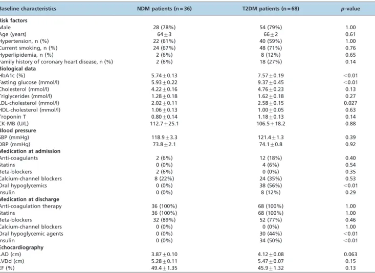
As shown in Table 1, all enrolled patients had high TnT and CK-MB levels; FBG and HbA1C levels were higher in patients with T2DM. As expected, the use of oral hypogly-cemic agents and/or insulin was more frequent among
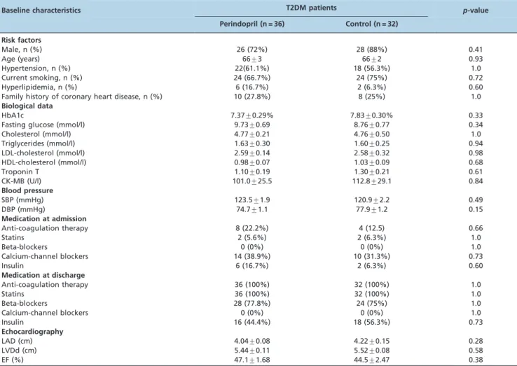
T2DM patients. Moreover, diabetic patients had slightly higher blood lipid levels, especially low-density lipoprotein (LDL)-cholesterol levels (p,0.05). There was a tendency for worse cardiac function in T2DM patients. Table 2 displays the baseline characteristics of the diabetic patients in the perindopril and control groups. The demographic charac-teristics and cardiovascular risk profiles were comparable between the two groups at admission. Irrespective of ACE inhibitor therapy, there was no difference in the medication used before and after hospital admission, as well as in cardiac structure and function.
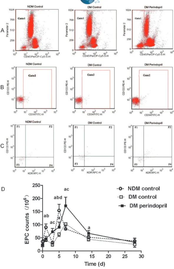
Circulating EPC levels post-AMI were increased in T2DM patients receiving ACE inhibitors
Figure 1 shows the changes in circulating CD452/low+
CD34+CD133+KDR+ EPC counts over time. In the NDM
patients, circulating EPCs were increased on day 1 after PCI and reached a peak on day 5, and these counts were significantly higher than the counts in the diabetic groups at the same time points (day 1: NDM controls: 90¡13/106
MNCs vs. T2DM controls: 43¡7/106 MNCs vs. treated
T2DM: 46¡10/106 MNCs, p,0.05 between the NDM and
T2DM groups; day 5: NDM controls: 154¡24/106MNCsvs. T2DM controls: 64¡9/106MNCsvs. treated T2DM: 91¡19/
106MNCs, p,0.05 between the NDM and two T2DM
Table 1 -Clinical characteristics of NDM and T2DM patients at admission.
Baseline characteristics NDM patients (n = 36) T2DM patients (n = 68) p-value
Risk factors
Male 28 (78%) 54 (79%) 1.00
Age (years) 64¡3 66¡2 0.61
Hypertension, n (%) 22 (61%) 40 (59%) 1.00
Current smoking, n (%) 24 (67%) 48 (71%) 0.76
Hyperlipidemia, n (%) 2 (6%) 8 (12%) 0.65
Family history of coronary heart disease, n (%) 2 (6%) 18 (27%) 0.14
Biological data
HbA1c (%) 5.74¡0.13 7.57¡0.19 ,0.01
Fasting glucose (mmol/l) 5.93¡0.22 9.37¡0.45 ,0.01
Cholesterol (mmol/l) 4.22¡0.16 4.76¡0.23 0.13
Triglycerides (mmol/l) 1.28¡0.18 1.62¡0.18 0.27
LDL-cholesterol (mmol/l) 2.02¡0.11 2.58¡0.15 0.027
HDL-cholesterol (mmol/l) 1.06¡0.13 1.00¡0.05 0.63
Troponin T 0.80¡0.14 1.18¡0.13 0.14
CK-MB (U/L) 112.7¡25.1 106.5¡18.2 0.88
Blood pressure
SBP (mmHg) 118.9¡3.3 121.4¡1.3 0.39
DBP (mmHg) 73.8¡2.1 74.1¡0.8 0.92
Medication at admission
Anti-coagulants 2 (6%) 12 (18%) 0.40
Statins 0 (0%) 4 (6%) 0.54
Beta-blockers 2 (6%) 0 (0%) 0.35
Calcium-channel blockers 8 (22%) 24 (35%) 0.53
Oral hypoglycemics 0 (0%) 38 (56%) ,0.01
Insulin 0 (0%) 8 (12%) 0.29
Medication at discharge
Anti-coagulation therapy 36 (100%) 68 (100%) 1.00
Statins 36 (100%) 68 (100%) 1.00
Beta-blockers 32 (89%) 52 (77%) 0.46
Calcium-channel blockers 0 (0%) 0 (0%) 1.00
Oral hypoglycemic agents 0 (0%) 30 (44%) ,0.01
Insulin 0 (0%) 34 (50%) ,0.01
Echocardiography
LAD (cm) 3.87¡0.10 4.12¡0.08 0.063
LVDd (cm) 5.28¡0.11 5.47¡0.07 0.15
EF (%) 49.4¡1.35 45.9¡1.32 0.13
groups). In patients with T2DM, the EPC peak was delayed to day 7, and the count was decreased in the T2DM controls (103¡23/106 MNCs). However, in perindopril-treated
T2DM patients, the EPC counts steadily increased from days 1 to 7 after AMI and were approximately 2.5-fold higher than the T2DM control counts on day 3, finally reaching the same level at a later time (day 7) compared with the peak of the NDM controls (day 5). Moreover, half a month after AMI, when circulating EPC counts fell quickly in all patients, counts in the perindopril group were still higher (67¡11/106MNCs) than those in controls (44¡13/ 106MNCs in the T2DM controls and 52¡7/106MNCs in the
NDM controls,p,0.05).
Plasma VEGF and SDF-1apost-AMI levels were
higher in diabetic patients receiving ACE inhibitor therapy, whereas the level of hsCRP was decreased
Before PCI, plasma VEGF and SDF-1a levels were
decreased in diabetic patients compared with the NDM subjects. Although the kinetics of VEGF over time were similar between NDM and T2DM controls, an apparent impairment in the kinetics on day 1 and day 5 (peak time) was observed in the T2DM controls. Perindopril treatment increased plasma VEGF from days 3 to 5, reaching a peak on day 7, which was much higher than the peak observed in
the T2DM controls (263.85¡39.75 vs. 147.24¡21.32 ng/l,
p,0.05) and similar to the peak of the NDM controls (209.48¡29.68 ng/l) (Figure 2A). Regarding plasma
SDF-1a levels, the NDM controls had the highest SDF-1a level
on day 5, but there was no significant difference in this level compared with that in the T2DM groups at the same time point. A notable decrease was observed on days 3 and 7 in the T2DM controls, but the SDF-1a levels were
increased by perindopril on day 7 (peak point) (treated T2DM: 2542.5¡103.7 ng/l on day 7 vs. NDM controls:
2114.9¡191.7 ng/l on day 5 vs. T2DM controls:
1784.7¡65.0 ng/l on day 5, p,0.05 between the treated T2DM and two control groups at the peak point) (Figure 2B). In addition, the T2DM controls exhibited a significant increase in plasma hsCRP levels from days 1 to 7, with a peak on day 3 after AMI. The changes in hsCRP levels in the NDM controls were almost the same as those in the T2DM controls, but the levels were much lower at all time points in the NDM controls. The beneficial effect of perindopril on this parameter was observed from day 3 (treated T2DM: 13.69¡2.86 mg/l vs. NDM: 23.84¡4.18 mg/l vs. T2DM controls: 78.50¡17.40 mg/l,
p,0.05 between the T2DM controls and the other two groups on day 3) and constantly decreased thereafter (Figure 2C).
Table 2 -Clinical characteristics of diabetic patients at admission.
Baseline characteristics T2DM patients p-value
Perindopril (n = 36) Control (n = 32)
Risk factors
Male, n (%) 26 (72%) 28 (88%) 0.41
Age (years) 66¡3 66¡2 0.93
Hypertension, n (%) 22(61.1%) 18 (56.3%) 1.0
Current smoking, n (%) 24 (66.7%) 24 (75%) 0.72
Hyperlipidemia, n (%) 6 (16.7%) 2 (6.3%) 0.60
Family history of coronary heart disease, n (%) 10 (27.8%) 8 (25%) 1.0
Biological data
HbA1c 7.37¡0.29% 7.83¡0.30% 0.33
Fasting glucose (mmol/l) 9.73¡0.69 8.76¡0.77 0.34
Cholesterol (mmol/l) 4.77¡0.21 4.76¡0.50 1.0
Triglycerides (mmol/l) 1.63¡0.30 1.60¡0.25 0.94
LDL-cholesterol (mmol/l) 2.59¡0.14 2.58¡0.32 0.98
HDL-cholesterol (mmol/l) 0.98¡0.07 1.03¡0.09 0.68
Troponin T 1.10¡0.19 1.30¡0.21 0.61
CK-MB (U/l) 101.0¡25.5 112.8¡29.1 0.84
Blood pressure
SBP (mmHg) 123.5¡1.9 120.9¡2.2 0.49
DBP (mmHg) 74.7¡1.1 77.9¡1.2 0.15
Medication at admission
Anti-coagulation therapy 8 (22.2%) 4 (12.5) 0.66
Statins 2 (5.6%) 2 (6.3%) 1.0
Beta-blockers 0 (0%) 0 (0%) 1.0
Calcium-channel blockers 14 (38.9%) 10 (31.3%) 0.73
Insulin 6 (16.7%) 2 (6.3%) 0.60
Medication at discharge
Anti-coagulation therapy 36 (100%) 32 (100%) 1.0
Statins 36 (100%) 32 (100%) 1.0
Beta-blockers 28 (77.8%) 24 (75%) 1.0
Calcium-channel blockers 0 (0%) 0 (0%) 1.0
Insulin 16 (44.4%) 18 (56.3%) 0.73
Echocardiography
LAD (cm) 4.04¡0.08 4.22¡0.15 0.28
LVDd (cm) 5.44¡0.11 5.52¡0.08 0.58
EF (%) 47.1¡1.68 44.5¡2.47 0.38
Figure 1 -Changes in circulating CD452/low+CD133+CD34+KDR+EPCs in NDM and T2DM patients with AMI. The EPC counts in the peripheral
blood were determined by flow cytometry before acute PCI and on days 1, 3, 5, 7, 14, and 28 after PCI. (A–C) Flow cytometric analysis of EPCs for each group. (A) CD452/low+EPC subset analysis was conducted on the Gate1 region using PerCP-Cy5.5-labeled antibodies against CD45. (B) The
proportion of CD34+cells was analyzed in the Gate2 region by flow cytometry using FITC-labeled antibodies against CD34. (C) The proportion
of CD133+KDR+cells was analyzed in the F2 region by two-color flow cytometry using PE-labeled antibodies against CD133 and APC-labeled
antibodies against KDR. (D) EPC kinetics over time for all three groups. a:p,0.05vs. the T2DM controls at the same time point; b:p,0.05vs. the
perindopril-treated T2DM group at the same time point; c:p,0.05vs.the NDM controls at the same time point; d:p,0.05vs. the T2DM
Post-AMI clinical outcomes were better in patients with T2DM receiving ACE inhibitor treatment
No patient was lost to follow-up during the 6-month period. There was no significant change in BP over time in the three groups (Figure 3A). The T2DM patients who did not receive ACE inhibitor treatment exhibited an increased left ventricular end-diastolic diameter (LVDd) and a decreased left ventricular EF (LVEF) at admission and follow-up compared with the NDM patients. The effect of perindopril on LVDd was limited, but LVEF was improved after 6 months of perindopril treatment (treated T2DM: 49.8¡1.5% vs. NDM controls:
50.7¡1.2% vs. T2DM controls: 43.3¡2.1%, p,0.01 between
the T2DM controls and the other two groups). There was no difference in left atrial diameter (LAD) and inter-ventricular septal thickness (IVST) between the three groups, but a change in the left ventricular posterior wall thickness (LVPWT) was observed in the T2DM patients receiving perindopril treat-ment. ACE inhibitor-treated subjects had an increased LVPWT over time, especially by month 6 (treated T2DM: 0.90¡0.03 cm
vs. T2DM controls: 0.81¡0.02 cm vs. NDM controls:
0.83¡0.02 cm,p,0.05 between the treated T2DM group and both control groups) (Figure 3B).
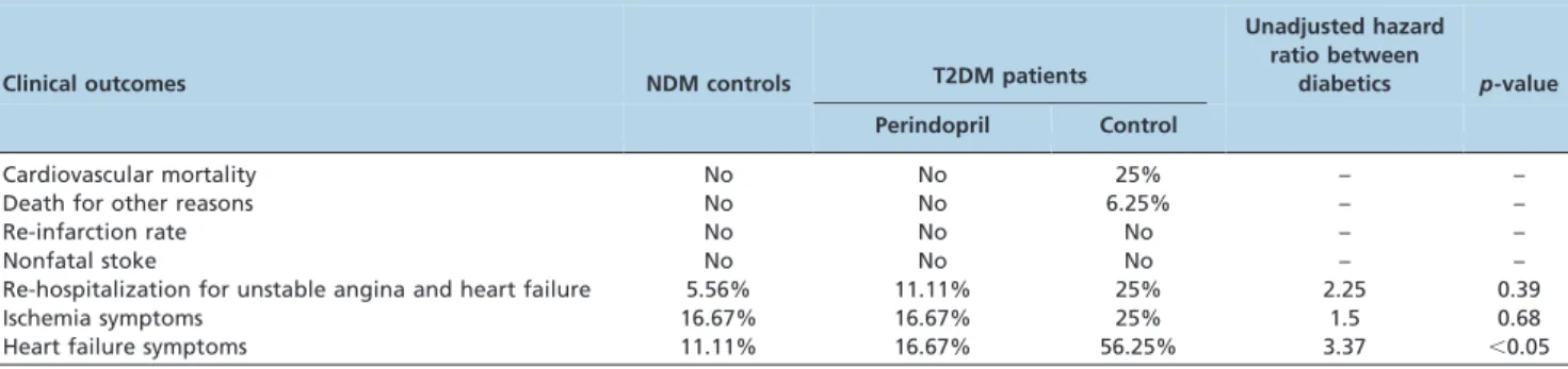
There were no deaths in the NDM control and perindo-pril-treated T2DM groups. The T2DM patients who did not receive ACE inhibitors had a higher mortality rate during follow-up (18.75%vs. 0%). Four of the patients died from acute congestive heart failure (two at 2 weeks, one at 5 weeks, and one at 8 weeks). Of the other two deaths, one was due to sudden death at 3 weeks, and the other was caused by a fatal subarachnoid hemorrhage 4 weeks post-AMI. No patient suffered a stroke or re-infarction. The control T2DM patients had higher rates of ischemia symptoms and re-hospitalization for unstable angina and heart failure, but the rate difference did not reach statistical significance. Most importantly, a higher proportion of T2DM controls compared with treated T2DM patients experienced heart failure symptoms (T2DM controls: 56.25%vs. treated T2DM: 16.67%,p,0.05) (Table 3).
& DISCUSSION
In this study, we observed that the circulating EPC levels were decreased in T2DM patients with AMI and that their
peak level was delayed compared with that in NDM patients. These observations are in accordance with the observed reduced plasma VEGF and SDF-1a levels,
increased plasma hsCRP level, and worse cardiac function and prognosis in T2DM patients compared with NDM patients. However, this impaired mobilization of bone-marrow EPCs was improved by ACE inhibition because perindopril-treated T2DM patients had an earlier and greater increase in bone-marrow EPCs compared with T2DM controls, which lasted up to 14 days post-AMI. Moreover, plasma VEGF, SDF-1a, and hsCRP levels were
also improved. Furthermore, perindopril-treated T2DM patients suffered a lower frequency of cardiovascular events, as demonstrated by a decreased cardiovascular mortality and occurrence of heart failure symptoms together with improved LVEF and left ventricular posterior wall hypertrophy.
Based on our results, increases in EPC counts due to perindopril administration in T2DM patients may play a pivotal role in improving prognosis after AMI. Because vessel endothelial cell injury induced by T2DM is regarded as the main reason for composite cardiovascular diseases, EPCs activated by perindopril could contribute to ongoing endothelial repairs by providing a circulating pool of cells that can home to injured parts of the artery or replace dysfunctional endothelial cells (22,23). Additionally, EPCs, once mobilized into the peripheral circulation, locate to the infarcted myocardium and are involved in the formation of new vessels (24,25), which will then improve the blood supply in ischemic penumbra, finally leading to decreased infarction area, improving LV function and reducing LV remodeling after AMI (26). As suggested by our results at even half a month after AMI, the numbers of circulating EPCs were significantly higher in the perindopril-treated T2DM group, contributing to a better EF and a decreased occurrence of heart failure symptoms.
Many studies suggest that ACE inhibition is an effective approach for the treatment of cardiovascular diseases in T2DM patients and in T2DM-associated vascular complica-tions, but the underlying mechanisms are not clearly understood (27,28). In our study, the significant EPC activation observed may be partly associated with the different effects of the ACE inhibitors themselves, including
Figure 2 -Changes in plasma VEGF, SDF-1a, and hsCRP levels in NDM and T2DM patients with AMI. Plasma samples were obtained from
1 ml of peripheral blood before acute PCI and on days 1, 3, 5, 7, 14, and 28 after PCI. VEGF, SDF-1a, and hsCRP analyses were performed
by an enzyme-linked immunosorbent assay. (A, B, and C) Plasma VEGF, SDF-1a, and hsCRP levels, respectively. a:p,0.05vs. the T2DM
inhibiting bradykinin degradation, increasing cGMP in vascular endothelial cells, restraining the renin-angiotensin system (RAS) activity, and decreasing the production of oxygen free radicals, which leads to sensitized NO synthase (NOS) and increased NO release. All these effects can in turn induce EPC mobilization from the bone marrow (29).
Furthermore, it has already been reported that circulating EPCs mainly originate from the bone marrow and that this mobilization involves a complex interaction between a number of cytokines and active molecules, such as the hypoxia inducible factor-1a (HIF-1a), VEGF, and SDF-1a.
These factors could participate in the regulation of EPC mobilization from the bone marrow and homing to the ischemic myocardium. Moreover, previous studies have also indicated that VEGF may be involved in ACE inhibitor-induced restoration of the myocardial structure (16,30,31). We observed that after taking perindopril, plasma VEGF and SDF-1alevels rose in post-AMI T2DM patients, and the
kinetics of these two proteins were very similar to the kinetics of the EPCs. Increased plasma VEGF can mobilize EPCs from the bone marrow for peripheral circulation through the activation of the KDR/Akt/eNOS pathway (32). SDF-1aalso plays a vital role in EPC mobilization and
homing. It has been reported that the insufficient release of
SDF-1a may lead to defective mobilization of EPCs in
diabetic rats (33). The only receptor of SDF-1a, CXCR4, is
highly expressed on the surface of CD34+hemopoietic stem
cells/progenitor cells. SDF-1acan then induce EPC
chemo-taxis in ischemic tissue via activation of the HIF pathway (18).
As a prototype marker of inflammation, CRP is now widely used to predict cardiac events in various cardiovas-cular diseases (34). In T2DM, insulin resistance is constantly associated with continuous, low-level inflammation, leading to increased serum hsCRP levels that finally results in impaired EPC levels and function (35). Recently, CRP has been shown to down-regulate endothelial NOS expression and to promote EPCs apoptosis through modulation of the receptor for advanced glycation end-products (RAGE) (36). Our previous study also demonstrated that following AMI, T2DM patients had low circulating EPC levels and increased plasma hsCRP levels, which may be due to the greater inflammation condition caused by more severe ischemia in T2DM. In addition, ACE inhibitors, as anti-inflammatory agents, have been found to decrease plasma hsCRP levels in patients with CAD (37). In the present study, our data revealed that post-AMI serum hsCRP levels were decreased by perindopril, thus decreasing
Figure 3 -Changes in blood pressure and cardiac function over time in NDM and T2DM patients after AMI. (A) Systolic and diastolic blood pressure; (B) Cardiac function examined by echocardiography, shown are LAD, LVDd, EF, IVST, and LVPWT.#p,0.05vs. the NDM
inflammation, which may be involved in improving ischemia or hypoxia conditions.
Study limitations
The present study has several limitations. First, because this was a single-center parallel controlled study with many exclusion criteria, this study had a small sample size of 104 subjects, and the results require further confirmation in larger cohorts. Second, the follow-up time was insufficient to observe cardiovascular events, especially congestive heart failure. However, the 6-month post-AMI period is the most important period for cardiac repair and cardiac function recovery. Third, in addition to circulating EPC counts, EPC function may also contribute to cardiovascular repair, but function was not measured in this study.
In this study, we demonstrated that perindopril treatment increased circulating EPC numbers from day 3 in post-AMI T2DM patients, and this increase lasted for at least half a month. These changes were accompanied by a similar increase in plasma VEGF and SDF-1alevels and a decrease
in serum hsCRP levels. As a result, higher levels of circulating EPCs may exert beneficial effects on cardiac function and clinical outcomes in T2DM patients. EPCs represent a novel treatment for post-AMI cardiovascular repair in T2DM patients.
& ACKNOWLEDGMENTS
This work was supported by grants from the National Natural Science Foundation of China (grant numbers 81070195, 81000055), the Jiangsu Key Laboratory for Molecular Medicine of Nanjing University (Research Grant 2008), and the Natural Science Foundation of Jiangsu (grant numbers GBK2009036, BK2010107). We thank Song Jie, Huang Wei, and Zhang Jing-mei for drawing blood before acute PCI and Liu Yong and Xue Jin-kui for conducting the flow cytometry analysis.
& AUTHOR CONTRIBUTIONS
Sun JY planned the study, selected patients, measured circulating EPC counts, performed the statistical analyses, and was involved in the preliminary writing of the manuscript. Zhai L was responsible for patient selection, clinical data collection, and patient follow-up. Li QL conducted the laboratory analyses and patient follow-up examinations. Ye JX was involved in the detection of circulating EPCs. Kang LN contributed to the study design and interpretation of data. Ye JX was involved in writing the manuscript. Xu B supervised the project and was involved in writing the manuscript. All the authors read and approved the final version of the manuscript.
& REFERENCES
1. Shihara M, Tsutsui H, Tsuchihashi M, Tada H, Kono S, Takeshita A. In-hospital and one-year outcomes for patients undergoing percutaneous
coronary intervention for acute myocardial infarction. Am J Cardiol. 2002;90(9):932-6, http://dx.doi.org/10.1016/S0002-9149(02)02656-5. 2. Morrow DA, Antman EM, Murphy SA, Qin J, Ruda M, Guneri S, et al.
Effect of enoxaparin versus unfractionated heparin in diabetic patients with ST-elevation myocardial infarction in the Enoxaparin and Thrombolysis Reperfusion for Acute Myocardial Infarction Treatment-Thrombolysis In Myocardial Infarction study 25 (ExTRACT-TIMI 25) trial. Am Heart J. 2007;154(6):1078-84, 84 e1, http://dx.doi.org/10.1016/j. ahj.2007.07.027.
3. Mak KH, Moliterno DJ, Granger CB, Miller DP, White HD, Wilcox RG, et al. Influence of diabetes mellitus on clinical outcome in the thrombolytic era of acute myocardial infarction. GUSTO-I Investigators. Global Utilization of Streptokinase and Tissue Plasminogen Activator for Occluded Coronary Arteries. J Am Coll Cardiol. 1997;30(1):171-9, http://dx.doi.org/10.1016/S0735-1097(97)00118-6.
4. Jaffe AS, Spadaro JJ, Schechtman K, Roberts R, Geltman EM, Sobel BE. Increased congestive heart failure after myocardial infarction of modest extent in patients with diabetes mellitus. Am Heart J. 1984;108(1):31-7, http://dx.doi.org/10.1016/0002-8703(84)90541-6.
5. Lamblin N, Fertin M, de Groote P, Bauters C. Cardiac remodeling and heart failure after a first anterior myocardial infarction in patients with diabetes mellitus. J Cardiovasc Med (Hagerstown). 2012;13(6):353-9, http://dx.doi.org/10.2459/JCM.0b013e328353694b.
6. Werner N, Priller J, Laufs U, Endres M, Bohm M, Dirnagl U, et al. Bone marrow-derived progenitor cells modulate vascular reendothelialization and neointimal formation: effect of 3-hydroxy-3-methylglutaryl coenzyme a reductase inhibition. Arterioscler Thromb Vasc Biol. 2002;22(10):1567-72, http://dx.doi.org/10.1161/01.ATV.0000036417.43987.D8.
7. Fadini GP, Miorin M, Facco M, Bonamico S, Baesso I, Grego F, et al. Circulating endothelial progenitor cells are reduced in peripheral vascular complications of type 2 diabetes mellitus. J Am Coll Cardiol. 2005;45(9):1449-57, http://dx.doi.org/10.1016/j.jacc.2004.11.067. 8. Massa M, Rosti V, Ferrario M, Campanelli R, Ramajoli I, Rosso R, et al.
Increased circulating hematopoietic and endothelial progenitor cells in the early phase of acute myocardial infarction. Blood. 2005;105(1):199-206, http://dx.doi.org/10.1182/blood-2004-05-1831.
9. Friedrich EB, Walenta K, Scharlau J, Nickenig G, Werner N. CD34-/ CD133+/VEGFR-2+ endothelial progenitor cell subpopulation with potent vasoregenerative capacities. Circ Res. 2006;98(3):e20-5, http:// dx.doi.org/10.1161/01.RES.0000205765.28940.93.
10. Ling L, Shen Y, Wang K, Jiang C, Fang C, Ferro A, et al. Worse clinical outcomes in acute myocardial infarction patients with type 2 diabetes mellitus: relevance to impaired endothelial progenitor cells mobilization. PLoS One. 2012;7(11):e50739, http://dx.doi.org/10.1371/journal.pone. 0050739.
11. You D, Cochain C, Loinard C, Vilar J, Mees B, Duriez M, et al. Combination of the angiotensin-converting enzyme inhibitor perindopril and the diuretic indapamide activate postnatal vasculogenesis in spontaneously hypertensive rats. J Pharmacol Exp Ther. 2008;325(3):766-73, http://dx.doi.org/10.1124/jpet.107.131532. 12. Muller P, Kazakov A, Jagoda P, Semenov A, Bohm M, Laufs U. ACE
inhibition promotes upregulation of endothelial progenitor cells and neoangiogenesis in cardiac pressure overload. Cardiovasc Res. 2009;83(1):106-14, http://dx.doi.org/10.1093/cvr/cvp123.
13. Cacciatore F, Bruzzese G, Vitale DF, Liguori A, de Nigris F, Fiorito C, et al. Effects of ACE inhibition on circulating endothelial progenitor cells, vascular damage, and oxidative stress in hypertensive patients. Eur J Clin Pharmacol. 2011;67(9):877-83.
14. Wang CH, Verma S, Hsieh IC, Chen YJ, Kuo LT, Yang NI, et al. Enalapril increases ischemia-induced endothelial progenitor cell mobilization through manipulation of the CD26 system. J Mol Cell Cardiol. 2006;41(1):34-43, http://dx.doi.org/10.1016/j.yjmcc.2006.03.006. 15. McConnell KJ, Humphries TL, Raebel MA, Merenich JA. Clinical
pharmacy specialist implementation of lisinopril therapy in patients with coronary artery disease and diabetes mellitus. Pharmacotherapy. 2003;23(12):1564-72, http://dx.doi.org/10.1592/phco.23.15.1564.31963.
Table 3 -Cardiovascular events during the 6 months of follow-up.
Clinical outcomes NDM controls T2DM patients
Unadjusted hazard ratio between
diabetics p-value
Perindopril Control
Cardiovascular mortality No No 25% – –
Death for other reasons No No 6.25% – –
Re-infarction rate No No No – –
Nonfatal stoke No No No – –
Re-hospitalization for unstable angina and heart failure 5.56% 11.11% 25% 2.25 0.39
Ischemia symptoms 16.67% 16.67% 25% 1.5 0.68
16. Toblli JE, Cao G, DeRosa G, Di Gennaro F, Forcada P. Angiotensin-converting enzyme inhibition and angiogenesis in myocardium of obese Zucker rats. Am J Hypertens. 2004;17(2):172-80.
17. Yamaguchi J, Kusano KF, Masuo O, Kawamoto A, Silver M, Murasawa S, et al. Stromal cell-derived factor-1 effects on ex vivo expanded endothelial progenitor cell recruitment for ischemic neovascularization. Circulation. 2003;107(9):1322-8, http://dx.doi.org/10.1161/01.CIR. 0000055313.77510.22.
18. Asahara T, Takahashi T, Masuda H, Kalka C, Chen D, Iwaguro H, et al. VEGF contributes to postnatal neovascularization by mobilizing bone marrow-derived endothelial progenitor cells. EMBO J. 1999;18(14):3964-72, http://dx.doi.org/10.1093/emboj/18.14.3964.
19. Moher D, Schulz KF, Altman D. The CONSORT statement: revised recommendations for improving the quality of reports of parallel-group randomized trials. JAMA. 2001;285(15):1987-91, http://dx.doi.org/10. 1001/jama.285.15.1987.
20. Case J, Mead LE, Bessler WK, Prater D, White HA, Saadatzadeh MR, et al. Human CD34+AC133+VEGFR-2+cells are not endothelial progenitor cells but distinct, primitive hematopoietic progenitors. Exp Hematol. 2007;35(7):1109-18, http://dx.doi.org/10.1016/j.exphem.2007.04.002. 21. Timmermans F, Van Hauwermeiren F, De Smedt M, Raedt R, Plasschaert
F, De Buyzere ML, et al. Endothelial outgrowth cells are not derived from CD133+ cells or CD45+ hematopoietic precursors. Arterioscler Thromb Vasc Biol. 2007;27(7):1572-9, http://dx.doi.org/10.1161/ ATVBAHA.107.144972.
22. Griese DP, Ehsan A, Melo LG, Kong D, Zhang L, Mann MJ, et al. Isolation and transplantation of autologous circulating endothelial cells into denuded vessels and prosthetic grafts: implications for cell-based vascular therapy. Circulation. 2003;108(21):2710-5, http://dx.doi.org/10. 1161/01.CIR.0000096490.16596.A6.
23. Hill JM, Zalos G, Halcox JP, Schenke WH, Waclawiw MA, Quyyumi AA, et al. Circulating endothelial progenitor cells, vascular function, and cardiovascular risk. N Engl J Med. 2003;348(7):593-600.
24. Thal MA, Krishnamurthy P, Mackie AR, Hoxha E, Lambers E, Verma S, et al. Enhanced angiogenic and cardiomyocyte differentiation capacity of epigenetically reprogrammed mouse and human endothelial progenitor cells augments their efficacy for ischemic myocardial repair. Circ Res. 2012;111(2):180-90, http://dx.doi.org/10.1161/CIRCRESAHA.112.270462. 25. Westenbrink BD, Oeseburg H, Kleijn L, van der Harst P, Belonje AM,
Voors AA, et al. Erythropoietin stimulates normal endothelial progenitor cell-mediated endothelial turnover, but attributes to neovascularization only in the presence of local ischemia. Cardiovasc Drugs Ther. 2008;22(4):265-74, http://dx.doi.org/10.1007/s10557-008-6094-y. 26. Davidson EP, Coppey LJ, Holmes A, Yorek MA. Effect of inhibition of
angiotensin converting enzyme and/or neutral endopeptidase on
vascular and neural complications in high fat fed/low dose streptozo-tocin-diabetic rats. Eur J Pharmacol. 2012;677(1-3):180-7.
27. Gerc V, Buksa M. Advantages of renin-angiotensin system blockade in the treatment of cardiovascular diseases. Med Arh. 2010;64(5):295-9. 28. Rahman S, Ismail AA, Rahman AR. Treatment of diabetic vasculopathy
with rosiglitazone and ramipril: Hype or hope? Int J Diabetes Dev Ctries. 2009;29(3):110-7, http://dx.doi.org/10.4103/0973-3930.54287.
29. Aicher A, Heeschen C, Mildner-Rihm C, Urbich C, Ihling C, Technau-Ihling K, et al. Essential role of endothelial nitric oxide synthase for mobilization of stem and progenitor cells. Nat Med. 2003;9(11):1370-6, http://dx.doi.org/10.1038/nm948.
30. Zoja C, Cattaneo S, Fiordaliso F, Lionetti V, Zambelli V, Salio M, et al. Distinct cardiac and renal effects of ETA receptor antagonist and ACE inhibitor in experimental type 2 diabetes. Am J Physiol Renal Physiol. 2011;301(5):F1114-23, http://dx.doi.org/10.1152/ajprenal.00122.2011. 31. Infanger M, Faramarzi S, Grosse J, Kurth E, Ulbrich C, Bauer J, et al.
Expression of vascular endothelial growth factor and receptor tyrosine kinases in cardiac ischemia/reperfusion injury. Cardiovasc Pathol. 2007;16(5):291-9, http://dx.doi.org/10.1016/j.carpath.2007.04.001. 32. Dong L, Kang L, Ding L, Chen Q, Bai J, Gu R, et al. Insulin modulates
ischemia-induced endothelial progenitor cell mobilization and neovas-cularization in diabetic mice. Microvasc Res. 2011;82(3):227-36. 33. Hattori K, Heissig B, Tashiro K, Honjo T, Tateno M, Shieh JH, et al.
Plasma elevation of stromal cell-derived factor-1 induces mobilization of mature and immature hematopoietic progenitor and stem cells. Blood. 2001;97(11):3354-60, http://dx.doi.org/10.1182/blood.V97.11.3354. 34. Ridker PM. Clinical application of C-reactive protein for cardiovascular
disease detection and prevention. Circulation. 2003;107(3):363-9, http:// dx.doi.org/10.1161/01.CIR.0000053730.47739.3C.
35. Verma S, Kuliszewski MA, Li SH, Szmitko PE, Zucco L, Wang CH, et al. C-reactive protein attenuates endothelial progenitor cell survival, differentiation, and function: further evidence of a mechanistic link between C-reactive protein and cardiovascular disease. Circulation. 2004;109(17):2058-67, http://dx.doi.org/10.1161/01.CIR.0000127577.63323. 24.
36. Chen J, Jin J, Song M, Dong H, Zhao G, Huang L. C-reactive protein down-regulates endothelial nitric oxide synthase expression and promotes apoptosis in endothelial progenitor cells through receptor for advanced glycation end-products. Gene. 2012;496(2):128-35, http://dx. doi.org/10.1016/j.gene.2011.12.039.