BASIC RESEARCH
Effects of diabetes on myocardial capillary density
and serum angiogenesis biomarkers in male rats
Majid Khazaei,IAli Reza Fallahzadeh,I Mohammad Reza Sharifi,INoushin Afsharmoghaddam,II Shaghayegh Haghjooy Javanmard,IIIEnsieh SalehiI
IDepartment of Physiology, Isfahan University of Medical Sciences, Isfahan, Iran.IIDepartment of Pathology, Isfahan University of Medical Sciences, Isfahan, Iran.IIIPhysiology Research Center, Department of Physiology, Isfahan University of Medical Sciences, Isfahan, Iran.
INTRODUCTION: Cardiovascular disease is one of the main causes of mortality and morbidity in diabetic patients. This study evaluated the effects of diabetes on myocardial capillary density and several serum angiogenic factors including nitric oxide, vascular endothelial growth factor, and soluble vascular endothelial growth factor receptors.
METHODS:Twelve male rats were divided into two groups: control and diabetic (n= 6 each). Diabetes was induced
with a single dose of streptozotocin (50 mg/kg). After 21 days, capillary density in the myocardial tissue was evaluated using immunohistochemical staining and is reported as capillaries per mm2. Blood samples were collected before and after the induction of diabetes.
RESULTS: In the diabetic group, serum nitric oxide and soluble vascular endothelial growth factor receptor 2 concentrations were lower than the levels in the control group, while the level of soluble vascular endothelial growth factor receptor 1 was significantly higher. There was no significant change in the serum vascular endothelial growth factor concentration between the diabetic and control groups; however, the ratio of vascular endothelial growth factor to vascular endothelial growth factor receptor 1 was significantly lower in the diabetic animals. The myocardial capillary density was also lower in the diabetic group compared with the control group (1549¡161vs.
2156¡202/mm2, respectively).
CONCLUSION:Reduced serum nitric oxide and vascular endothelial growth factor receptor 2 levels, increased serum vascular endothelial growth factor receptor 1 levels and a lower vascular endothelial growth factor to vascular endothelial growth factor receptor 1 ratio may be responsible for the decreased myocardial capillary density in diabetic rats.
KEYWORDS: Diabetes; Capillary Density; Vascular Endothelial Growth Factor; Nitric Oxide; Myocardium.
Khazaei M, Fallahzadeh AR, Sharifi MR, Afsharmoghaddam N, Javanmard SH, Salehi E. Effects of diabetes on myocardial capillary density and serum angiogenesis biomarkers in male rats. Clinics. 2011;66(8):1419-1424.
Received for publication onDecember 23, 2010;First review completed onFebruary 21, 2011;Accepted for publication onApril 26, 2011 E-mail: [email protected]
Tel.: 98 3117922417
INTRODUCTION
Cardiovascular disease is one of the main causes of mortality and morbidity in diabetic subjects, and several long-term complications of diabetes involve impaired angiogenesis. Angiogenesis is the growth of new capillaries from pre-existing vessels1 and occurs during physiological conditions such as wound healing. However, abnormal angiogenesis may occur in some pathological conditions, including diabetic retinopathy.1
Nitric oxide (NO) and vascular endothelial growth factor (VEGF) are important factors affecting angiogenesis.2,3 NO has important roles in multiple physiological processes and pathological states,4,5and VEGF and its receptors have essen-tial roles in pathological and physiological angiogenesis.2,6
The two VEGF receptor subtypes that are primarily involved in angiogenesis are VEGF receptor type 1 (VEGFR-1) and VEGF receptor type 2 (VEGFR-2). Soluble VEGFR-1 (sVEGFR-1) binds with a high affinity to VEGF and reduces VEGF activity and angiogenesis,6-8 whereas VEGFR-2 has proangiogenic effect in the angiogenesis process.9 Increased VEGF-mediated angiogenesis in dia-betic retinopathy and nephropathy and a decreased angiogenic response in wound healing and ulcers have been documented in diabetic subjects.10,11
This study aimed to evaluate the effect of diabetes on the serum concentrations of VEGF, NO and the soluble forms of VEGFR-1 and VEGFR-2 (sVEGFR-1 and sVEGFR-2) as well as myocardial capillary density in type I diabetic rats.
METHODS
Animals
Male Wistar rats (aged 10–12 weeks, weighing 230¡30 g) were used in the present study. The animals were kept two Copyrightß2011CLINICS– This is an Open Access article distributed under
per cage at room temperature (20–25
˚
C) with a 12 h light/ dark cycle and ad libitum access to food and water. The experimental protocols were approved by the ethics committee of Isfahan University of Medical Sciences.Induction of Diabetes
After one week of adaptation to the animal room, the rats were intraperitoneally injected with a single 50 mg/kg dose of streptozotocin (STZ, Sigma) to induce diabetes.12,13 STZ was dissolved in cold, normal saline and administered immediately. Two days after injection, the fasting blood glucose levels were determined. Animals with blood glucose greater than 300 mg/dl were considered dia-betic.12,13In the control group, normal saline was injected.
Experimental Design
The rats were divided into normal and diabetic groups (n= 6 each). Blood samples were taken before and after the induction of diabetes. The samples were centrifuged, and
the serum was kept at -70
˚
C until the serum VEGF, NO, sVEGFR-1, and sVEGFR-2 concentrations were measured. After 21 days, the animals were euthanized. The apex of the heart was removed, washed with normal saline and fixed in formalin solution for immunohistochemical evaluation. Body weight was measured before induction of diabetes and 21 days after the experiment.Serum VEGF, sVEGFR-1, sVEGFR-2, and NO Measurements
Serum VEGF, sVEGFR-1, and sVEGFR-2 concentrations were measured using the quantitative sandwich enzyme immunoassay technique with the appropriate kits (R&D Systems, Minneapolis, USA).14,15The serum nitrite concen-tration, one of the main metabolites of NO, was determined using the Griess reagent method (Promega, Madison, USA).16
Immunohistochemistry
Capillary density was measured using immunohistochem-istry with a rat monoclonal antibody directed against mouse CD31 (Abcam, Cambridge UK).17 Fifteen fields of three different sections from each cardiac tissue sample were randomly examined using a light microscope (4006) by two blinded observers. The number of capillaries in each tissue sample is reported as the number of capillaries per mm2.
Statistical Analysis
All values are reported as the mean¡SE. Student’st-test
was used to evaluate differences between the two groups. Paired data were used for the data analysis before and after
Figure 1 -Serum nitrite concentration in control and diabetic rats before and after the induction of diabetes. *p,0.05.
Table 1 -Fasting blood glucose level (mg/dl) and body weight (g) before and after the induction of diabetes.
Group n Body Weight (g)
Fasting Blood Glucose Level (mg/dl)
Day 0 Day 21 Day 0 Day 21
control 6 245¡4.8 285.71¡7.4* 122.57¡3.5 117.43¡5.4 diabetic 6 240¡7.6 173¡9.6* 125¡6.0 508.6¡25.4*
*p,0.05 versus day 0.
Effects of diabetes on myocardial capillary density
the induction of diabetes. Bivariate correlations were calculated using Pearson’s correlation coefficient. Ap-value less than 0.05 was considered statistically significant.
RESULTS
Blood glucose and body weight
Table 1 shows the fasting blood glucose levels and body weight in the two groups. Body weight significantly increased over the course of the experiment in the control group, whereas the body weight of diabetic rats decreased over time (p,0.05). The fasting blood glucose level in the diabetic animals was higher than the level in the controls and remained elevated throughout the experiment.
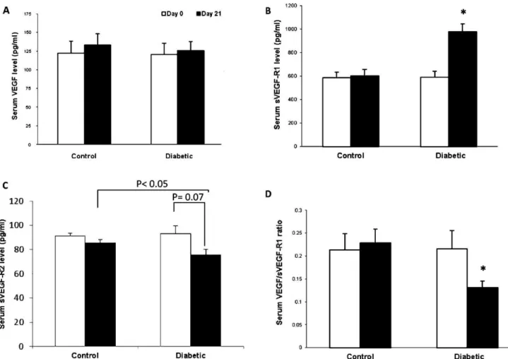
Effects of diabetes on serum angiogenic factors Figure 1 illustrates the serum NO concentration in the two groups before and after the induction of diabetes. In the diabetic group, the serum NO level was significantly decreased at the end of experiment when compared with the level prior to the induction of diabetes (p,0.05; Figure 1). While there was no significant difference in the serum VEGF concentration between the groups (Figure 2A), the serum sVEGFR-1 concentration was increased and the sVEGFR-2 level was decreased in the diabetic group compared with the control group (p,0.05; Figure 2B and C). In addition, the VEGF:sVEGFR-1 ratio in diabetic animals was lower than the ratio in the controls (p,0.05; Figure 2D).
Effect of diabetes on myocardial capillary density The capillary density in the myocardial tissue (expressed as the number of capillaries per mm2) was significantly reduced in the diabetic group compared with the control group (p,0.05) (Figure 3B). Representative images of the immunohistochemical staining are presented in figure 3A.
Correlation analysis
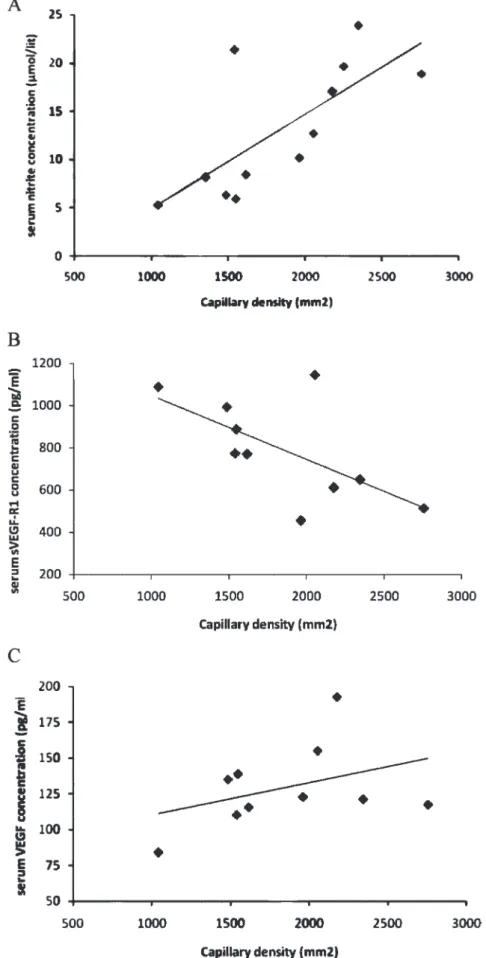
In the correlation analysis, we found that capillary density in myocardial tissue was positively correlated with the serum nitrite level (r= 0.70), the VEGF level (r= 0.40), the sVEGFR-1 level (r= -0.69) and the VEGF:sVEGFR-1 ratio (r= 0.71) (Figure 4).
DISCUSSION
angiogenic growth factors affect angiogenesis as well by increasing NO production.19A decrease in NO production in endothelial NO synthase–deficient mice disrupts the development of the coronary vessels and angiogenesis.20In the present study, the serum NO concentration in diabetic rats was lower than the concentration in the control group, which supports the results of previous studies.21-24 Some possible mechanisms responsible for decreased NO produc-tion under high glucose condiproduc-tions include the suppression of endothelial NO synthase expression and activity,25 overproduction of superoxides,26and activation of protein kinase C.23
VEGF has been shown to be an important inducer of angiogenesis in a variety of in vivo models.6 Several experimental and clinical reports examining the effect of diabetes on the serum or plasma VEGF level have been published, and the results are contradictory: an increase,27-29 decrease,30,31and no change32,33in the VEGF levels in type I and II diabetes have been reported. In the present study, although the capillary density was lower in diabetic animals, the serum VEGF concentration was similar to that in control animals. It has been suggested that even with an increase or no change in the VEGF level, the VEGF signaling pathway is defective during diabetes, and diabetes is considered VEGF resistant.29,34 In a recent study, Hazarika et al. showed that in the absence of ischemia, VEGF signaling in diabetic mice was lower than that in control mice.35 They also showed that in the absence of
ischemia, diabetic mice had increased VEGF and sVEGFR-1 levels and decreased AKT/AKT and phospho-endothelial NO synthase/phospho-endothelial NO synthase levels. Lu et al. also found that the post-ischemic capillary density and revascularization in the limbs of diabetic rats was decreased, with reduced mRNA and protein expression of eNOS, VEGF, and bFGF.31However, despite the similarity in serum VEGF levels between the groups, we found that diabetic animals had higher sVEGFR-1 and lower sVEGFR-2 levels compared with the controls. sVEGFR-1 binds with a high affinity to VEGF9and reduces VEGF activity, endothe-lial cell proliferation, angiogenesis and tumor growth.6-8 Thus, sVEGFR-1 acts as an antagonist of VEGF.36 In contrast, VEGF-R2 is an effector of proangiogenic signaling in the angiogenesis process.9Therefore, reduced sVEGFR-2 levels and increased sVEGFR-1 levels in the serum may be responsible for the lower capillary density in the myocardial tissue of diabetic rats. We also investigated the VEGF to sVEGFR-1 ratio, as some studies have used the VEGF:sVEGFR-1 ratio as an indicator of angiogenesis status.37,38 In the present study, we found that the VEGF:sVEGFR-1 ratio was reduced in diabetic rats, which suggests decreased angiogenesis in the diabetic group.
In conclusion, myocardial capillary density was lower in the diabetic group compared with the control group. Furthermore, the lower serum levels of the proangiogenic factors NO and sVEGFR-2, the higher level of the antiangiogenic factor sVEGFR-1 and the reduced VEGF:sVEGFR-1 ratio may Figure 3 -Effect of diabetes on the myocardial capillary density. A. Representative images of the immunohistochemical staining (4006)
with an anti-CD31 monoclonal antibody. B. Myocardial capillary density in the control and diabetic groups.n= 6 per group. *p,0.05. Effects of diabetes on myocardial capillary density
Figure 4 -Correlation between the capillary density in myocardial tissue and the serum nitrite (r= 0.70) (A), sVEGFR-1 (r = -0.69) (B), and
explain the reduced coronary angiogenesis observed in diabetic animals.
ACKNOWLEDGMENTS
This study was supported by a grant from Isfahan University of Medical Sciences (grant number: 188103).
REFERENCES
1. Carmeliet P. Angiogenesis in health and disease. Nat Med. 2003;9:653-60, doi: 10.1038/nm0603-653.
2. Folkman J. Angiogenesis. Annu Rev Med. 2006;57:1-18, doi: 10.1146/ annurev.med.57.121304.131306.
3. Milkiewicz M, Hudlicka O, Brown MD, Silgram H. Nitric oxide, VEGF, and VEGFR-2: interactions in activity-induced angiogenesis in rat skeletal muscle. Am J Physiol Heart Circ Physiol. 2005;289:H336-H343, doi: 10.1152/ajpheart.01105.2004.
4. Cengel A, Sahinarslan A. Nitric oxide and cardiovascular system. Anadolu Kardiyol Derg. 2006;6:364-8.
5. Tuck ML. Nitric oxide in diabetes mellitus. J Hypertens. 2003;21:1081-3, doi: 10.1097/00004872-200306000-00005.
6. Olsson AK, Dimberg A, Kreuger J, Claesson-Welsh L. VEGF receptor signalling - in control of vascular function. Nat Rev Mol Cell Biol. 2006;7:359-71, doi: 10.1038/nrm1911.
7. Hasumi Y, Mizukami H, Urabe M, Kohno T, Takeuchi K, Kume A, et al. Soluble FLT-1 expression suppresses carcinomatous ascites in nude mice bearing ovarian cancer. Cancer Res. 2002;62:2019-23.
8. Roberts DM, Kearney JB, Johnson JH, Rosenberg MP, Kumar R, Bautch VL. The vascular endothelial growth factor (VEGF) receptor Flt-1 (VEGFR-1) modulates Flk-1 (VEGFR-2) signaling during blood vessel formation. Am J Pathol. 2004;164:1531-5, doi: 10.1016/S0002-9440(10) 63711-X.
9. Wu FT, Stefanini MO, Mac GF, Kontos CD, Annex BH, Popel AS. A systems biology perspective on sVEGFR1: its biological function, pathogenic role and therapeutic use. J Cell Mol Med. 2010;14:528-52. 10. Brem H, Tomic-Canic M. Cellular and molecular basis of wound healing
in diabetes. J Clin Invest. 2007;117:1219-22, doi: 10.1172/JCI32169. 11. Folkman J. Angiogenesis in cancer, vascular, rheumatoid and other
disease. Nat Med. 1995;1:27-31, doi: 10.1038/nm0195-27.
12. Movahedian A. Antihyperlipidemic effect of peucedanum pastinacifo-lium extract in streptozotocin-induced diabetic rats. Clinics. 2010;65:629-33.
13. Budin SB. The effects of palm oil tocotrienol-rich fraction supplementa-tion on biochemical parameters, oxidative stress and the vascular wall of streptozotocin-induced diabetic rats. Clinics. 2009;64:235-44, doi: 10. 1590/S1807-59322009000300015.
14. Carlstrom M. Angiogenesis inhibition causes hypertension and placental dysfunction in a rat model of preeclampsia. J.Hypertens. 2009;27:829-37, doi: 10.1097/HJH.0b013e328324f8ce.
15. Pratheeshkumar P. Vernolide-A inhibits tumour specific angiogenesis by regulating proinflammatory cytokines, VEGF, MMPs and TIMP. Eur J Pharmacol. 2011;656:10-8, doi: 10.1016/j.ejphar.2010.12.041. 16. Nematbakhsh M. The effect of estrogen on serum nitric oxide
concentra-tions in normotensive and DOCA Salt hypertensive ovariectomized rats. Clin Chim Acta. 2004;344:53-7, doi: 10.1016/j.cccn.2004.01.019.
17. Li P. Role of bradykinin, nitric oxide, and angiotensin II type 2 receptor in imidapril-induced angiogenesis. Hypertension. 2008;51:252-58, doi: 10. 1161/HYPERTENSIONAHA.107.097394.
18. Cooke JP. NO and angiogenesis. Atheroscler Suppl. 2003;4:53-60, doi: 10. 1016/S1567-5688(03)00034-5.
19. Hood JD, Meininger CJ, Ziche M, Granger HJ. VEGF upregulates ecNOS message, protein, and NO production in human endothelial cells. Am J Physiol. 1998;274(3 Pt 2):H1054-H1058.
20. Zhao X, Lu X, Feng Q. Deficiency in endothelial nitric oxide synthase impairs myocardial angiogenesis. Am J Physiol Heart Circ Physiol. 2002;283:H2371-H2378.
21. Lash JM, Nase GP, Bohlen HG. Acute hyperglycemia depresses arteriolar NO formation in skeletal muscle. Am J Physiol. 1999;277:H1513-H1520. 22. Brodsky SV, Morrishow AM, Dharia N, Gross SS, Goligorsky MS.
Glucose scavenging of nitric oxide. Am J Physiol Renal Physiol. 2001;280:F480-F486.
23. Bohlen HG, Nase GP. Arteriolar nitric oxide concentration is decreased during hyperglycemia-induced betaII PKC activation. Am J Physiol Heart Circ Physiol. 2001;280:H621-H627.
24. Silva AM, Penno Lde M, Bertoluci MC, Irigoyen MC, Schaan BD. Insulin therapy does not interfere with venous endothelial function evaluation in patients with type 2 diabetes mellitus. Clinics. 2010;65:1139-42, doi: 10. 1590/S1807-59322010001100015.
25. Ding Y, Vaziri ND, Coulson R, Kamanna VS, Roh DD. Effects of simulated hyperglycemia, insulin, and glucagon on endothelial nitric oxide synthase expression. Am J Physiol Endocrinol Metab. 2000; 279:E11-E17.
26. Bagi Z, Toth E, Koller A, Kaley G. Microvascular dysfunction after transient high glucose is caused by superoxide-dependent reduction in the bioavailability of NO and BH(4). Am J Physiol Heart Circ Physiol. 2004;287:H626-H633, doi: 10.1152/ajpheart.00074.2004.
27. Blann AD, Belgore FM, McCollum CN, Silverman S, Lip PL, Lip GY. Vascular endothelial growth factor and its receptor, Flt-1, in the plasma of patients with coronary or peripheral atherosclerosis, or Type II diabetes. Clin Sci (Lond). 2002;102:187-94, doi: 10.1042/CS20010178. 28. Li Y, Hazarika S, Xie D, Pippen AM, Kontos CD, Annex BH. In mice with
type 2 diabetes, a vascular endothelial growth factor (VEGF)-activating transcription factor modulates VEGF signaling and induces therapeutic angiogenesis after hindlimb ischemia. Diabetes. 2007;56:656-65, doi: 10. 2337/db06-0999.
29. Sasso FC, Torella D, Carbonara O, Ellison GM, Torella M, Scardone M, et al. Increased vascular endothelial growth factor expression but impaired vascular endothelial growth factor receptor signaling in the myocardium of type 2 diabetic patients with chronic coronary heart disease. J Am Coll Cardiol. 2005;46:827-34, doi: 10.1016/j.jacc.2005.06.007. 30. Chou E, Suzuma I, Way KJ, Opland D, Clermont AC, Naruse K, et al. Decreased cardiac expression of vascular endothelial growth factor and its receptors in insulin-resistant and diabetic States: a possible explana-tion for impaired collateral formaexplana-tion in cardiac tissue. Circulaexplana-tion. 2002;105:373-9, doi: 10.1161/hc0302.102143.
31. Gao L, Yu DM. Molecular mechanism of limbs’ postischemic revascular-ization improved by perindopril in diabetic rats. Chin Med J (Engl). 2008;121:2129-33.
32. Kivela R, Silvennoinen M, Lehti M, Jalava S, Vihko V, Kainulainen H. Exercise-induced expression of angiogenic growth factors in skeletal muscle and in capillaries of healthy and diabetic mice. Cardiovasc Diabetol. 2008;7:13, doi: 10.1186/1475-2840-7-13.
33. Larger E, Marre M, Corvol P, Gasc JM. Hyperglycemia-induced defects in angiogenesis in the chicken chorioallantoic membrane model. Diabetes. 2004;53:752-61, doi: 10.2337/diabetes.53.3.752.
34. Waltenberger J. VEGF resistance as a molecular basis to explain the angiogenesis paradox in diabetes mellitus. Biochem Soc Trans. 2009;37:1167-70.
35. Hazarika S, Dokun AO, Li Y, Popel AS, Kontos CD, Annex BH. Impaired angiogenesis after hindlimb ischemia in type 2 diabetes mellitus: differential regulation of vascular endothelial growth factor receptor 1 and soluble vascular endothelial growth factor receptor 1. Circ Res. 2007;101:948-56, doi: 10.1161/CIRCRESAHA.107.160630.
36. Ambati BK, Nozaki M, Singh N, Takeda A, Jani PD, Suthar T, et al. Corneal avascularity is due to soluble VEGF receptor-1. Nature. 2006;443:993-7, doi: 10.1038/nature05249.
37. Chang YT, Chang MC, Wei SC, Tien YW, Hsu C, Liang PC, et al. Serum vascular endothelial growth factor/soluble vascular endothelial growth factor receptor 1 ratio is an independent prognostic marker in pancreatic cancer. Pancreas. 2008;37:145-50, doi: 10.1097/MPA.0b013e318164548a. 38. Bando H, Weich HA, Brokelmann M, Horiguchi S, Funata N, Ogawa T,
et al. Association between intratumoral free and total VEGF, soluble VEGFR-1, VEGFR-2 and prognosis in breast cancer. Br J Cancer. 2005;92:553-61.
Effects of diabetes on myocardial capillary density