CASE REPORT
Scapular winging in Poland syndrome
Murat Uludag,I,IIHasan Cece,III Serap Incebiyik,II Ahmet Demirkol,IIEkrem Karakas,IVKenan AkgunI IDepartment of Physical Medicine and Rehabilitation, I˙stanbul University Cerrahpas¸a Medical Faculty, Istanbul, Turkey.IIDepartment of Physical Medicine
and Rehabilitation, Harran University Medical Faculty, Sanliurfa, Turkey.IIIDepartment of Radiology, Harran University Medical Faculty, Harran, Turkey. IVDepartment of Radiology, Sanliurfa Education and Research Hospital, Sanliurfa, Turkey.
Email: [email protected] Tel.: 90 50 59247871
INTRODUCTION
Poland syndrome (PS), which was first described in 1841 by Sir Alfred Poland, is characterized by the unilateral absence of the sternal head of the pectoralis major muscle, hypoplasia of the rib cage and upper extremities, breast and nipple hypoplasia or aplasia, and scoliosis.1
The majority of the reported cases are sporadic; however, the disease may be inherited as an autosomal dominant trait.2This disease tends to occur more frequently in males
and most often involves the right side of the body.3 Scapular winging is a rare but potentially debilitating condition that impairs a person’s ability to perform his or her activities of daily living.4Postural deformities can also
negatively impact one’s quality of life during childhood and adulthood.5
The current work presents the case study of an 18-year-old youth with scapular winging that was associated with PS. In this work, we also describe the pain that was associated with PS, which is rarely mentioned in cases of PS.
Case Report
An 18-year-old youth with no history of trauma presented with complaints of pain and limited left shoulder activity over the previous two years.
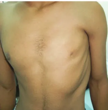
Upon physical examination, the left nipple and areola were observed to be hypoplastic and lightly pigmented (Fig. 1). In addition, thoracic vertebral scoliosis and an elevated scapula were observed (Fig. 2). Active myofascial trigger points were detected in the left upper trapezius, levator scapula, and infraspinatus muscles. The left shoulder exhibited a limited range of motion and was painful during flexion and abduction when compared to the right shoulder.
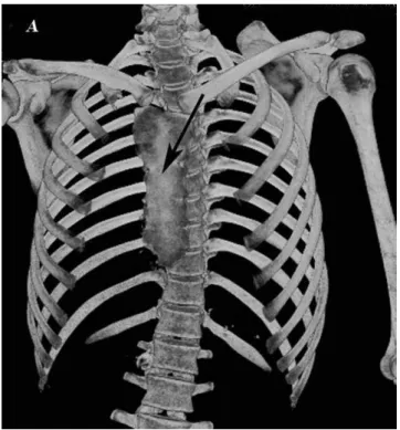
The laboratory findings were normal. Three-dimensional thoracic (3-D) computed tomography (CT) revealed a sternal rotational anomaly, thoracic scoliosis, and an elevated scapula (Fig. 3). An axial sequence in the thoracic CT revealed the absence of the left pectoralis major and minor muscles (Fig. 4).
The patient was treated with an analgesic, a non-steroidal anti-inflammatory drug, and physical therapy (including exercises for the scapular stabilizer muscles and the shoulder girdle muscles), which improved his clinical signs.
At his two-month follow-up, the patient reported no recurrence of pain or physical limitations.
DISCUSSION
The present case study was a case of undiagnosed PS despite two years of severe shoulder and back pain and functional limitations in the patient’s left upper extremity. The thoracic anomalies of PS (an absent pectoral muscles, a hypoplastic or absent breast and nipple, rib hypoplasia, pectus excavatum, pectus carinatum, elevated scapula, and scoliosis) have been previously reported in the absence of hand anomalies.6Isolated deformities occur with a greater frequency than the entire collection of anomalies that are observed in PS patients.7 The patient in the present case exhibited an absence of the pectoral muscles, breast and nipple hypoplasia, an elevated scapula, thoracic scoliosis, and a winging scapula due to serratus anterior muscle hypoplasia.
3-D thoracic CT is the modality of choice in cases for which a correction of the chest wall deformity is necessary, especially in patients with missing ribs or sternal rotational
Copyrightß2011CLINICS– This is an Open Access article distributed under
the terms of the Creative Commons Attribution Non-Commercial License (http:// creativecommons.org/licenses/by-nc/3.0/) which permits unrestricted non-commercial use, distribution, and reproduction in any medium, provided the original work is properly cited.
Figure 1 - The absence of the pectoralis muscles and nipple hypoplasia on the left side. Also note the hypoplasia of the left serratus anterior muscle.
CLINICS 2011;66(5):929-930 DOI:10.1590/S1807-59322011000500037
anomalies.8In the present case, the absence of the pectoralis major and minor muscles, serratus anterior muscle hypoplasia, sternal rotational abnormalities, an elevated
scapula, and thoracic scoliosis were demonstrated using 3-D thoracic CT.
The coexistence of PS and scapular winging with severe shoulder and back pain has not been previously reported. In conclusion, the early detection of PS in clinical practice is important because this disease can be associated with musculoskeletal pain, functional limitations, and develop-mental defects.
REFERENCES
1. Allam SR, Yadav R, Meziane M, Mehta AC. A middle-aged man with asymptomatic chest wall asymmetry. Cleve Clin J Med. 2006;73:754–6, doi: 10.3949/ccjm.73.8.754.
2. Fokin AA, Robicsek F. Poland’s syndrome revisited. Ann Thorac Surg. 2002;74:2218–25, doi: 10.1016/S0003-4975(02)04161-9.
3. Assadi FK, Salem M. Poland syndrome associated with renal agenesis. Pediatr Nephrol. 2002;17:269–71, doi: 10.1007/s00467-001-0804-z. 4. Martin RM, Fish DE. Scapular winging: anatomical review, diagnosis,
and treatments. Curr Rev Musculoskeletal Med. 2008;1:1–11, doi: 10. 1007/s12178-007-9000-5.
5. Penha PJ, Joa˜o SM, Casarotto RA, Amino CJ, Penteado DC. Postural assessment of girls between 7 and 10 years of age. Clinics. 2005;60:9-16, doi: 10.1590/S1807-59322005000100004
6. Karnak I, Tanyel FC, Tuncbilek E, Unsal M, Buyukpamukcu N. Bilateral Poland anomaly. Am J Med. Genet. 1998;75:505–7, doi: 10.1002/ (SICI)1096-8628(19980217)75:5,505::AID-AJMG9.3.0.CO;2-L. 7. Ireland DC, Takayama N, Flatt AE. Poland’s syndrome. J Bone Joint Surg
Am. 1976;58:52–8.
8. Mentzel HJ, Seidel J, Sauner D, Vogt S, Fitzek C, Zintl F. Radiological aspects of the Poland syndrome and implications for treatment: a case study and review. Eur J Pediatr. 2002;161:455–9, doi: 10.1007/s00431-002-0974-0.
Figure 2 -The scapula is displaced medially and superiorly during a push-up motion against a wall. This displacement represents the loss of serratus anterior muscle function.
Figure 3 -A three-dimensional thoracic CT scan reveals a sternal rotational abnormality (black arrow), thoracic scoliosis, and an elevated left scapula.
Figure 4 -An axial thracic CT scan reveals a defect in major and minor muscles of left pectoralis (white arrow).
Scapular Winging in Poland Syndrome
Uludag M et al. CLINICS 2011;66(5):929-930