Differentiation by Targeting the PI3K/Akt Pathway:
Implications for Therapy of Fibrosis
Ajit A. Kulkarni1,4, Thomas H. Thatcher1,4, Keith C. Olsen1,2,4, Sanjay B. Maggirwar2, Richard P. Phipps1,2,3,4, Patricia J. Sime1,2,3,4*
1The Division of Pulmonary and Critical Care Medicine, Department of Medicine, University of Rochester School of Medicine and Dentistry, Rochester, New York, United States of America,2Department of Microbiology and Immunology, University of Rochester School of Medicine and Dentistry, Rochester, New York, United States of America,3Department of Environmental Medicine, University of Rochester School of Medicine and Dentistry, Rochester, New York, United States of America,4Lung Biology and Disease Program, University of Rochester School of Medicine and Dentistry, Rochester, New York, United States of America
Abstract
Transforming growth factor beta (TGFb) induced differentiation of human lung fibroblasts to myofibroblasts is a key event in the pathogenesis of pulmonary fibrosis. Although the typical TGFb signaling pathway involves the Smad family of transcription factors, we have previously reported that peroxisome proliferator-activated receptor-c(PPAR-c) ligands inhibit TGFb-mediated differentiation of human lung fibroblasts to myofibroblasts via a Smad-independent pathway. TGFbalso activates the phosphatidylinositol 3 kinase/protein kinase B (PI3K/Akt) pathway leading to phosphorylation of AktS473. Here, we report that PPAR-c ligands, 2-cyano-3,12-dioxooleana-1,9-dien-28-oic acid (CDDO) and 15-deoxy-(12,14)-15d-prostaglandin J2 (15d-PGJ2), inhibit human myofibroblast differentiation of normal and idiopathic pulmonary fibrotic (IPF) fibroblasts, by blocking Akt phosphorylation at Ser473 by a PPAR-c-independent mechanism. The PI3K inhibitor LY294002 and a dominant-negative inactive kinase-domain mutant of Akt both inhibited TGFb-stimulated myofibroblast differentiation, as determined by Western blotting fora-smooth muscle actin and calponin. Prostaglandin A1 (PGA1), a structural analogue of 15d-PGJ2 with an electrophilic center, also reduced TGFb-driven phosphorylation of Akt, while CAY10410, another analogue that lacks an electrophilic center, did not; implying that the activity of 15d-PGJ2and CDDO is dependent on their electrophilic properties. PPAR-cligands inhibited TGFb-induced Akt phosphorylation via both post-translational and post-transcriptional mechanisms. This inhibition is independent of MAPK-p38 and PTEN but is dependent on TGFb-induced phosphorylation of FAK, a kinase that acts upstream of Akt. Thus, PPAR-cligands inhibit TGFbsignaling by affecting two pro-survival pathways that culminate in myofibroblast differentiation. Further studies of PPAR-cligands and small electrophilic molecules may lead to a new generation of anti-fibrotic therapeutics.
Citation:Kulkarni AA, Thatcher TH, Olsen KC, Maggirwar SB, Phipps RP, et al. (2011) PPAR-cLigands Repress TGFb-Induced Myofibroblast Differentiation by Targeting the PI3K/Akt Pathway: Implications for Therapy of Fibrosis. PLoS ONE 6(1): e15909. doi:10.1371/journal.pone.0015909
Editor:Ramy K. Aziz, Cairo University, Egypt
ReceivedOctober 12, 2010;AcceptedNovember 26, 2010;PublishedJanuary 6, 2011
Copyright:ß2011 Kulkarni et al. This is an open-access article distributed under the terms of the Creative Commons Attribution License, which permits unrestricted use, distribution, and reproduction in any medium, provided the original author and source are credited.
Funding:This research was supported by National Institutes of Health Grants: HL075432, HL075432-04S1, HL075432-04S2, HL095402-02, T32 HL66988, F30 HL097596, T32 ES07026 and P30 ES01247. The funders had no role in study design, data collection and analysis, decision to publish, or preparation of the manuscript.
Competing Interests:The authors have declared that no competing interests exist. * E-mail: [email protected]
Introduction
Idiopathic Pulmonary Fibrosis (IPF) is a progressive disease of unknown etiology that can result in respiratory failure [1,2]. IPF is anatomically characterized by scarring of lung tissues owing to excessive deposition of extracellular matrix proteins (ECM). This excessive and uncontrolled deposition of ECM compro-mises normal lung function and structure [1,3]. Fibroblasts are structural cells that show plasticity and ability to differentiate into myofibroblasts upon tissue injury or inflammation [4,5]. Myofibroblasts are characterized by expression of alpha smooth muscle actin (aSMA), calponin and extracellular matrix (ECM) proteins including Type I and III collagen (Col1A1 and Col3A1), fibronectin and proteoglycan [4]. Deposition of ECM and other proteins produced by myofibroblasts plays an important role in normal physiologic processes such as wound healing. However, in pathologic conditions such as IPF,
myofibroblasts accumulation and matrix deposition is excessive leading to scarring [1,2].
Activated Protein Kinase (MAPK) and Focal Adhesion Kinase (FAK) [16,17]. FAK is a non-receptor protein tyrosine kinase that is phosphorylated in response to integrin clustering and growth factor-mediated migration [18]. FAK is rapidly recruited to the focal adhesion upon integrin clustering [19], and is subsequently activated by phosphorylation at Tyr397. Increase in phosphory-lation of FAKY397 correlates with its increased catalytic activity
[20,21] and is required for the recruitment of p85, a regulatory subunit of PI3K [22]. In fetal lung fibroblasts, FAK is involved in myofibroblast differentiation via TGFb, adhesion andb1-integrin mediated pathways [17,23]. Moreover, it has been implicated as an upstream activator of Akt and may thus contribute to fibrogenesis [24,25,26].
Designing any effective therapy for pulmonary fibrosis requires precise understanding of the signaling events that are responsible for myofibroblast differentiation. We have previously reported that ligands of peroxisome proliferator-activated receptor-c(PPAR-c) suppress TGFb-induced myofibroblast differentiation [27,28] in a Smad-independent manner. PPAR-cis a ligand-activated nuclear receptor which has been extensively studied for its involvement in adipogenesis, insulin sensitization, differentiation, proliferation [29], and more recently, for its anti-inflammatory and anti-fibrotic activities [29,30,31,32,33]. Typically upon ligand binding,
PPAR-cheterodimerizes with Retinoid Acid Receptor (RXR) and binds to PPAR-Response-Elements (PPRE) on target genes resulting in a transcriptional response [29]. Three of the main classes of PPAR-c
ligands include; Thiazolidinediones or TZDs (e.g. Rosiglitazone), Prostaglandins (e.g. 15d-PGJ2: 15-deoxy-D12, 14 -Prostaglandin
J2) and, Triterpenoids (e.g. CDDO:
2-cyano-3,12-dioxoolean-1,9-dien-28-oic-acid). We and others have shown that PPAR-cligands inhibit TGFb-mediated trans-differentiation of human lung fibroblasts to myofibroblasts, and this mechanism is largely PPAR-c independent [32,33]. The exact molecular mechanism of action of PPAR-cligands remains poorly understood.
Here, we report that the PI3K/Akt and FAK pathways are crucial for fibroblast to myofibroblast differentiation of normal and diseased primary human lung fibroblasts. Our data identify a novel mechanism by which PPAR-cligands inhibit TGFb-induced fibroblast to myofibroblast differentiation and suggest that it may be possible to develop small molecule Akt and FAK inhibitors for use as novel anti-fibrotic therapeutics.
Results
TGFb-Stimulated Myofibroblast Differentiation of Primary Human Lung Fibroblasts Requires the
Phosphatidylinositol 3-Kinase Pathway
TGFb is known to induce cell-type specific actions that are context-specific and microenvironment-dependent [9]. We previ-ously reported that inhibition of TGFb-stimulated myofibroblast differentiation of primary human lung fibroblast (HLF) by PPAR-c
ligands was largely Smad-independent [33]. To investigate potential non-Smad signaling pathways, we examined whether TGFb drives myofibroblast differentiation via the PI3K/Akt pathway. Primary HLF cells were treated with TGFbin presence or absence of LY294002, a highly selective and potent inhibitor of PI3K. Following treatment, whole-cell lysates were subjected to specific Western blot analyses. As evident in Fig 1A, TGFb
potently induced phosphorylation of Akt. TGFbwas also able to induce myofibroblast differentiation as determined either by Western blot analysis (Fig 1A) or indirect immunofluorescence (Fig 1B) for aSMA and calponin. Pretreatment of cells with LY294002 markedly reduced TGFb-induced phosphorylation of Akt (Fig 1A) and almost completely inhibited myofibroblast
differentiation (Fig 1A and 1B). LY294002 treatment alone significantly reduced basal level of Akt phosphorylation and expression of calponin (Fig 1A). These results indicate that TGFb
induces phosphorylation of Akt and myofibroblast differentiation via the PI3K pathway in primary HLF.
To investigate if functional Akt kinase is required for TGFb -induced myofibroblast differentiation, primary HLF cells trans-fected with either an empty vector or a dominant negative kinase-dead Akt (KD-AktK179A) plasmid that encodes a mutant form of Akt [34] and lacks kinase activity [35]. Overexpression of the KD-AktK179Amarkedly inhibited TGFb-induced expression ofaSMA and calponin (Fig 1C) indicating that the intact kinase domain of Figure 1. Inhibition of PI3K-Akt pathway by LY294002 inhibits myofibroblast differentiation.Primary HLFs were treated with the PI3K inhibitor LY294002 (50mM) followed by TGFb(5ng/ml) for 48 hours and A, immunoblots were performed to detect expression of the
indicated proteins, andB, immunofluorescence foraSMA (green) was
performed to assess the effects of PI3K inhibition on TGFb-induced myofibroblast differentiation. DAPI (blue) was used to visualize nuclei.C,
HLF cells were transfected with an empty vector or a dominant negative kinase-dead (KD) Akt construct, treated with TGFb, and assayed for myofibroblast differentiation by Western blot. Protein lysates from all the indicated samples were electrophoretically separated on the same gel, and representative lanes from a single experiment are shown here. These data indicate that a functional PI3K-Akt pathway is essential for the TGFb-induced myofibroblast differentiation in primary human lung fibroblast.
Akt is essential for TGFb-induced myofibroblast differentiation. These results firmly establish that TGFb induces myofibroblast differentiation of primary human lung fibroblasts via a PI3K-Akt-dependent mechanism.
PPAR-cLigands Block TGFb-induced Phosphorylation of Akt in a Dose-Dependent Manner
After establishing that Akt activity is essential for TGFb-induced myofibroblast differentiation, we examined whether PPAR-c
ligands CDDO and 15d-PGJ2 inhibit Akt phosphorylation.
Primary HLF cells were treated with TGFb alone or in combination with varying pharmacological concentrations of PPAR-c ligands. The efficacy of PPAR-c ligands to repress TGFb-induced Akt phosphorylation at various concentrations was assessed by Western blot analysis.
Both CDDO and 15d-PGJ2 inhibited Akt phosphorylation as
well as myofibroblast differentiation (Fig 2), but CDDO was about five times more potent than 15d-PGJ2 (IC50 of 0.5mM and 2.5mM, respectively). The potency of PPAR-cligands to inhibit TGFb-induced myofibroblast differentiation correlates extremely
well with their relative efficacy in inhibiting Akt phosphorylation under the same physiological conditions (Fig 2 and [33]).Taken together, our results obtained thus far demonstrate that a functional PI3K-Akt pathway is essential for TGFb-driven myofibroblast differentiation, and PPAR-c ligands target this pathway through inhibition of AktS473phosphorylation.
Since Akt is involved in the cell survival pathway next, we examined if 15d-PGJ2 and CDDO are cytotoxic at the
concentrations used in these experiments. Neither 15d-PGJ2
([27] and Fig 2B) nor CDDO (Fig 2B) were found to be cytotoxic as measured by LDH release assay. Additionally, we confirmed that 15d-PGJ2 and CDDO both were able to induce PPAR-c
-dependent transcription as measured by their ability to induce PPRE-luciferase (PPAR-c Response Elements-luciferase) (Fig 2C and [27]).
Inhibition of TGFb-Stimulated Activation of Akt by
PPAR-cLigands is Independent of PPAR-cActivity
Typically, PPAR-cagonists bind to the ligand binding site of PPAR-c, causing its nuclear translocation and resulting in a
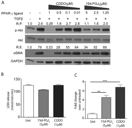
Figure 2. PPAR-cligands inhibit TGFb-induced phosphorylation of Akt and myofibroblast differentiation in a dose-dependent manner.A, Primary HLFs were grown until 70–80% confluent, serum starved for 24 hours and treated with the indicated concentrations of PPAR-c
ligands for 48 hours. Total cell lysates were prepared, and subjected to SDS-PAGE followed by immunoblotting. The blot was probed with antibodies against phospho-AktS473, stripped and probed to detect total Akt,aSMA and loading control GAPDH. The relative changes in the ratio of
phospho-AktS473/total Akt (R.P.) and relative changes in the expression ofaSMA/GAPDH (R.E.) are as indicated in the figure. The experiment was performed in triplicate and a representative blot is shown here.B, LDH release does not increase in response to 15d-PGJ2or CDDO. Primary human lung fibroblasts
were treated with either 5mM 15d-PGJ2or 1mM CDDO for 72 hours and LDH release was measured (nmol/min/mL).C, Primary human lung fibroblasts
were transfected with a PPRE luciferase reporter and a CMVb-galactosidase construct. Cells were treated with either 5mM 15d-PGJ2or 1mM CDDO for
48 hrs and luciferase activity was measured. Background was subtracted and data normalized tob-galactosidase transfection efficiency and reported as fold induction of luciferase units over the untreated samples. These data represent three independent experiments (mean6S.E. shown, **p#0.01, *** p#0.001, compared to untreated).
transcriptional response at target genes [10]. However, we [27,28,33] and others [32], have shown that PPAR-cligands also have effects that are independent of PPAR-cactivity. To examine if inhibition of Akt phosphorylation by PPAR-c ligands was dependent on the functional activity of PPAR-c, we used a pharmacological approach involving a PPAR-c antagonist GW9662, an irreversible PPAR-cantagonist that covalently binds to the active site of PPAR-c[36].
Primary HLF were pretreated with GW9662 followed by treatment with PPAR-cligands and TGFb. After 48 hours, total protein lysate was subjected to Western blot analysis to assess the level of changes in Akt phosphorylation. TGFbalone increased phosphorylation of Akt while addition of CDDO or 15d-PGJ2blocked TGFb-induced Akt
phosphorylation (Fig 3A). However, treatment with GW9662 did not restore either TGFb-induced phospho-Akt levels in PPAR-c -ligand-treated cultures or myofibroblast differentiation (Fig 3A) suggesting that CDDO and 15d-PGJ2block TGFb-induced Akt phosphorylation
in a PPAR-c-independent mechanism.
The Electrophilic Center Present in PPAR-c Ligands is Critical to Their Ability to Block TGFb-induced Phosphorylation of Akt
CDDO and 15d-PGJ2contain electrophilic carbons (Fig 3B) that
can modify sulfhydryl groups in proteins via the ‘Michael addition reaction’ [37,38]. To determine whether the ability of PPAR-c
ligands to inhibit TGFb-induced Akt phosphorylation is dependent on the presence of an electrophilic carbon, we used two different structural analogues of 15d-PGJ2, prostaglandin A1(PGA1), which
has an electrophilic center, and CAY10410, which does not have an electrophilic carbon but binds to PPAR-c and activates ligand-dependent transcription. Additionally, we used another potent electrophilic compound, diphenyl diselenide (DSPS), which, like CDDO, is structurally different from 15d-PGJ2 and has two
electrophilic carbons (Fig 3B). Interestingly, only the compounds with electrophilic centers, PGA1and DSPS, reduced TGFb-driven
phosphorylation of Akt, while CAY10410, did not (Fig 3C). Based on these results, we concluded that the electrophilic carbons present in the structures of CDDO and 15d-PGJ2have an essential role in
blocking TGFb-mediated activation of Akt.
PPAR-cLigands Inhibit TGFb-induced Phosphorylation of Akt in a Transcription-Independent Manner
Because the effects of CDDO and 15d-PGJ2 are PPAR-c
independent, we hypothesized that their effect would be indepen-dent of transcription as well. To investigate if PPAR-c ligands requirede novotranscription for their inhibition of Akt phosphor-ylation we treated primary HLF cells with a transcription inhibitor Actinomycin D (ActD), followed by the PPAR-cligands and TGFb. Western blot analysis was performed to detect phospho-AktS473and total Akt and the ratio of phospho-AktS473/Akt was calculated. ActD partly inhibited TGFb-induced phosphorylation of Akt (Fig 4). This suggests that TGFbactivates Akt kinase in a transcriptionally-dependent mechanism. However, inhibition by CDDO and 15d-PGJ2remained unaltered even in the presence of ActD (Fig 4A, and
Fig 4B, black bars) suggesting that these compounds directly inhibit Akt kinase activity or upregulate a phosphatase in a post-translational mechanism that is independent ofde novotranscription.
PPAR-cLigands Block TGFb-Stimulated Phosphorylation of Akt by Inhibiting FAK but not PTEN and MAPK-p38 Phosphorylation
To investigate proteins that could potentially phosphorylate or dephosphorylate Akt, we performed a time-course of action of
PPAR-c ligands and TGFb on the phosphorylation of PTEN, p38-MAPK and FAK.
First, we investigated the time-course of TGFb-mediated phosphorylation of Akt and its repression by CDDO and 15d-PGJ2(Fig 5A). Next, we examined changes in phosphorylation of
PTEN, which is a negative regulator of Akt phosphorylation. PTEN is more stable but less active when it is phosphorylated at T308, and less stable but more active when is dephosphorylated at the same site [39]. If PPAR-cligands inhibit Akt phosphorylation via upregulation of phosphatase activity of PTEN, we would expect to see a decrease in the ratio of phospho-PTENT308to total PTEN. We found that although TGFbslightly increased levels of phospho-PTEN after two hours (Fig 5B, filled squares), treatment with PPAR-cligands was unable to cause any significant deviation in the ratio of phospho-PTEN to total PTEN as compared to the TGFbalone-treated samples (Fig 5B, open squares and circles).
It has been reported that TGFbinduces p38-MAPK in some cell types [17]. We also observed an increase in the phospho-p38T180/Y182 to total p38 ratio in response to TGFb but, phosphorylation of TGFb-induced p38 was not reduced upon treatment with either CDDO or 15d-PGJ2 (Fig 5C), indicating
that the mode of action of PPAR-cligands is likely independent of p38-MAPK activity. Finally, we examined the effect of PPAR-c
ligands on FAKY397phosphorylation, which is required for FAK kinase activity. We observed TGFb-induced phosphorylation of FAK repressed by both the PPAR-c ligands 24 hours after the treatment (Fig 5D). CDDO inhibited FAKY397 phosphorylation more potently than 15d-PGJ2 (Fig 5D). Since PPAR-c ligands
inhibit FAK phosphorylation but do not change either p38-MAPK or PTEN phosphorylation, we suggest that PPAR-c
ligands inhibit Akt pathway by inhibiting the upstream FAK kinase.
Pharmacological Inhibition of FAK Activity Inhibits the PI3K-Akt Pathway and Myofibroblast Differentiation
To determine whether TGFb-mediated myofibroblast differen-tiation mediated through PI3K-Akt signaling also requires FAK activity in our cell strain, we treated primary HLF cells with a FAK-kinase inhibitor (AG1879) and its inactive analogue, PP3. Western blot analysis was used to measure phospho-FAKY397, total FAK, phospho-AktS473, total Akt, aSMA, calponin and GAPDH. We observed that inhibition of FAK inhibited not only Akt phosphorylation but also myofibroblast differentiation (Fig 6). Although, inhibition of FAK inhibitedaSMA expression, we did not observe complete inhibition of expression ofaSMA under the conditions tested. These results indicate that FAK activity is important for TGFb-mediated Akt activation and myofibroblast differentiation of primary HLF.
Next, we used a complimentary genetic approach to ascertain involvement of FAK in myofibroblast differentiation of primary HLFs. We found that over-expression of FAK resulted in marked up-regulation of Akt phosphorylation and myofibroblast differen-tiation (Fig 6B).
PPAR-c Ligands Inhibit Myofibroblast Differentiation of Primary IPF Fibroblasts by Inhibiting FAK and PI3K-Akt Pathways
To ascertain the therapeutic potential of PPAR-c ligands we examined their ability to suppress Akt and FAK in bona fide
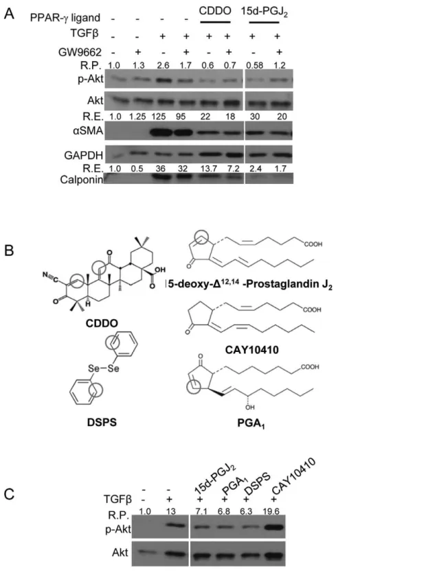
Figure 3. PPAR-cligands inhibit TGFb-induced Akt phosphorylation and myofibroblast differentiation in a PPAR-c-independent but electrophilic carbon-dependent manner.HLF cells were treated with indicated compounds and/or TGFb(5ng/ml) for 48 hours. Immunoblots were performed to assess the expression of indicated proteins. Protein lysates from all the indicated samples were electrophoretically separated on the same gel, and representative lanes from a single experiment are shown here.A, the ability of PPAR-cligands (CDDO (1mM) and 15d-PGJ2(5mM)) to
reduce p-Akt was not altered upon GW9662-mediated inhibition of PPAR-c. GW9662 (5mM) inhibits PPAR-cactivity by a covalent bond formation with PPAR-cprotein [36]. R.P. indicates relative changes in Akt phosphorylation compared to control sample, and R.E., relative changes in expression compared to control sample.B, PPAR-cligands contain electrophilic carbons. Here, positions of the electrophilic carbons in the structures of the
compounds are marked. CAY10410 and PGA1are structural analogues of 15-d-PGJ2. PGA1has an electrophilic center but CAY10410 does not. DSPS,
like CDDO, has two electrophilic centers. Cells were pre-treated with CAY10410 (5mM), PGA1(10mM) and DSPS (10mM) for 30 minutesC, only
selective inhibitors of PI3K pathway, LY294002 and wortmannin (Fig 7A) or increasing concentrations of AG1879 (PP2), a FAK inhibitor (Fig 7B) followed by TGFb. Inhibition of both the pathways potently blocked Akt phosphorylation and myofibroblast differentiation (Fig 7A and B). Finally, we treated IPF fibroblasts with CDDO and 15d-PGJ2followed by TGFb. These data firmly
establish that CDDO and 15d-PGJ2are both capable of blocking
myofibroblast differentiation via Akt and FAK pathways (Fig 7C) as measured by the expression ofaSMA and calponin.
Discussion
Idiopathic pulmonary fibrosis (IPF) can lead to respiratory failure and death due to deteriorating respiratory function [1,4]. There are currently few if any effective therapies. Therefore, novel
antifibrotic drugs are urgently needed for the treatment of IPF and other scarring diseases.
PPAR-cligands are emerging as exciting potential therapeutics for inflammatory and fibrotic and other diseases [29,40,41]. PPAR-cactivation induces adipogenesis and differentiation, and represses inflammation [27,29,33]. PPAR-c ligands including rosiglitazone and other members of TZD family are used for the treatment of type II diabetes [29]. Clinical trials for CDDO are in progress, and it has been found to be orally active for the treatment of solid tumors and lymphoma [42]. However, the mechanisms of anti-fibrotic actions of PPAR-c ligands remain poorly understood. Therefore, we investigated the underlying molecular pathways targeted by distinct PPAR-c ligands to explore the potential of PPAR-cagonists as anti-fibrotic therapies. Our previous work identified CDDO, a novel PPAR-cligand, as a potent anti-fibrotic agent of TGFb-driven pro-fibrotic activity
in vitro[27,33]. TGFb induces lung fibrosisin vivo [43] and also stimulates phosphorylation of Akt in animal models [44] and other human organs [10,45]. Studies in fetal lung fibroblasts demon-strate the role of TGFb-induced Akt pathway in myofibroblast differentiation [13]. Here, we report that in both normal and IPF primary human lung fibroblasts, PPAR-cligands potently block myofibroblast differentiation via a PPAR-c-independent mecha-nism by targeting the TGFb-induced PI3K-Akt pathway involving FAK.
We investigated the role of PI3K-Akt pathway in TGFb -stimulated myofibroblast differentiation using LY294004, a specific PI3K activity inhibitor, and by using a kinase-dead (KD-Akt) construct of Akt. Both LY294002 and the Akt mutant strongly blocked TGFb-stimulated myofibroblast differentiation, confirming the central role of PI3K-Akt pathway in TGFb -mediated myofibroblast differentiation in adult human normal and ‘‘diseased’’ IPF lung fibroblasts (Fig 1 and 7). Although, CDDO [46,47] and 15d-PGJ2[48] have been reported to reduce
Akt phosphorylation in some studies, their mechanism of reduction of TGFb-induced myofibroblast differentiation through Akt pathway is not yet reported. Here, we show that the suppression of TGFb-induced phosphorylation of AktS473is the central mechanism of action of CDDO and 15d-PGJ2that
leads to their anti-fibrotic activity. CDDO suppresses TGFb -induced phospho-Akt more potently than 15d-PGJ2, which
correlates very well with the abilities of CDDO and 15d-PGJ2
to reduce TGFb-induced myofibroblast differentiation (Fig 2). Compared to CDDO and 15d-PGJ2, rosiglitazone was relatively
poorly effective at inhibiting TGFb-induced Akt phosphorylation (data not shown). Interestingly, Kilter et al. reported that rosiglitazone facilitates rephosphorylation of Akt in rat myocar-diocytes [49]. We and others have reported that rosiglitazone has some anti-fibrotic effectsin vitro[27,32,33,50], but is much less potent than either CDDO or 15d-PGJ2. If rosiglitazone indeed
facilitates re-phosphorylation of Akt, then it would result in a pro-fibrotic response that undercuts its anti-pro-fibrotic effects, suggesting that rosiglitazone is not an optimal choice for treating fibrotic lung diseases. We did not investigate rosiglitazone further in this study.
CDDO and 15d-PGJ2 have electrophilic properties that
rosiglitazone does not, and we have previously identified the electrophilic centers as important in the antifibrotic activity of these compounds. Building on these observations, here we demonstrate that the PPAR-c-independent effects of CDDO and 15d-PGJ2 on Akt phosphorylation are dependent on the
electrophilic properties (Fig 3). These observations offer an additional possibility that CDDO and 15d-PGJ2 could directly
bind to the active site of a signaling molecule involved in the Akt Figure 4. Actinomycin D partially inhibits TGFb-induced
phosphorylation of Akt but not PPAR-c ligand-mediated inhibition of Akt phosphorylation.Primary HLFs were pre-treated with transcription inhibitor ActD (1mg/ml), followed by PPAR-cligands (CDDO (1mM) and 15d-PGJ2(5mM)) and TGFb(5ng/ml).A, Immunoblots
were performed to detect levels of indicated proteins upon inhibition of transcription. The experiment was performed with triplicate samples. Protein lysates from all the indicated samples were electrophoretically separated on the same gel, and representative lanes from a single experiment are shown here.B, The triplicate samples were measured by
densitometry and the ratio of phospho to total Akt was determined and normalized to untreated and bar graphs were plotted. The significance was calculated by one way ANOVA. For samples without ActD treatment (open bars), * indicates difference (P#0.05) over untreated and ** indicates difference (P#0.05) over TGFb-treated samples. For ActD-treated samples (black bars),#indicates difference (P#0.05) over untreated and ## indicates difference (P#0.05) over TGFb-treated samples.
pathway. One study has shown that biotinylated CDDO is capable of binding and thus inactivating the active site of PTEN [37], and 15d-PGJ2is capable of inhibiting the activities of proteins through
direct covalent modification [51,52]. Further studies involving biochemical approaches should help us understand the exact nature of action of compounds involving electrophilic carbon. To generate newer anti-fibrotic therapeutics, further investigation of the electrophilic properties of PPAR-c ligands and similar compounds is necessary.
Because CDDO and 15d-PGJ2 block Akt phosphorylation in
the absence of new transcription (Fig 4), this suggests that CDDO and 15d-PGJ2 act to directly inhibit a kinase or activate a
phosphatase that acts on Akt. We examined the role of three important upstream regulators of Akt phosphorylation: MAPK, PTEN and FAK (Fig 8). We chose to examine p38-MAPK because it is previously known to be a part of the PI3K/ Akt pathway [13] while other MAP kinases are involved in separate [53] or even antagonistic pathways [54]. TGFbactivates a number of signaling pathways including Smads, Akt and the MAPK-ERK, and while our data does not completely rule out that PPAR-c ligands may act via other pathways, the ability of LY294002 and KD-Akt to almost completely block myofibroblast differentiation shows that the PI3K/Akt pathway is the most important pathway. Since TGFb stimulates phosphorylation of p38-MAPK to activate myofibroblast differentiation by up-regulating Akt phosphorylation we examined involvement of p38-MAPK in primary HLF.
In agreement with a previous report [13], we determined that p38-MAPK is phosphorylated following TGFbtreatment of HLF (Fig. 5C), and MAPK inhibitor SB203580 reduced phosphoryla-tion of Akt (data not shown). However, neither CDDO nor 15d-PGJ2 altered TGFb-induced p38-MAPK phosphorylation,
indi-cating that MAPK is likely not involved in inhibition of Akt by CDDO and 15d-PGJ2. We were also able to exclude PTEN as a
major mediator of the effects of PPAR-cligands in HLFs. PTEN can inhibit the Akt pathway by dephosphorylating Akt, and PTEN phosphatase is itself activated by dephosphorylation at Thr308. Thus, if CDDO or 15d-PGJ2 blocked Akt phosphorylation via
PTEN, we would expect increased PTEN phosphatase activity associated with increase in its dephosphorylation at T308. In fact, CDDO or 15d-PGJ2 do not change PTEN phosphorylation
(Fig 5B). Interestingly, in human retinal epithelial cells, biotiny-lated CDDO (CDDO-Bt) binds to Cys124 within the active site of PTEN and inhibits the lipid phosphatase activity of PTENin vitro
[37]. If PTEN activity inhibition was an important mechanism in our system, we would expect levels of phospho-Akt to increase in presence of CDDO; instead we observed the opposite.
Multiple reports show that TGFbstimulates autophosphoryla-tion-dependent activation of focal adhesion kinase (FAK) [23,55]. For example in CCL20 lung fibroblasts, Xia et al demonstrated thatb1-integrin signaling upregulates FAK phosphorylation and its physical interaction with PI3K-p85 resulting in phosphorylation of Akt [24]. Although FAK is a widely accepted upstream regulator of Akt phosphorylation, it has been reported that FAK Figure 5. PPAR-cligands inhibit TGFb-induced phosphorylation of Akt and FAK but not MAPK-p38 and PTEN.Primary HLFs were pretreated with PPAR-c ligands; CDDO (1mM) and 15d-PGJ2(5mM) followed by TGFb(5ng/ml). Cells were harvested and lysates analyzed by
immunoblots at the indicated time. The ratio of phospho-protein to total protein was measured by densitometric analysis and normalized to untreated cells (untreated = 1.0). TGFb-induced phosphorylation ofA, AktS473andD, FAKY397was inhibited significantly by the PPAR-cligands but the
phosphorylation ofB, PTENT308andC, p38-MAPKT180/Y182was not affected. The statistical significance over TGFb-treatment alone was calculated
does not act upstream of Akt during TGFb signaling in IMR90 human fetal lung fibroblasts [17]. However, it is widely accepted that integrins are able to activate TGFb[56] and FAK [57,58], both of which are involved in myofibroblast differentiation. Our results demonstrate that PPAR-c ligands are able to inhibit phosphorylation of FAK and limit the TGFb-mediated fibrotic response in adult primary HLFs (Fig 5D). We confirmed both in our normal (Fig 6) and IPF (Fig 7) primary human cell strains that blocking FAK activity inhibited not only phosphorylation of Akt, but also expression of aSMA, confirming that FAK indeed regulates myofibroblast differentiation under normal and diseased conditions through activation of Akt pathway. To establish the therapeutic potential of PPAR-c ligands we treated fibroblasts obtained from IPF patients, in parallel, with PPAR-cligands, two PI3K inhibitors and a FAK inhibitor and confirmed that CDDO and 15d-PGJ2potently block myofibroblast differentiation of not
only normal HLF but also diseased IPF fibroblasts (Fig 7) via PI3K-Akt and FAK pathways. Although by using a complimen-tary genetic approach we confirmed that overexpression of FAK is capable of upregulating Akt phosphorylation and myofibroblast
differentiation (Fig 6B), at this point, we cannot rule out an additional mechanism of action of PPAR-c ligands that would result in reduction of Akt phosphorylation (Fig 8).
Current models of pulmonary fibrosis suggest that TGFb, mechanical stress, or adhesion and integrin mediated activation of myofibroblast differentiation all contribute to upregulation of a fibrotic response. One very critical, central and shared event in all of these pathways involves upregulation of FAK activity, defined by its phosphorylation at Tyr397. It is conceivable that once TGFb activates myofibroblast differentiation, the increased deposition of extracellular matrix proteins would cause additional mechanical stress on the cell surface leading to sustained and continual activation of FAK. Since FAK itself upregulates myofibroblast differentiation, once TGFb initiates this process, sustained activation of FAK would be able to perpetuate the fibrotic response even in the absence of active TGFb. Our work is the first report in any biological system demonstrating that
PPAR-cligands reduce FAK activity by reducing FAK phosphorylation at Tyr397. Since FAK plays a cardinal role in myofibroblast differentiation, drugs that target the catalytic activity of FAK could be very valuable in the treatment of pulmonary fibrosis.
This study highlights a very important mechanism of action of CDDO and 15d-PGJ2that involves down-regulation of PI3K-Akt
pathway in both normal and IPF fibroblasts. Knowing that Akt is a central regulator of multiple cellular pathways including cell proliferation, cell cycle progression, inflammation and apoptosis [7,10], interfering with the Akt pathway can have multiple cellular and organ-wide effects. Although, we have noted sustained basal activity of Akt in untreated cells, the nature of Akt activation is largely inducible and dependent on upstream signaling molecules. Therefore, the use of Akt-inhibition as a potential therapy for pulmonary fibrosis is a very novel and exciting concept.
Overall, we propose that certain PPAR-c ligands have tremendous translational potential as therapeutics for pulmonary fibrosis by not only inhibiting Akt but also FAK activation. Future
in vivostudies involving PPAR-cligands will be pivotal in exploring the promising potential of PPAR-c ligands as therapeutics for pulmonary fibrosis as well as other scarring diseases.
Materials and Methods
Cells and reagents
Normal primary human lung fibroblasts (HLFs) were derived as previously described [33], grown in MEM supplemented with 10%FBS, L-Glutamine, antibiotic and antimycotic (Gibco, Carlsbad, CA) and used between passages 7–10. They were grown until 70–80% confluent and serum-starved for 24 hrs before treatment, unless otherwise mentioned. Primary human idiopathic pulmonary fibrotic (IPF) fibroblasts were derived from lung tissues obtained from patients with IPF undergoing wedge biopsy. A written informed consent was obtained from all the subjects in accordance with the University of Rochester Medical Center Institutional Review Board. Explant technique was used to isolate primary fibroblasts as described previously [33] and fibroblasts were used between passages 5–9 and maintained as described above.
Treatments
PPAR-c agonists were used at the following concentrations; 1mM of CDDO (NIH-RAID Program and Reata Pharmaceuti-cals, Dallas, TX), 5mM 15d-PGJ2 and 9,10-dihydro-15-deoxy-D12,14-PGJ2 (CAY10410) (Cayman Pharmaceuticals, Ann Arbor, MI). Ligands were dissolved in DMSO to make 10mM stock solution and diluted in serum-free media before treatment. Figure 6. Pharmacological inhibition of FAK activity inhibits
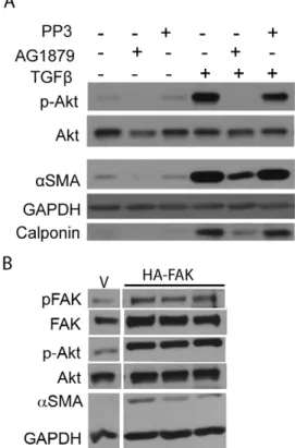
the PI3K-Akt pathway and myofibroblast differentiation. A,
Primary HLFs were treated in presence or absence of TGFband 10mM specific Src-FAK kinase inhibitor AG1879 (PP2) or its analogue, PP3, that does not inhibit FAK activity and immunoblots were performed to analyze expression of the indicated proteins. The FAK inhibitor AG1879, but not its analogue PP3, inhibited TGFb-induced phosphorylation of FAKY397 and AktS473 and reduced myofibroblast differentiation as
determined by expression of aSMA and calponin.B, HLF cells were
transfected with the empty vector (V) or FAK overexpressing construct (HA-FAK) and assayed for myofibroblast differentiation by Western blot. Protein lysates from all the indicated samples were electrophoretically separated on the same gel, irrelevant lanes excluded and representative lanes from a single experiment are shown here. These data indicate that FAK overexpression induces myofibroblast differentiation of primary human lung fibroblasts.
GW9662 (Sigma, St. Louis, MO), 15-deoxy-D12,14-Prostaglandin A1 (PGA1) (Cayman Pharmaceuticals, Ann Arbor, MI) and
diphenyl diselenide (DSPS) (Sigma, St. Louis, MO) were prepared in the same manner as described above. GW9662 (5mM) was added two hours prior to any other treatment. Human recombinant TGFb1 (R&D Systems, Minneapolis, MN) was used at a final concentration of 5ng/ml. 50mM LY294002 (Cell Signaling Technology, Danvers, MA), 10 or 20mM AG1879 (4-Amino-5-(4-chlorophenyl)-7-(t-butyl)pyrazolo[3,4-d]pyrimidine, (PP2)) and its analogue, 4-Amino-7-phenylpyrazol[3,4-d]pyrimidine (PP3) (EMD Chemicals Inc. Gibbstown, NJ), 100nM wortmannin and, 1mg/ml Actinomycin D (Sigma, St. Louis, MO) were used to pretreat cells 30 min prior to TGFbtreatment.
Western blots
Primary human lung fibroblasts were plated (16105cells/well)
in six-well plates (Falcon/Becton Dickson, Franklin Lakes, NJ) for all experiments and allowed to grow for 48 hours prior to any treatment. Crude cellular protein lysates were prepared using NP-40 lysis buffer supplemented with protease inhibitor, phosphatase inhibitor and 1mM PMSF (Sigma, St. Louis, MO). Total proteins (5 to 10mg) were resolved by 10% SDS-PAGE, electrophoretically transferred to nitrocellulose membranes, and specific proteins were
detected by standard Western blotting and chemiluminescence (Western Lightning, Perkin-Elmer, Wellesley, MA). Kodak Molecular Imaging Software (Rochester, NY) was used to perform densitometry on Western blot films and the band intensities were normalized to the loading control. The following primary antibodies were used: phospho-AktS473, total Akt, phospho-PTENS380, total PTEN, phospho-p38 MAPKT180/Y182, p38 MAPK, total FAK (Cell Signaling Technologies, Danvers, MA),
aSMA (Sigma, St. Louis MO), calponin (DAKO, Carpinteria, CA), GAPDH (Abcam, Cambridge, MA) and phospho-FAKY397 (Invitrogen Corporation Camarillo, CA). The secondary antibod-ies used were; goat anti-rabbit (sc-2004), goat anti-mouse (sc-2031, Santa Cruz Biotechnology, Inc. Santa Cruz, CA. 95060 U.S.A) and donkey anti-rabbit (NA 934, Amersham/GE Health Care Life Sciences Piscataway, NJ 08855-1327).
Cell cytotoxicity assay
Cell cytotoxicity was measured by lactate dehydrogenase release assay (LDH5 assay) using an optimized LDH assay kit (Sigma, Cat #DG1340-K). Briefly, fibroblasts were plated in triplicate at a density of 16105cells per well in 6 well plates and treated with either CDDO (1mM) or 15d-PGJ2 (5mM) or left untreated for 72 hrs. Release of LDH (nmol/min/mL) was measured at 340 nm Figure 7. PPAR-cligands block myofibroblast differentiation of primary human IPF fibroblasts.Primary IPF fibroblasts were treated with eitherA, two PI3K inhibitors LY294002 (50mM) or wortmannin (100nM) orB, a Src-FAK inhibitor AG1879 (10 and 20mM) orC, CDDO (1mM) or 15d-PGJ2
(5mM) followed by TGFb(5ng/ml) for 48 hours. Protein lysates were prepared and immunoblots were performed to detect expression levels of the indicated proteins. The experiment was performed in triplicate on three independent IPF fibroblast strains. Protein lysates from all the indicated samples were electrophoretically separated on the same gel, and representative lanes from one representative set of data are shown here. These data confirm that CDDO and 15d-PGJ2inhibit both, FAK and PI3K-Akt pathways to inhibit TGFb-induced myofibroblast differentiation of primary IPF
fibroblasts.
using a spectrophotometer as per the manufacturer’s protocol. The results were normalized to untreated control samples and plotted as fold change over untreated samples.
PPRE luciferase reporter assay
Primary lung fibroblasts cultured in 6-well plates were co-transfected using Fugene6 (Roche Applied Science, Indianapolis, IN) with a PPAR-c-luciferase reporter construct containing three PPREs (a gift from Dr. Brian Seed, Harvard University) [59]. A CMV-b-galactosidase control construct was included as control. After 24 h, the cells were washed and then treated with 15d-PGJ2
(5mM) or CDDO (1mM) in medium and harvested after a further 48-hr incubation. Luciferase activity was measured using a luciferase assay system (Promega, Madison, WI) in a luminometer
(Packard Instruments, Meriden, CT) and normalized to b -galactosidase activity, determined by a colorimetric assay (Pro-mega). The experiments were carried out in triplicate wells.
Transfections
Primary HLF cells were plated (56104cells/well) in 12 well
plates (Falcon/Becton Dickson, Franklin Lakes, NJ) and Fugene 6 transfection kit was used as per the manufacturer’s protocol (Roche Applied Science, Indianapolis, IN) for transfection. Transfection reactions were carried out using either empty vector pcDNA3.1 or a dominant negative kinase dead Akt (KD-Akt) (a kind gift from Dr. Robert Freeman, University of Rochester, NY USA [34]). Upon transfection, cells were allowed to grow for 16– 24 hours, and were then supplemented with 10% FBS for 24 hours followed by the treatment. Cells were lysed using NP-40 lysis buffer and subjected to further analysis as described above. Transfections with HA-FAK (a kind gift from Dr. William Cance, Roswell Park Cancer Institute, Buffalo, NY USA) were performed in a similar manner.
Indirect immunofluorescence assay
Cells were grown in four well chamber slides and treated as outlined above. Cells were fixed in methanol and stained with an antibody toa-SMA (St. Louis, MO, USA) and with anti-mouse AlexaFluor 488 (Invitrogen Corporation Carlsbad, CA, USA). Slides were mounted with Prolong Gold supplemented with DAPI (Invitrogen Corporation Carlsbad, CA, USA) to visualize the nuclei and analyzed by fluorescence-microscopy using a Zeiss Axio Imager Z.1 Microscope.
Acknowledgments
We would like to thank Dr. Robert Freeman (University of Rochester, Rochester NY) for providing a KD-Akt construct and Dr. William Cance (Roswell Park Cancer Institute, Buffalo, NY) for providing a HA-FAK construct, Dr. Heather Ferguson, Katherine Smolnycki and Marta Ekstrom for their technical help, and Dr. R.M. Kottmann for critically reading the manuscript.
Author Contributions
Conceived and designed the experiments: AAK THT RPP PJS. Performed the experiments: AAK KLO. Analyzed the data: AAK THT KLO SBM PJS. Wrote the paper: AAK.
References
1. Meltzer EB, Noble PW (2008) Idiopathic pulmonary fibrosis. Orphanet J Rare Dis 3: 8.
2. Wilson MS, Wynn TA (2009) Pulmonary fibrosis: pathogenesis, etiology and regulation. Mucosal Immunol 2: 103–121.
3. Selman M, Pardo A (2002) Idiopathic pulmonary fibrosis: an epithelial/ fibroblastic cross-talk disorder. Respir Res 3: 3.
4. Sime PJ, O’Reilly KM (2001) Fibrosis of the lung and other tissues: new concepts in pathogenesis and treatment. Clin Immunol 99: 308–319.
5. Baglole CJ, Ray DM, Bernstein SH, Feldon SE, Smith TJ, et al. (2006) More than structural cells, fibroblasts create and orchestrate the tumor microenviron-ment. Immunol Invest 35: 297–325.
6. Vallance BA, Gunawan MI, Hewlett B, Bercik P, Van Kampen C, et al. (2005) TGF-beta1 gene transfer to the mouse colon leads to intestinal fibrosis. Am J Physiol Gastrointest Liver Physiol 289: G116–128.
7. Guo X, Wang XF (2009) Signaling cross-talk between TGF-beta/BMP and other pathways. Cell Res 19: 71–88.
8. Khalil N (1999) TGF-beta: from latent to active. Microbes Infect 1: 1255–1263. 9. Moustakas A, Heldin CH (2009) The regulation of TGFbeta signal transduction.
Development 136: 3699–3714.
10. Assinder SJ, Dong Q, Kovacevic Z, Richardson DR (2009) The TGF-beta, PI3K/Akt and PTEN pathways: established and proposed biochemical integration in prostate cancer. Biochem J 417: 411–421.
11. Danielpour D (2005) Functions and regulation of transforming growth factor-beta (TGF-factor-beta) in the prostate. Eur J Cancer 41: 846–857.
12. Yamada KM, Araki M (2001) Tumor suppressor PTEN: modulator of cell signaling, growth, migration and apoptosis. J Cell Sci 114: 2375–2382. 13. Horowitz JC, Lee DY, Waghray M, Keshamouni VG, Thomas PE, et al. (2004)
Activation of the pro-survival phosphatidylinositol 3-kinase/AKT pathway by transforming growth factor-beta1 in mesenchymal cells is mediated by p38 MAPK-dependent induction of an autocrine growth factor. J Biol Chem 279: 1359–1367. 14. Parsons CJ, Takashima M, Rippe RA (2007) Molecular mechanisms of hepatic
fibrogenesis. J Gastroenterol Hepatol 22 Suppl 1: S79–84.
15. Li JD (2003) Exploitation of host epithelial signaling networks by respiratory bacterial pathogens. J Pharmacol Sci 91: 1–7.
16. Thomas PE, Peters-Golden M, White ES, Thannickal VJ, Moore BB (2007) PGE(2) inhibition of TGF-beta1-induced myofibroblast differentiation is Smad-independent but involves cell shape and adhesion-dependent signaling. Am J Physiol Lung Cell Mol Physiol 293: L417–428.
17. Horowitz JC, Rogers DS, Sharma V, Vittal R, White ES, et al. (2007) Combinatorial activation of FAK and AKT by transforming growth factor-beta1 confers an anoikis-resistant phenotype to myofibroblasts. Cell Signal 19: 761–771.
18. Parsons JT, Martin KH, Slack JK, Taylor JM, Weed SA (2000) Focal adhesion kinase: a regulator of focal adhesion dynamics and cell movement. Oncogene 19: 5606–5613.
19. Parsons JT, Parsons SJ (1997) Src family protein tyrosine kinases: cooperating with growth factor and adhesion signaling pathways. Curr Opin Cell Biol 9: 187–192.
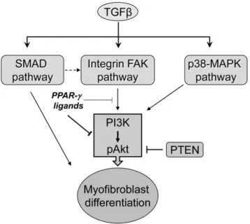
Figure 8. A proposed model showing the mechanism of action of electrophilic PPAR-cligands on TGFb-induced myofibroblast differentiation.TGFbinduces myofibroblast differentiation by acti-vating SMAD, FAK and PI3K-Akt pathways. However, PPAR-c ligands inhibit the TGFb-induced PI3K-Akt pathway, partly by targeting FAK induced activation of Akt.
20. Lipfert L, Haimovich B, Schaller MD, Cobb BS, Parsons JT, et al. (1992) Integrin-dependent phosphorylation and activation of the protein tyrosine kinase pp125FAK in platelets. J Cell Biol 119: 905–912.
21. Calalb MB, Polte TR, Hanks SK (1995) Tyrosine phosphorylation of focal adhesion kinase at sites in the catalytic domain regulates kinase activity: a role for Src family kinases. Mol Cell Biol 15: 954–963.
22. Chen HC, Appeddu PA, Isoda H, Guan JL (1996) Phosphorylation of tyrosine 397 in focal adhesion kinase is required for binding phosphatidylinositol 3-kinase. J Biol Chem 271: 26329–26334.
23. Thannickal VJ, Lee DY, White ES, Cui Z, Larios JM, et al. (2003) Myofibroblast differentiation by transforming growth factor-beta1 is dependent on cell adhesion and integrin signaling via focal adhesion kinase. J Biol Chem 278: 12384–12389.
24. Xia H, Nho RS, Kahm J, Kleidon J, Henke CA (2004) Focal adhesion kinase is upstream of phosphatidylinositol 3-kinase/Akt in regulating fibroblast survival in response to contraction of type I collagen matrices via a beta 1 integrin viability signaling pathway. J Biol Chem 279: 33024–33034.
25. Verma A, Guha S, Wang H, Fok JY, Koul D, et al. (2008) Tissue transglutaminase regulates focal adhesion kinase/AKT activation by modulating PTEN expression in pancreatic cancer cells. Clin Cancer Res 14: 1997–2005. 26. Chiu YC, Lin CY, Chen CP, Huang KC, Tong KM, et al. (2009) Peptidoglycan
enhances IL-6 production in human synovial fibroblasts via TLR2 receptor, focal adhesion kinase, Akt, and AP-1- dependent pathway. J Immunol 183: 2785–2792.
27. Burgess HA, Daugherty LE, Thatcher TH, Lakatos HF, Ray DM, et al. (2005) PPARgamma agonists inhibit TGF-beta induced pulmonary myofibroblast differentiation and collagen production: implications for therapy of lung fibrosis. Am J Physiol Lung Cell Mol Physiol 288: L1146–1153.
28. Ferguson HE, Thatcher TH, Olsen KC, Garcia-Bates TM, Baglole CJ, et al. (2009) Peroxisome proliferator-activated receptor-gamma ligands induce heme oxygenase-1 in lung fibroblasts by a PPARgamma-independent, glutathione-dependent mechanism. Am J Physiol Lung Cell Mol Physiol 297: L912–919. 29. Tontonoz P, Spiegelman BM (2008) Fat and beyond: the diverse biology of
PPARgamma. Annu Rev Biochem 77: 289–312.
30. Lakatos HF, Thatcher TH, Kottmann RM, Garcia TM, Phipps RP, et al. (2007) The Role of PPARs in Lung Fibrosis. PPAR Res 2007: 71323.
31. Sime PJ (2008) The antifibrogenic potential of PPARgamma ligands in pulmonary fibrosis. J Investig Med 56: 534–538.
32. Milam JE, Keshamouni VG, Phan SH, Hu B, Gangireddy SR, et al. (2008) PPAR-gamma agonists inhibit profibrotic phenotypes in human lung fibroblasts and bleomycin-induced pulmonary fibrosis. Am J Physiol Lung Cell Mol Physiol 294: L891–901.
33. Ferguson HE, Kulkarni A, Lehmann GM, Garcia-Bates TM, Thatcher TH, et al. (2009) Electrophilic peroxisome proliferator-activated receptor-gamma ligands have potent antifibrotic effects in human lung fibroblasts. Am J Respir Cell Mol Biol 41: 722–730.
34. Crowder RJ, Freeman RS (1998) Phosphatidylinositol 3-kinase and Akt protein kinase are necessary and sufficient for the survival of nerve growth factor-dependent sympathetic neurons. J Neurosci 18: 2933–2943.
35. Franke TF, Yang SI, Chan TO, Datta K, Kazlauskas A, et al. (1995) The protein kinase encoded by the Akt proto-oncogene is a target of the PDGF-activated phosphatidylinositol 3-kinase. Cell 81: 727–736.
36. Huang JT, Welch JS, Ricote M, Binder CJ, Willson TM, et al. (1999) Interleukin-4-dependent production of PPAR-gamma ligands in macrophages by 12/15-lipoxygenase. Nature 400: 378–382.
37. Pitha-Rowe I, Liby K, Royce D, Sporn M (2009) Synthetic triterpenoids attenuate cytotoxic retinal injury: cross-talk between Nrf2 and PI3K/AKT signaling through inhibition of the lipid phosphatase PTEN. Invest Ophthalmol Vis Sci 50: 5339–5347.
38. Straus DS, Pascual G, Li M, Welch JS, Ricote M, et al. (2000) 15-deoxy-delta 12,14-prostaglandin J2 inhibits multiple steps in the NF-kappa B signaling pathway. Proc Natl Acad Sci U S A 97: 4844–4849.
39. Vazquez F, Ramaswamy S, Nakamura N, Sellers WR (2000) Phosphorylation of the PTEN tail regulates protein stability and function. Mol Cell Biol 20: 5010–5018.
40. Hart CM, Roman J, Reddy R, Sime PJ (2008) PPARgamma: a novel molecular target in lung disease. J Investig Med 56: 515–517.
41. Roman J (2008) Peroxisome proliferator-activated receptor gamma and lung cancer biology: implications for therapy. J Investig Med 56: 528–533. 42. Petronelli A, Pannitteri G, Testa U (2009) Triterpenoids as new promising
anticancer drugs. Anticancer Drugs 20: 880–892.
43. Sime PJ, Xing Z, Graham FL, Csaky KG, Gauldie J (1997) Adenovector-mediated gene transfer of active transforming growth factor-beta1 induces prolonged severe fibrosis in rat lung. J Clin Invest 100: 768–776.
44. Vinals F, Pouyssegur J (2001) Transforming growth factor beta1 (TGF-beta1) promotes endothelial cell survival during in vitro angiogenesis via an autocrine mechanism implicating TGF-alpha signaling. Mol Cell Biol 21: 7218–7230. 45. Kattla JJ, Carew RM, Heljic M, Godson C, Brazil DP (2008) Protein kinase B/
Akt activity is involved in renal TGF-beta1-driven epithelial-mesenchymal transition in vitro and in vivo. Am J Physiol Renal Physiol 295: F215–225. 46. Gao X, Deeb D, Jiang H, Liu Y, Dulchavsky SA, et al. (2007) Synthetic
triterpenoids inhibit growth and induce apoptosis in human glioblastoma and neuroblastoma cells through inhibition of prosurvival Akt, NF-kappaB and Notch1 signaling. J Neurooncol 84: 147–157.
47. Deeb D, Gao X, Dulchavsky SA, Gautam SC (2008) CDDO-Me inhibits proliferation, induces apoptosis, down-regulates Akt, mTOR, NF-kappaB and NF-kappaB-regulated antiapoptotic and proangiogenic proteins in TRAMP prostate cancer cells. J Exp Ther Oncol 7: 31–39.
48. Han H, Shin SW, Seo CY, Kwon HC, Han JY, et al. (2007) 15-Deoxy-delta 12,14-prostaglandin J2 (15d-PGJ 2) sensitizes human leukemic HL-60 cells to tumor necrosis factor-related apoptosis-inducing ligand (TRAIL)-induced apoptosis through Akt downregulation. Apoptosis 12: 2101–2114.
49. Kilter H, Werner M, Roggia C, Reil JC, Schafers HJ, et al. (2009) The PPAR-gamma agonist rosiglitazone facilitates Akt rephosphorylation and inhibits apoptosis in cardiomyocytes during hypoxia/reoxygenation. Diabetes Obes Metab 11: 1060–1067.
50. Tan X, Dagher H, Hutton CA, Bourke JE () Effects of PPAR gamma ligands on TGF-beta1-induced epithelial-mesenchymal transition in alveolar epithelial cells. Respir Res 11: 21.
51. Kim DH, Kim EH, Na HK, Sun Y, Surh YJ (2010) 15-Deoxy-Delta(12,14)-prostaglandin J(2) stabilizes, but functionally inactivates p53 by binding to the cysteine 277 residue. Oncogene 29: 2560–2576.
52. Kalantari P, Narayan V, Henderson AJ, Prabhu KS (2009) 15-Deoxy-Delta12,14-prostaglandin J2 inhibits HIV-1 transactivating protein, Tat, through covalent modification. FASEB J 23: 2366–2373.
53. Hong KM, Belperio JA, Keane MP, Burdick MD, Strieter RM (2007) Differentiation of human circulating fibrocytes as mediated by transforming growth factor-beta and peroxisome proliferator-activated receptor gamma. J Biol Chem 282: 22910–22920.
54. Berra E, Diaz-Meco MT, Moscat J (1998) The activation of p38 and apoptosis by the inhibition of Erk is antagonized by the phosphoinositide 3-kinase/Akt pathway. J Biol Chem 273: 10792–10797.
55. Mimura Y, Ihn H, Jinnin M, Asano Y, Yamane K, et al. (2005) Constitutive phosphorylation of focal adhesion kinase is involved in the myofibroblast differentiation of scleroderma fibroblasts. J Invest Dermatol 124: 886–892. 56. Goodwin A, Jenkins G (2009) Role of integrin-mediated TGFbeta activation in
the pathogenesis of pulmonary fibrosis. Biochem Soc Trans 37: 849–854. 57. Greenberg RS, Bernstein AM, Benezra M, Gelman IH, Taliana L, et al. (2006)
FAK-dependent regulation of myofibroblast differentiation. FASEB J 20: 1006–1008.
58. Mitra SK, Schlaepfer DD (2006) Integrin-regulated FAK-Src signaling in normal and cancer cells. Curr Opin Cell Biol 18: 516–523.