Should fetal growth be a matter of concern in isolated single
umbilical artery?
LORENA MESQUITA CALDAS1, ADOLFO LIAO1, MÁRIO HENRIQUE CARVALHO1, ROSSANA PULCINELI VIEIRA FRANCISCO1, MARCELO ZUGAIB1
1Department of Obstetrics and Gynecology, Hospital das Clínicas, São Paulo University Medical School, São Paulo, SP, Brazil
SUMMARY
Study conducted at Hospital das Clínicas, São Paulo University Medical School, São Paulo, SP
Article received: 04/04/13
Accepted for publication: 08/30/13
Correspondence:
Departamento de Ginecologia e Obstetrícia Hospital das Clínicas Faculdade de Medicina da USP Address: Av. Dr. Enéas de Carvalho Aguiar,
255 – 10º. Andar, cj. 10037 ZIP Code: 05403-000 São Paulo – SP – Brazil Phone: +55 11 2661-6209
Fax +55 11 2661- 8183 [email protected]
http://dx.doi.org/10.1590/1806-9282.60.02.009
Conflict of interest: none
Objective: To examine birth weight in pregnancies with isolated single
umbili-cal artery (ISUA).
Methods: Case control study with retrospective review of 131 singleton
pregnan-cies with isolated single umbilical artery diagnosed before birth. Control group consisted of 730 singleton pregnancies recruited prospectively, that had histolo-gical conirmation of a 3 vessels cord. Pregnancies were classiied as uncomplica-ted or high-risk according to the presence of diseases that increase the risk of pla-cental insuficiency during pregnancy. Mean birth weight and frequency of low birth weight (< 2.500 g), very low birth weight (< 1.500 g) and fetal growth restric-tion below the 5th and 10th centiles were compared between groups.
Results: Mean birth weight difference between ISUA (n=131, 2840±701g) and
control (n=730, 2.983 ± 671g) pregnancies was 143g (95% CI= 17-269; p=0.04) and birth weight below the 5th centile was signiicantly more common in ISUA group [28/131 (21.4%) versus 99/730 (13.6%), p=0.02]. When only
uncomplica-ted pregnancies were considered in both groups, no birth weight differences were observed. Amongst high-risk subgroups, birth weight below the 5th centile re-mained signiicantly more common in ISUA compared to control pregnancies [10/35 (28.6%) versus 53/377 (14.1%), p=0.04].
Conclusion: Isolated single umbilical artery does not increase the risk of fetal
growth restriction in uncomplicated singleton pregnancies.
Key words: single umbilical artery, fetal growth retardation, birth weight,
ultra-sonography.
I
NTRODUCTIONNormally, the human umbilical cord contains two arteries that transport blood from the fetus to the placenta, and oxygenated blood returns to the fetus via a single umbili-cal vein. However, a single umbiliumbili-cal artery (SUA) can be diagnosed antenatally by ultrasound in up to 2% of preg-nancies1 and, when it is associated with other fetal struc-tural defects, there is an increased risk of chromosomal ab-normalities and adverse pregnancy outcome.2
Nevertheless, the clinical relevance of an isolated SUA remains controversial. Compensatory mechanisms allow increase in blood low through the single artery in order to meet the demands of fetal growth and development throughout pregnancy.3 Although several studies have reported diminished fetal growth in these pregnancies,4-12
others have not shown differences.13-15 These contradic-tory indings may be attributed to diverse methodologi-cal differences in the published literature.
The present study examines birth weight in pregnan-cies with isolated single umbilical artery diagnosed ante-natally in a tertiary care teaching hospital.
M
ETHODSIsolated single umbilical artery study group
A computer database search was performed to retrospec-tively identify all singleton pregnancies in which an iso-lated single umbilical artery (ISUA) was diagnosed ante-natally by ultrasound scan between 1998 and 2010. The diagnosis was based on the visualization of two vessels in a cross section view of a free loop of umbilical cord in a fetus without structural abnormalities. All cases were conirmed after birth by clinical and/or pathological exa-mination of the cord. Live born infants with no phenoty-pic features of a chromosomal defect were assumed to be euploid.
Control group
It was based on a cohort of 759 unselected singleton preg-nancies prospectively examined between 2007 and 2009 – that had already been included in a previous publica-tion1. In this group, a three vessels cord was conirmed by two ultrasound examinations (carried out at 11-13 weeks and 17-24 weeks) and placental examination after delivery. Seven hundred and thirty (96.2%) women deli-vered phenotypically normal live born infants.
Data collection
Hospital records were reviewed for pregnancy and outco-me information. Wooutco-men who delivered their babies in other hospitals were contacted by telephone.
Maternal characteristics including age, ethnic group and presence of clinical or obstetrical complications were recorded. Pregnancies were classiied as uncomplicated or high-risk according to the presence of diseases that in-crease the risk of placental insuficiency during pregnancy: hypertension, cardiomyopathy, asthma, diabetes, hemo-globinopathy, thrombophilia, thromboembolism, cirrho-sis, kidney failure or auto immune diseases.
Gestational age was calculated based on the irst day of the last menstrual period when available and conir-med by early ultrasound examination. When menstrual dates were uncertain, or there was discrepancy greater than 7 days between clinical and ultrasound dates, ges-tational age was established according to the earliest ul-trasound scan indings.
Birth weight was examined according to previously published reference values16 and primary outcomes were deined as birth weight below the 5th and 10th centiles.
Statistical analysis
All data was entered in an Excel spreadsheet (Microsoft Corporation, USA). Maternal demographics and frequency of birth weight below the 5th and 10th centiles were
com-pared between ISUA and control groups using unpaired Student t test and chi square tests or Fisher’s exact test,
when appropriate. Statistical calculations were perfor-med using Statsdirect (StatsDirect Ltd, UK). Signiicance
level was set as 0.05.
R
ESULTSThe database search identiied 134 singleton pregnancies in which an isolated single umbilical artery was diagno-sed prenatally by ultrasound. Mean gestational age at diagnosis was 25.1 ± 5.8 weeks and 18 (13.4%) cases were referred before 20 weeks. 131 (97.8%) pregnancies resul-ted in live births, and the diagnosis of ISUA was conir-med in all cases.
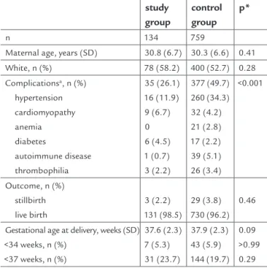
No differences were observed regarding maternal age, proportion of white women and gestational age at deli-very between ISUA and control groups. Nevertheless, com-plications were observed signiicantly more often in con-trol pregnancies (377/759, 49.7%) compared to the ISUA group (35/134, 26.1%, p<0.001, table 1).
TABLE 1 Characteristics and perinatal outcome in isolated
single umbilical artery and control pregnancies.
study group
control group
p*
n 134 759
Maternal age, years (SD) 30.8 (6.7) 30.3 (6.6) 0.41 White, n (%) 78 (58.2) 400 (52.7) 0.28 Complicationsa, n (%)
hypertension cardiomyopathy anemia diabetes
autoimmune disease thrombophilia
35 (26.1) 16 (11.9) 9 (6.7) 0 6 (4.5) 1 (0.7) 3 (2.2)
377 (49.7) 260 (34.3) 32 (4.2) 21 (2.8) 17 (2.2) 39 (5.1) 26 (3.4)
<0.001
Outcome, n (%) stillbirth live birth
3 (2.2) 131 (98.5)
29 (3.8) 730 (96.2)
0.46
Gestational age at delivery, weeks (SD) <34 weeks, n (%)
<37 weeks, n (%)
37.6 (2.3) 7 (5.3) 31 (23.7)
37.9 (2.3) 43 (5.9) 144 (19.7)
0.09 >0.99 0.29 GA: gestational age at delivery, n: number of cases, SD: standard deviation.
*Mann-Whitney U test or chi2 test/Fisher´s exact test.
a defined in the present study as the occurrence of diseases that increase the risk of placental
in-sufficiency during pregnancy.
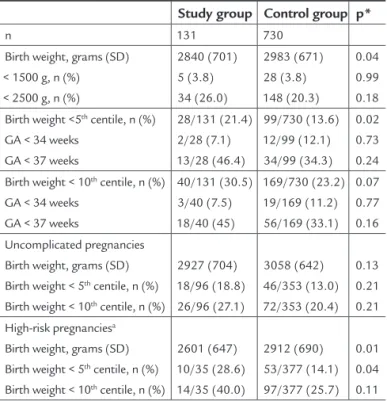
p=0.02, table 2]. However, when only uncomplicated preg-nancies were considered in both groups, no birth weight differences were observed. Amongst high-risk subgroups, birth weight below the 5th centile remained signiicantly more common in ISUA compared to control pregnancies [10/35 (28.6%) versus 53/377 (14.1%), p=0.04].
D
ISCUSSIONSeveral studies have previously shown the association between single umbilical artery and less fetal growth. Ba-sed on these indings, some authors have recommended that fetal growth should be monitored throughout preg-nancy after prenatal diagnosis of a single umbilical ar-tery.7-9,11,12 As a matter of fact, throughout the study period, our management protocol for pregnancies diagnosed with ISUA included serial follow up fetal growth scans. And when fetal growth restriction was diagnosed additional fe-tal well being tests were performed. Our current indings suggest that close fetal growth follow up is only necessary when ISUA is associated with the occurrence of maternal disease or pregnancy complication.
Moreover, gestational age at delivery and the rates of preterm delivery (before 34 and 37 weeks) were not sta-tistically different between ISUA and control
pregnan-0 1000 2000 3000 4000
25 30 35 40
Gestacional age (weeks)
birth weight (g)
FIGURE 1 Scatter plot of birth weight according to gestational age at delivery. Open circles: control group (n=730), full circles: isolated single
umbilical artery (n=131), continuous line: 50th percentile, dashed lines: 10th and 90th percentiles, dotted lines: 5th and 95th percentiles.16
TABLE 2 Birth weight in isolated single umbilical artery and
control pregnancies
Study group Control group p*
n 131 730
Birth weight, grams (SD) < 1500 g, n (%) < 2500 g, n (%)
2840 (701) 5 (3.8) 34 (26.0)
2983 (671) 28 (3.8) 148 (20.3)
0.04 0.99 0.18 Birth weight <5th centile, n (%)
GA < 34 weeks GA < 37 weeks
28/131 (21.4) 2/28 (7.1) 13/28 (46.4)
99/730 (13.6) 12/99 (12.1) 34/99 (34.3)
0.02 0.73 0.24 Birth weight < 10th centile, n (%)
GA < 34 weeks GA < 37 weeks
40/131 (30.5) 3/40 (7.5) 18/40 (45)
169/730 (23.2) 19/169 (11.2) 56/169 (33.1)
0.07 0.77 0.16 Uncomplicated pregnancies
Birth weight, grams (SD) Birth weight < 5th centile, n (%)
Birth weight < 10th centile, n (%)
2927 (704) 18/96 (18.8) 26/96 (27.1)
3058 (642) 46/353 (13.0) 72/353 (20.4)
0.13 0.21 0.21 High-risk pregnanciesa
Birth weight, grams (SD) Birth weight < 5th centile, n (%)
Birth weight < 10th centile, n (%)
2601 (647) 10/35 (28.6) 14/35 (40.0)
2912 (690) 53/377 (14.1) 97/377 (25.7)
0.01 0.04 0.11 GA: gestational age at delivery, n: number of cases, SD: standard deviation.
*Mann-Whitney U test or chi2 test/Fisher´s exact test.
a defined in the present study as the occurrence of diseases that increase the risk of placental
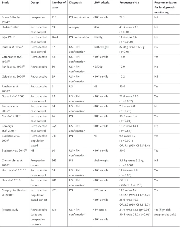
before birth, these pregnancies were more likely to have reached full term despite the presence of growth deviation. Overall, twelve of these studies have made statements regarding the need for fetal growth follow up scans in pregnancies with ISUA: nine recommended routine scans and three considered them unnecessary (table 3).
The present study demonstrates that the likelihood of giving birth to a growth restricted neonate is not in-creased in uncomplicated pregnancies with an isolated single umbilical artery. However, in complicated pregnan-cies, this likelihood seems increased. Nevertheless, due to the limited sample size, and possible additional confoun-ding factors that have not been addressed, this inconfoun-ding needs to be further evaluated.
Due to the tertiary nature of our hospital, our data presents an opportunity to examine the effect of ISUA on fetal growth in uncomplicated pregnancies, and a consi-derable number of cases with preexisting maternal medi-cal and/or pregnancy complications. In the latter sub-group, the odds for fetal growth restriction seem enhanced and speciic antenatal follow up and surveil-lance is advisable after the diagnosis of an ISUA. To the best of our knowledge, this is the irst paper that presents speciic data on the impact of ISUA on birth weight in high-risk pregnancies.
Nevertheless, due to the lack of robust prospective an-tenatal series, the true magnitude and long term impact of an ISUA on perinatal results and posterior childhood deve-lopment are still not clear in these pregnancies, especially in otherwise uncomplicated ones. Prospective collaborative collection of data may help determine the best antenatal management protocols in these cases.
C
ONCLUSIONIsolated single umbilical artery does not increase the risk of fetal growth restriction in uncomplicated singleton pregnancies.
R
ESUMOArtéria umbilical única isolada e restrição do crescimen-to fetal
Objetivo: Examinar a frequência de peso ao nascer
abai-xo dos percentis 5 e 10, em gestações únicas com artéria umbilical única isolada (AUUI), de acordo com a presen-ça de complicações maternas.
Métodos: Estudo caso-controle. De acordo com a
pre-sença de doenças maternas prévias à gestação, ou ocor-rência de complicações obstétricas, as gestações foram classiicadas em “não complicadas” ou de “alto risco”. As cies. This inding suggests that prenatal diagnosis of
sin-gle umbilical artery does not prompt anticipation of delivery, nor adds additional clinically relevant morbidity besides fetal growth deviation.
As a matter of fact, Wiegand et al.17 evaluated fetal growth by repeated ultrasound examinations in 138 preg-nancies following the diagnosis of ISUA. Intrauterine growth restriction was observed in 3% of the cases and the authors concluded that the risk of growth impair-ment is not different from the rest of their population.
Additional clinical relevance of ISUA was also evalua-ted after birth in a prospective cohort by Chetty-John et al.10 No signiicant differences in physical growth and neurological development were observed. There was also no evidence of increased need for admission to neonatal intensive care units (Bombrys et al.).14
In fetuses with two vessels cord, compensatory me-chanisms are in place to allow the single artery to meet the demands for fetal development during late pregnancy. One of these mechanisms is an increase of the arterial diameter. Sepulveda et al. have described that in fetuses
with ISUA, the diameter of the umbilical artery was grea-ter than 50% of the umbilical vein, resulting in an umbi-lical vein to umbiumbi-lical artery ratio ≤ 2.3
Additional adaptive mechanism was suggested by the observation that birth weight centile correlates with both umbilical vein to artery perimeter ratio and impedance to low in the uterine arteries.18 The authors hypothesized that fetal growth restriction associated with ISUA is not based on deicient trophoblast invasion but could be due to chan-ges in the maternal uterine circulation and morphological characteristics of the vessels in the umbilical cord.
Moreover, a recent publication showed that birth weight was lower in left side absent umbilical artery sin-gleton pregnancies compared to controls, but not in right side absent artery cases.19 The authors suggest that diffe-rences between left and right side vascular anatomy pos-sibly also play a role in single umbilical artery fetal adap-tive mechanisms.
Table 3 summarizes the studies that have evaluated the association between ISUA and low birth weight. Ele-ven series are based on cases diagnosed prenatally and signiicant differences in birth weight were demonstra-ted in four out of six studies that included control groups.6,7,11,13-15,18,20-23
TABLE 3 Studies evaluating the association between isolated single umbilical artery and low birth weight
Stud y Design Number of cases
Diagnosis LBW criteria Frequency (% ) Recommendation
for fetal growth monitoring
Bryan & Kohler 197424
prospective 113 PN examination <10th centile 22.1 NS
Heifetz 19844 Retrospective
case-control
69 Autopsy SGA 43.5 versus 23.8 (p<0.01)
NS
Lilja 19915 Retrospective
case-control
1674 PN examination <2500g 11.4 versus 3.6 (p <0.0001)
NS
Jones et al. 19936 Retrospective
case-control
37 US + PN confirmation
Birth weight 2750 g versus 3170 g (p<0.01)
NS
Catanzarite et al. 199520
Retrospective 38 US + PN confirmation
<10th centile 18.0 Yes
Parilla et al. 199521 Retrospective 50 US + PN
confirmation
<2500g 12.0 No
Geipel et al. 200022 Retrospective 59 US + PN
confirmation
<10th centile 10.2 NS
Rinehart et al. 200023
Retrospective 6 US NS 50.0 Yes
Gornall et al. 20037 Retrospective
case-control
83 US + PN confirmation
<10th centile 22.0 versus 12.0
(p <0.007)
Yes
Predanic et al. 200513
Retrospective case-control
84 US + PN confirmation
<10th centile 7.1 versus 4.8 (p= 0.75)
No
Mu et al. 20088 Retrospective
case-control
14 PN <10th centile 35.7 versus 3.6 (p= 0.01)
Yes
Bombrys et al. 200814
Retrospective case-control
255 US + PN confirmation
<10th centile 13.7 versus 13.1
(p= 0.84)
No
Burshtein et al. 20099
Retrospective population based
243 PN NS 9.5 versus 1.9 (p <0.001)
OR 5.4 (95% CI 3.5-8.4) Yes
Bugatto et al. 201018 NS 60 US + PN
confirmation
<10th centile 30.0 Yes
Chetty-John et al. 201010
Prospective cohort
263 PN birth weight 3.1 kg versus 3.2 kg (p <0.0001)
NS
Horton et al. 201015 Retrospective
case-control
68 US + PN confirmation
<10th centile 17.6 versus 8.8
(p= 0.06)
Yes
Hua et al. 201011 Retrospective
cohort
281 US + PN confirmation
<10th centile OR 1.9
(95% CI: 1.4 - 2.5)
Yes
Murphy-Kaulbeck et al. 201012
Retrospective population based-cohort
725 PN <3rd centile
<10th centile
11.1 versus 3.7
OR 2.5 (95% CI 1.9-3.2) 25.0 versus 10.9 OR 2.2 (95% CI 1.8-2.7)
Yes
Present study Retrospective cases and prospective controls
131 US + PN confirmation
<5th centile
<10th centile
21.4 versus 13.6 (p=0.03) 30.5 versus 23.2 (p=0.06)
Yes (high-risk pregnancies only)
frequências de peso ao nascer abaixo dos percentis 5 e 10 foram comparadas entre os subgrupos.
Resultados: O peso ao nascer foi signiicativamente
me-nor em gestações com AUUI (n=134, 2840 ± 701 g) quan-do comparaquan-do com o grupo controle (n= 730, 2983 ± 671 g, p= 0,04; média da diferença=143 g, IC 95% = 17-269). Em gestações de alto risco, peso ao nascer abai-xo do 5º percentil foi signiicativamente mais frequente no subgrupo com AUUI [10/35 (28,6%) versus 53/377
(14,1%), p= 0,04; razão de chances= 2.45 (IC 95% = 1,11-5,38)]; não foi observada diferença em relação ao peso abaixo do 10º percentil (p= 0,11). Em gestações não com-plicadas, não foram observadas diferenças na frequência de peso ao nascer abaixo do 5º e 10º percentis entre os subgrupos com AUUI e cordão com 3 vasos (p= 0,21).
Conclusão: Em gestações de alto risco, a frequência de
peso ao nascer abaixo do percentil 5 é signiicativamente aumentada.
Unitermos: artéria umbilical única, retardo do
cresci-mento intrauterino, peso ao nascer, ultrassonograia.
R
EFERENCES1. Lamberty CO, Burlacchini de Carvalho MH, Miguelez J, Liao AW, Zugaib M. Ultrasound detection rate of single umbilical artery in the irst trimester of pregnancy. Prenatal Diagnosis. 2011;31(9):865-8.
2. Rembouskos G, Cicero S, Longo D, Sacchini C, Nicolaides KH. Single umbilical artery at 11-14 weeks’ gestation: relation to chromosomal defects. Ultrasound Obstet Gynecol. 2003;22(6):567-70.
3. Sepulveda W, Peek MJ, Hassan J, Hollingsworth J. Umbilical vein to artery ratio in fetuses with single umbilical artery. Ultrasound Obstet Gynecol. 1996;8(1):23-6.
4. Heifetz SA. Single umbilical artery. A statistical analysis of 237 autopsy cases and review of the literature. Perspect Pediatr Pathol. 1984;8(4):345-78. 5. Lilja M. Infants with single umbilical artery studied in a national registry.
General epidemiological characteristics. Paediatr Perinat Epidemiol. 1991;5(1):27-36.
6. Jones TB, Sorokin Y, Bhatia R, Zador IE, Bottoms SF. Single umbilical artery: accurate diagnosis? Am J Obstet Gynecol. 1993;169(3):538-40.
7. Gornall AS, Kurinczuk JJ, Konje JC. Antenatal detection of a single umbilical artery: does it matter? Prenat Diagn. 2003;23(2):117-23.
8. Mu SC, Lin CH, Chen YL, Sung TC, Bai CH, Jow GM. The perinatal outcomes of asymptomatic isolated single umbilical artery in full-term neonates. Pediatr Neonatol. 2008;49(6):230-3.
9. Burshtein S, Levy A, Holcberg G, Zlotnik A, Sheiner E. Is single umbilical artery an independent risk factor for perinatal mortality? Arch Gynecol Obstet. 2011;283(2):191-4.
10. Chetty-John S, Zhang J, Chen Z, Albert P, Sun L, Klebanoff M, et al. Long-term physical and neurologic development in newborn infants with isolated single umbilical artery. Am J Obstet Gynecol. 2010;203(4):368.e1-7. 11. Hua M, Odibo AO, Macones GA, Roehl KA, Crane JP, Cahill AG. Single
umbilical artery and its associated indings. Obstet Gynecol. 2010;115(5):930--4.
12. Murphy-Kaulbeck L, Dodds L, Joseph KS, Van den Hof M. Single umbilical artery risk factors and pregnancy outcomes. Obstet Gynecol. 2010;116(4):843--50.
13. Predanic M, Perni SC, Friedman A, Chervenak FA, Chasen ST. Fetal growth assessment and neonatal birth weight in fetuses with an isolated single umbilical artery. Obstet Gynecol. 2005;105(5 Pt 1):1093-7.
14. Bombrys AE, Neiger R, Hawkins S, Sonek J, Croom C, McKenna D, et al. Pregnancy outcome in isolated single umbilical artery. Am J Perinatol. 2008;25(4):239-42.
15. Horton AL, Barroilhet L, Wolfe HM. Perinatal outcomes in isolated single umbilical artery. Am J Perinatol. 2010;27(4):321-4.
16. Alexander GR, Kogan M, Martin J, Papiernik E. What are the fetal growth patterns of singletons, twins, and triplets in the United States? Clin Obstet Gynecol. 1998;41(1):114-25.
17. Wiegand S, McKenna DS, Croom C, Ventolini G, Sonek JD, Neiger R. Serial sonographic growth assessment in pregnancies complicated by an isolated single umbilical artery. Am J Perinatol. 2008;25(3):149-52.
18. Bugatto F, Quintero-Prado R, Melero-Jiménez V, Fajardo-Expósito MA, Hervías-Vivancos B, Bartha JL. Ultrasound predictors of birth weight in euploid fetuses with isolated single umbilical artery. Ultrasound Obstet Gynecol. 2010;36(6):724-7.
19. Jiang Y, Li XH, Yang TZ. The impact of different sides of the absent umbilical artery on fetal growth in an isolated single umbilical artery. Arch Gynecol Obstet. 2013;288(3):531-6.
20. Catanzarite VA, Hendricks SK, Maida C, Westbrook C, Cousins L, Schrimmer D. Prenatal diagnosis of the two-vessel cord: implications for patient counselling and obstetric management. Ultrasound Obstet Gynecol. 1995;5(2):98-105.
21. Parilla BV, Tamura RK, MacGregor SN, Geibel LJ, Sabbagha RE. The clinical signiicance of a single umbilical artery as an isolated inding on prenatal ultrasound. Obstet Gynecol. 1995;85(4):570-2.
22. Geipel A, Germer U, Welp T, Schwinger E, Gembruch U. Prenatal diagnosis of single umbilical artery: determination of the absent side, associated anomalies, Doppler indings and perinatal outcome. Ultrasound Obstet Gynecol. 2000;15(2):114-7.
23. Rinehart BK, Terrone DA, Taylor CW, Isler CM, Larmon JE, Roberts WE. Single umbilical artery is associated with an increased incidence of structural and chromosomal anomalies and growth restriction. Am J Perinatol. 2000;17(5):229-32.