MUELLER WEISSSYNDROME – CASEREPORT
REV ASSOC MED BRAS 2014; 60(2):103-104 103
Image In medIcIne
Mueller Weiss syndrome – case report
S
ÍNDROME DEM
UELLER-W
EISS– R
ELATO DE CASOSergio eliaS NaSSar De Marchi1*, Soraya Silveira MoNteiro2, eloy De avila FerNaNDeS3
1 3rd-year Specializing Physician in Radiology and Imaging diagnosis at Hospital do Servidor Público estadual (Iamspe), São Paulo, SP, Brazil.
2 Radiology Physician at Iamspe’s Radiology Unit, Hospital do Servidor Público estadual, and at the department of Imaging diagnosis of the Federal University of São Paulo (Unifesp), São Paulo, SP, Brazil. 3 Radiology Physician employed by the Hospital do Servidor Público estadual (Iamspe), and physician employed by the department of Imaging diagnosis of the Federal University of São Paulo (Unifesp), São Paulo, SP,
Brazil.
Study carried out by the Hospital do Servidor Público estadual – Francisco morato de Oliveira HSPe/FmO, São Paulo, SP, Brazil.
*Correspondence: address: R. Oswaldo martins, 165 Sorocaba, São Paulo, SP - Brazil Zip code: 18045-490 Phone: +55 15 99790-8977 [email protected]
http://dx.doi.org/10.1590/1806-9282.60.02.005
Conflict of interest: none
C
ASE REPORTMale patient, 51 years old, followed up at the rheumato-logy outpatient clinic of the Hospital do Servidor Público
Es-tadual (Institute of Medical Assistance to Public Servants
of the State – Iamspe-SP), complaining of pain in his feet for 13 years and previous diagnosis of valgus flat foot, fi-bromyalgia, bilateral calcaneocuboid-talonavicular joint osteoarthrosis, and acromioclavicular osteoarthrosis bi-laterally, with no osteonecrosis-related diseases. Now re-ports burning pain in his left foot that worsens with am-bulation in recent weeks. The patient underwent magnetic resonance imaging (MRI) of both feet for diag-nostic investigation.
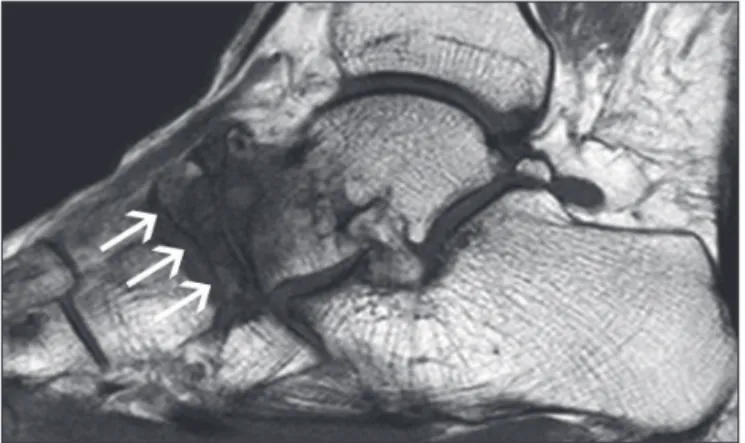
FIGURE 1 T2-weighted sagittal MRI of the left foot weighted in T2 fat sat, demonstrating flattening with deformity similar to a “comma” of the navicular bone (arrows), a pattern of bone edema and subchondral cysts in the talus and navicular bones.
MARCHI SEN ETAL.
104 REV ASSOC MED BRAS 2014; 60(2):103-104
FIGURE 3 T1-weighted sagittal MRI of the right foot, showing thinning with deformity similar to a “comma” of the navicular bone (arrows), a pattern of bone edema and subchondral cysts in the talus and navicular bones.
D
ISCUSSIONThe Mueller-Weiss syndrome is a rare disease that affects adults, usually between 40 and 60 years of age, is more common in women, and is characterized by compression of the navicular bone between the talus and the lateral cuneiform bone, leading to spontaneous osteonecrosis
of the navicular bone.¹ Symptoms are chronic, with seve-re pain in the midfoot and progseve-ressive deformity.²
Not to be confused with Koehler disease (osteonecro-sis of the navicular bone in children).³ When diagnosed early, disease progression can be prevented through non--surgical treatments, producing an improvement in the
patient’s quality of life.¹
The radiographic stages range from minimal chan-ges to the navicular bone, medial or dorsal deviation of part or the entire navicular bone, and a comma-shaped deformity, due to the collapse of the lateral portion un-til complete fragmentation of the navicular bone, for-ming a talocuneiform joint in more severe cases.³ Bilate-ral findings, asymmetric involvement and association with pathological fractures are common.²
R
EFERENCES1. Quintella DC, Calmon TR, Guimarães MF, Dos Santos AASMD, Reis M. Síndrome de Müeller-Weiss: aspectos radiográficos. Radiol Bras 2009; 42(Supl 1):1-115.
2. Rosenberg ZS, Beltran J, Bencardino JT. From the RSNA Refresher Courses. Radiological Society of North America. MR imaging of the ankle and foot. Radiographics 2000; 20(Spec No.):S153-79.