ABSTRACT
BACKGROUND AND OBJECTIVES: Amelogenesis
imper-fecta is characterized by enamel structural defects, which may severely afect dental structure in both dentitions. When accom-panied by severe decay, it may impair the pulp complex requiring endodontic treatment and in case of incomplete root formation treatment becomes more complex due to pulp volume and ana-tomic conditions. his study aimed at reporting a clinical case of patient with amelogenesis imperfecta and with multiple incom-plete root formation as a consequence.
CASE REPORT:Female patient, 12 years old, leucodermic, with painful symptoms, who has looked for dental assistance. At intraoral clinical evaluation teeth presented with shape and size changes, yellowish color, covered by a thin enamel layer with roughened surface and absent in some areas, with anterior ves-tibular sulcus istula and without edema. At radiographic eva-luation, both dentitions were afected by the abnormality with delayed chronology of permanent teeth eruption. At the end of all evaluations, patient was diagnosed with amelogenesis imper-fecta, and periodontal treatment was started, followed by endo-dontic treatment.
CONCLUSION: Amelogenesis treatment is complex, especially when in more advanced stages of dental structure destruction. However, it is possible to reestablish patient’s functionality and esthetics with good planning and multidisciplinary approach. Keywords: Amelogenesis imperfecta, Endodontic treatment, In-complete root formation, Mineral trioxide aggregate, Pain.
Intracanal pain remission in child with amelogenesis imperfecta. Case
report*
Remissão de dor intracanal em criança com amelogênese imperfeita. Relato de caso
Armiliana Soares Nascimento1, Criseuda Maria Benício Barros2, Renata de Souza Coelho Soares3, Luciana de Barros Correia
Fontes4, Rodivan Braz1
*Received from State University of Paraíba, Campina Grande, PB, Brazil.
1. Faculdade de Odontologia de Pernambuco, Recife, PE, Brasil. 2. Universidade Estadual da Paraíba, Campina Grande, PB, Brasil. 3. Universidade Federal de Campina Grande, Campina Grande, PB, Brasil. 4. Universidade Federal de Pernambuco, Recife, PE, Brasil.
Submitted in July 26, 2014.
Accepted for publication in July 31, 2015.
Conlict of interests: none – Sponsoring sources: none.
Correspondence to: Armiliana Soares Nascimento Rua Baraúnas, 351 - Bairro Universitário 58429-500 Campina Grande, PB, Brasil. E-mail: [email protected]
© Sociedade Brasileira para o Estudo da Dor
RESUMO
JUSTIFICATIVA E OBJETIVOS: A amelogênese imperfei-ta é caracterizada por defeitos estruturais do esmalte, os quais podem acometer gravemente a estrutura dentária, em ambas as dentições. Quando acometida por lesões cariosas graves, pode comprometer o complexo pulpar, necessitando de tratamento endodôntico, e em se tratando de dentes com RI, o tratamento torna-se mais complexo, em virtude do volume pulpar e das con-dições anatômicas. O objetivo deste estudo foi relatar um caso clínico de paciente com AI e que teve como consequência múl-tiplos casos de RI.
RELATO DO CASO: Paciente do gênero feminino, 12 anos, leucoderma, com sintomas dolorosos procurou atendimento odontológico. Ao exame clinico intraoral, apresentava dentes com alteração de forma e tamanho, coloração amarelada, reco-bertos por uma ina camada de esmalte com superfície rugosa e em algumas áreas ausente, com fístula na região do sulco ves-tibular anterior e sem edema. Ao exame radiográico ambas as dentições mostraram-se acometidas por anomalia, com retardo na cronologia de erupção dos dentes permanentes. Ao inal de todos os exames, recebeu o diagnóstico de amelogênese imper-feita, dando inicio ao tratamento, que começou pelo tratamento periodontal, seguido pelo endodôntico.
CONCLUSÃO: O tratamento da amelogênese é complexo, principalmente quando em estágios mais avançados de destrui-ção da estrutura dentária. Contudo é possível restabelecer a fun-cionalidade e a estética do paciente quando se faz um bom pla-nejamento do caso, com interação multidisciplinar.
Descritores: Agregado trióxido mineral, Amelogênese imperfei-ta, Dor, Rizogênese incompleimperfei-ta, Tratamento endodôntico.
INTRODUCTION
In spite of oral health quality improvements seen in recent years, toothache is still a public health problem and is re-ported as major reason for individuals, including children, to look for dental assistance1. Major pain causes are dental
trauma and infectious diseases, being caries the most com-mon2. Other abnormalities, such as amelogenesis imperfecta
(AI) may severely affect dental structure in both dentitions and, when affected by severe caries, it impairs pulpar com-plex preventing complete apical closing and characterizing a
presentation of incomplete root formation (IRF)3.
Endodontic treatment of ICF is complex due to teeth ana-tomic characteristics. Root canal is wide, with little thi-ckness of dentinal walls, lack of constriction and apical divergence4.
During obturation process, the most important factor is apical constriction, since it limits control over the exten-sion of filling materials. With this, root canal lacks an ade-quate retention factor to prevent filling material shift and leakage at apical level5.
Mineral trioxide aggregate (MTA), powder material made of tricalcium silicate particles, tricalcium oxide and silicate oxide, has been broadly indicated by the literature6,7. It has
low solubility, good biological action, stimulates cell repair and cell adhesion, has antimicrobial action, in addition to being hydrophilic with ability to become a colloidal gel crystallizing with expansion, favoring marginal sealing ca-pacity, in addition to inducing the formation of an apical barrier of hard tissue in IRF teeth8. MTA adapts to canal
shape as portions are gradually inserted in it9.
This study aimed at reporting a clinical case of patient with AI having as consequence multiple IRF.
CASE REPORT
Female patient, 12 years old, leukodermatous, who looked for dental assistance in the dental-pediatric clinic of the State University of Paraíba accompanied by the mother. Mother complained of daughter’s teeth appearance, stating that they were yellowish, misshapen and with abnormal size, in addition to painful symptoms reported by the child. During history, no relevant data indicating some cor-relation with the clinical presentation were reported. In addition, the mother did not know whether some other family member had the same problem. At intraoral clinical evaluation, we have observed teeth with changes in shape
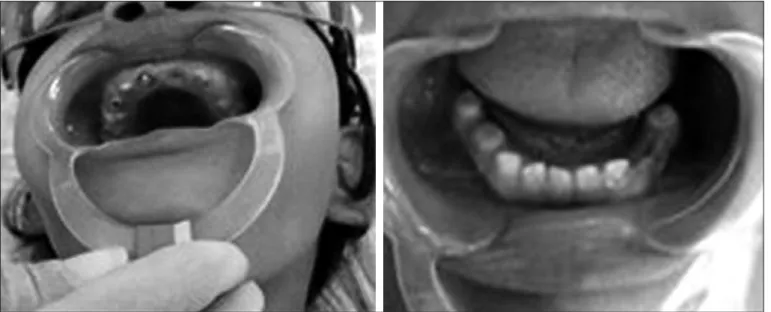
and size (upper anterior teeth crowns totally destroyed by caries, gingival hyperplasia and exposure of pulpar canal), yellowish color, covered by a thin layer of enamel with rou-gh surface and absent in some areas (Figure 1) and presen-ce of fistula in the anterior vestibular groove region with no edema.
At occlusion evaluation child had no proximal contact be-tween teeth and anterior open bite was observed with seve-re loss of occlusion vertical dimension (OVD).
X-rays have revealed lack of dental enamel in some sites and a thin layer of tissue in others, presence of radiolucent area suggestive of extensive periapical injury, incomplete root formation and open apex. After pulpar vitality tests, pulpar necrosis was diagnosed. We could also observe by panoramic X-rays that both dentitions were affected by the abnormality, as well as that there was delay in eruption of permanent teeth (Figure 2).
In light of was observed, and based on history, clinical and radiographic evaluations, and in the absence of sys-temic changes which could justify enamel malformation, we have confirmed the diagnosis of the abnormality called
Figure 1. Initial intraoral aspect
“hypoplastic-type amelogenesis imperfecta”10. Before
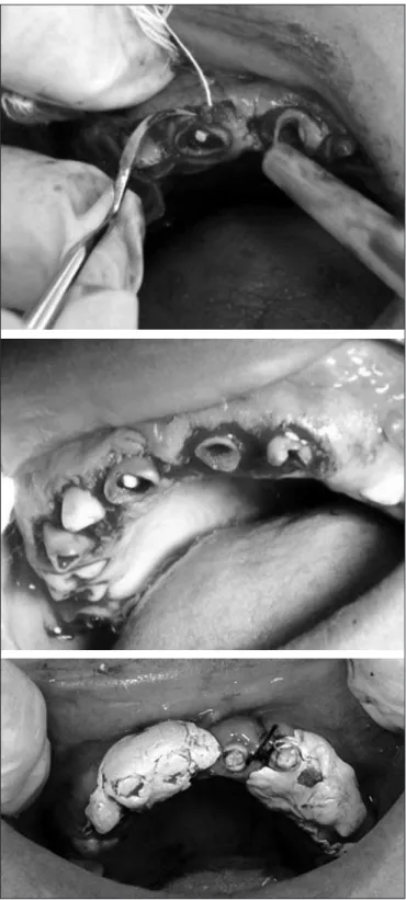
star-ting endodontic treatment, clinical crown from canine to canine was increased so that the rubber drape could be adapted for absolute insulation, since the crown had been destroyed due to both enamel brittleness and the caries process (Figure 3).
All compromised teeth were chemically-mechanically pre-pared, which was made difficult by thin and brittle walls,
since larger tools would circulate freely preventing conven-tional canal conformation. The canal was cleaned with file 80 and abundant irrigation with 0.5% sodium hypochlori-te. After preparation, root canal was dried with sterile ab-sorbent paper, intracanal drug with tricresolformalin and temporary sealing with glass ionomer. In the second ses-sion, biomechanics, canal drying and filling with calcium hydroxide were performed, being these clinical procedures performed every 15 days for four months, due to bleeding when renewing calcium hydroxide, fact which has preven-ted the obturation of root canals in a shorter clinical time. Finally, obturation was performed with MTA apical patch, aiming at establishing effective apical sealing and tissue repair with posterior obturation by the lateral condensa-tion technique using gutta-percha cone and endodontic cement – endofill (Figure 4). Preservation was carried out in 3 months, 6 months and 1 year, aiming at following up the treatment.
Figure 4. Radiographic control six month after
After adequate tissue healing, upper prosthesis was ma-nufactured to recover lost DVO due to excessive corona-ry destruction. Added to this, oral hygiene guidance was constantly given to better control gingival inflammatory process.
Acute pain due to dentinal hypersensitivity, to gengivitis and pulpitis was controlled during treatment and at the end patient had no pain whatsoever, both during brushing and chewing as well as spontaneous pain. An important factor for the success of pain remission were oral hygiene guidance and patient’s cooperation in the maintenance of periodontal health and caries prevention.
DISCUSSION
Toothache is a public health problem11 due to its high
pre-valence and its social, psychological and economic conse-quences for individuals and the community, with quality of life impairment. So, family characteristics, such as pa-rents perception of their oral health and of their children, oral hygiene and diet knowledge and habits are identified as indicators of oral health and, probably, of toothache12,13.
Among major causes of pain, caries has been the most stron-gly associated variable and although there has been
cant decrease in caries prevalence in Brazil in recent years in deciduous dentition, this decrease was lower than that on permanent dentition14. On the other hand, patients with
teeth mineralization disorders, such as AI, in addition to negative cosmetic effects, very often report significantly hi-gher levels of distress and are more sensitive and anxious to dental treatment-related pain15.
AI is a dental enamel change affecting both deciduous and permanent dentition, with several clinical variations in terms of severity16. It is present in the absence of systemic
charac-teristics, having several phenotypic variations which affect dental enamel. Clinically, enamel is hypoplastic (thin layer of enamel) hypomineralized (subdivided in hypomaturation and hypocalcification), or with combined phenotype10.
This is a case of a child with clinical aspects characteris-tic of generalized AI. This is an autossomal, recessive and rare disease, characterized by mild gingival hyperplasia and dental abnormalities, including generalized hypoplastic AI, intrapulpar calcifications and delayed dental eruption. A similar case was described by Martelli-Junior et al.17 where
four patients of the same family had the same abnormality and one patient had intellectual deficit.
AI diagnosis is slightly complex, because it presents a set of problems, such as rampant caries, impairment of ver-tical occlusion dimension, eruption abnormalities, dental sensitivity, in addition to psychosocial problems18. So, the
treatment depends on type and severity of the disorder, on factors as age, socioeconomic level and oral health of the patient at planning moment.
Among treatment options there are multiple dental extrac-tions, cosmetic restoraextrac-tions, steel or compound resin cro-wns, splints to reestablish vertical dimension, control of dentinal sensitivity and oral hygiene guidance, being the multiprofessional approach a critical factor for the success of the treatment19.
When teeth are affected by caries impairing pulpar plex before total root formation, we have a still more com-plex process, considering that these, by themselves, are brittle structures due to anatomic peculiarities. Factors such as less pulpar cavity volume, loss of dental structure, little thickness of root walls, wide open apices, brittle root walls and diverging to periapical tissues, give brittleness to teeth with incomplete root formation (IRF). And when necrotized, the treatment is apexification, which is induc-tion of apical closing to produce favorable condiinduc-tions for conventional obturation of the root canal20. Apexification
assures complete and functional healing of involved too-th or teetoo-th21, by preventing or decreasing extrusion of the
material to periodontal tissues, decreasing inflammatory process persistence22.
For a long time, preferred drug was calcium hydroxide, for having high success rates, in spite of the risk of reinfec-tion and tooth fracture. With the introducreinfec-tion of MTA as apical sealer, for its capacity of inducing the formation of mineralized tissue, of attaching well and being used in wet environment or in the presence of blood, it was possible
to treat IRF teeth with apical plug and treatment conclu-sion in a short period of time23. MTA has several clinical
applications due to its adequate physical properties, good sealing capacity and biocompatibility24
According to Witherspoon et al.25, success rate for
apexi-fication procedures with calcium hydroxide is 79 to 96%, while with MTA it is 81 to 100%. So, the apical barrier with MTA is an effective procedure for the apexification process26, due to its inherent properties. On the other
hand, Torabinejad et al.27 have reported antimicrobial
pro-perties of MTA for 5 out of 9 facultative bacteria more commonly found in infected root canals, however with no effect on strictly anaerobic bacteria. Annamalai & Munga-ra28 have also reported 100% clinical and radiological
suc-cess rate with MTA in the 12th month of follow-up, while
root extremity closing was observed in 86.6% of cases with root growth in just 30% of cases.
In our case, the combination of calcium hydroxide was used as intracanal drug for 15 days to obtain a dry and infection-free canal. In line with Bondanezi et al.29, who
state that calcium hydroxide apical barrier improves the quality of filling and sealing of canals of IRF teeth obtu-rated with mineral trioxide aggregate. The use of calcium hydroxide in the short term does not negatively affect too-th resistance30.
In our case, X-rays after six months of treatment has shown significant decrease in periapical lesions, being in agree-ment with studies of Annamalai & Mungara28 and Sarris et
al.31. However, the latter suggest broader clinical trials to
evaluate long term success. In our case, apical barrier had 5 mm for being, according to some authors, stronger than that of 2 mm32,33.
To induce apexification, MTA acts converting tricalcium oxide into calcium hydroxide when in contact with tissue fluids28. Calcium hydroxide is then dissociated in calcium
and hydroxyl ions. This separation increases pH and releases calcium ions. The latter reacts with tissue carbon dioxide, forming calcium carbonate as calcite crystals. On the other hand, fibronectin is associated to such crystals, providing cell adhesion and subsequent differentiation which results in mineralized tissue barrier. Released calcium also plays im-portant role in decreasing inflammation and differentiating and mineralizing pulpar cells. In this process, gingival fibro-blasts induce bone repair and cementogenesis34.
MTA for apexification has advantages as compared to cal-cium hydroxide since its attachment is immediate. So, soon after placement, the canal may be definitely obturated with endodontic cement and gutta-percha cones, decreasing tre-atment time35.
CONCLUSION
REFERENCES
1. Schuch HS, Correa MB, Torriani DD, Demarco FF, Goettems ML. Perceived dental pain: determinants and impact on Brazilian schoolchildren. J Oral Facial Pain Heada-che. 2015;29(2):168-76.
2. Boeira, GF, Correa MB, Peres KG, Peres MA, Santos IS, Matijasevich A, et al. Caries is the main cause for dental pain in childhood: indings from a birth cohort. Caries Res. 2012;46(5):488-95.
3. de Oliveira DM, de Souza Andrade ES, da Silveira MM, Camargo IB. Correlation of the radiographic and morphological features of the dental follicle of third molars with incomplete root formation. Int J Med Sci. 2008;5(1):36-40.
4. Vale MS, Silva PM. Endodontic conduct post trauma in teeth with incomplete root formation. Rev Odontol UNESP. 2011;40(1):47-52.
5. Moore A, Howley MF, O’Connell AC. Treatment of open apex teeth using two types of white mineral trioxide aggregate after initial dressing with calcium hydroxide in children. Dent Traumatol. 2011;27(3):166-73.
6. Hayashi M, Shimizu A, Ebisu S. MTA for obturation of mandibular central incisors with open apices: case report. J Endod. 2004;30(2):120-2.
7. Castro AN, Oliveira DC, Diniz LN, Eulalia AS, Paulillo LA, Pereira GD. Avaliação da utilização de MTA como plug apical em dentes com ápices abertos. Rev Bras Odontol. 2011;68(1):59-63.
8. Shabahang S, Torabinejad M, Boyne PP, Abedi H, McMillan P. A comparative study of root-end induction using osteogenic protein-1, calcium hydroxide, and mineral trioxide aggregate in dogs. J Endod. 1999;25(1):1-5.
9. Hachmeister DR, Schindler WG, Walker WA 3rd, homas DD. he sealing ability and retention characteristics of mineral trioxide aggregate in a model of apexiication. J Endod. 2002;28(5):386-90.
10. Morgado CL, Azul AC. A amelogénese imperfeita – Uma revisão da literatura. Rev Port Estomatol Cir Maxilofac. 2009;50(4):243-50.
11. Pau A, Baxevanos KG, Croucher R. Family structure is associated with oral pain in 12-year-old Greek schoolchildren. Int J Paediatr Dent. 2007;17(5):345-51. 12. Shearer DM, homson WM. Intergenerational continuity in oral health: a review.
Community Dent Oral Epidemiol. 2010;38(6):479-86.
13. Dye BA, Vargas CM, Lee JJ, Magder L, Tinanof N. Assessing the relationship between children’s oral health status and that of their mothers. J Am Dent Assoc. 2011;142(2):173-83.
14. Brasil: Primeiros Resultados do Projeto SBBrasil. nuppiim-uefs.blogspot.com/.../ sintese-dos-primeiros-resultados-do.html.
15. Pousette Lundgren G, Morling Vestlund GI, Trulsson M, Dahllöf G. A Randomized Controlled Trial of Crown herapy in Young Individuals with Amelogenesis Imper-fecta. J Dent Res. 2015;94(8):1041-7.
16. Marquezin MC, Zancopé BR, Pacheco LF, Gavião MB, Pascon FM. Aesthetic and functional rehabilitation of the primary dentition afected by amelogenesis imperfecta. Case Rep Dent. 2015;2015:790890.
17. Martelli-Júnior H, Bonan PR, Dos Santos LA, Santos SM, Cavalcanti MG, Coletta RD. Case reports of a new syndrome associating gingival ibromatosis and dental abnormalities in a consanguineous family. J Periodontol. 2008;79(7):1287-96.
18. Lourenço Neto L, Paschoal MA, Kobayashi TY, Rios D, Silva SM. Early oral rehabili-tation of a child with amelogenesis imperfecta. J Health Sci Inst. 2010;28(3):246-8. 19. Seow KW. Clinical diagnosis and management strategies of amelogenesis imperfecta
variants. Pediat Dent. 1993;15(6):384-93.
20. Rafter M. Apexiication: a review. Dent Traumatol. 2005;21(1):1-8.
21. Pawar AM, Kokate SR, Shah RA. Management of a large periapical lesion using Bio-dentine (™) as retrograde restoration with eighteen months evident follow up. J Con-serv Dent. 2013;16(6):573-5.
22. Rudagi KB, Rudagi B. One-step apexiication in immature tooth using grey mineral trioxide aggregate as an apical barrier and autologus platelet rich ibrin membrane as an internal matrix. J Conserv Dent. 2012;15(2):196-9.
23. Batista A, Sydney GB, Deonizio MD. Analise “in vitro” da viabilidade do uso do MTA e do hidróxido de cálcio como plug apical em dentes com rizogênese incompleta. ROBRAC. 2007;16(42).
24. Malik G, Bogra P, Singh S, Samra RK. Comparative evaluation of intracanal sealing ability of mineral trioxide aggregate and glass ionomer cement: an in vitro study. J Conserv Dent. 2013;16(6):540-5.
25. Witherspoon DE, Small JC, Regan JD, Nunn M. Retrospective analysis of open apex teeth obturated with mineral trioxide aggregate. J Endod. 2008;34(10):1171-6. 26. Brito-Júnior M, Ferreira A, Oliveira GL, Xavier LR, Xavier LA, Guerra PN, et al.
Evidências clínicas da técnica de apiciicação utilizando barreira apical com agregado trióxido mineral – uma revisão crítica. RFO. 2011;16(1):54-8.
27. Torabinejad M, Hong CU, McDonald F, Pitt Ford TR. Physical and chemical proper-ties of a new root-end illing material. J Endod. 1995;21(7):349-53.
28. Annamalai S, Mungara J. Eicacy of mineral trioxide aggregate as an apical plug in non-vital young permanent teeth: preliminary results. J Clin Pediatr Dent. 2010;35(2):149-55.
29. Bondanezi A, Munhoz EA, Cornejo AD, Bernardineli N, Moraes IG, Bramante CM. Efeito tampão apical selador date obturações com agregado trióxido mineral em den-tes com rizogênese incompleta. R Clin Pesq Odont. 2009;5(3):263-6.
30. Hasheminia SM, Norozynasab S, Feizianfard M. he efect of three diferent calcium hydroxide combinations on root dentin microhardness. Res J Biol Sci. 2009;4(1):121-5. 31. Sarris S, Tahmassebi JF, Duggal MS, Cross IA. A clinical evaluation of mineral trioxide aggregate for root-end closure of non-vital immature permanent incisors in children-a pilot study. Dent Traumatol. 2008;24(1):79-85.
32. Matt GD, horpe JR, Strother JM, McClanahan SB. Comparative study of white and gray material trioxide aggregate (MTA) simulating a one- or two-step apical barrier technique. J Endod. 2004;30(12):876-9.
33. Chhabra N, Singbal KP, Kamat S. Successful apexiication with resolution of the pe-riapical lesion using mineral trioxide aggregate and demineralized freeze-dried bone allograft. J Conserv Dent. 2010;13(2):106-9.
34. Guven G, Cehreli ZC, Ural A, Serdar MA, Basak F. Efect of mineral trioxide ag-gregate cements on transforming growth factor 1 and bone morphogenetic protein production by human ibroblasts in vitro. J Endod. 2007;33(4):447-50.