517
IMAGE IN MEDICINE
Rev Assoc Med Bras 2012; 58(5):517-518
Phantom tumor of the lung
BÁRBARA SANTANA D´AVILA MELO1, ANA CAROLINA MARTINS SERRA1, MÁRCIA TERESA CARREIRA TEIXEIRA BELO2, EPAMINONDAS BELO NETO3,
SAULO MAIA D’AVILA MELO4
1 Intern, Internal Medicine, Santa Casa da Misericórdia do Rio de Janeiro, Rio de Janeiro, RJ, Brazil
2 MSc in Gastroenterology; PhD Student, Postgraduate Course in Internal Medicine, Universidade Federal do Rio de Janeiro (UFRJ); Assistant Professor, Internal Medicine Clinic,
Universidade Gama Filho (UGF) and Fundação Técnico-Educacional Souza Marques, Rio de Janeiro, RJ, Brazil
3 Pneumologist; Assistant Professor, Internal Medicine, UGF and Fundação Técnico-Educacional Souza Marques, Rio de Janeiro, RJ, Brazil
4 MSc and PhD in Medical Sciences, Universidade Federal de Sergipe (UFS); Assistant Professor, Medical School, Universidade Tiradentes, Aracaju, SE, Brazil
Study conducted at Hospital Geral da Santa Casa da Misericórdia do Rio de Janeiro (SCMRJ) – 7ª Enfermaria – Serviço do Prof. André Filipe Marcondes Vieira, Rio de Janeiro, RJ, Brazil
Correspondence to: Bárbara Santana D´Avila Melo – Rua Piauí, 43/13, Higienópolis, São Paulo, SP, Brazil, CEP: 01241-001 – [email protected]
©2012 Elsevier Editora Ltda. All rights reserved.
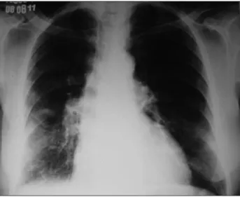
Figure 1 – PA (A) and lateral (B) chest X-rays show a well-delineated phantom tumor of the lung in the right horizontal fissure, enlarged cardiac silhouette and heterogeneous opacities in the lower thirds bilaterally, with opacification of the costophrenic sinuses, especially to the right.
he patient, S.B.E., was a 79-year-old black male, wid-owed, retired metallurgical employee from Espírito Santo, who had been living in Nova Iguaçu-RJ for 40 years, with uncontrolled hypertension and diabetes. He was admitted with a history of progressive dyspnea over the previous two months and currently had dyspnea at rest, orthopnea, paroxysmal nocturnal dyspnea, dry cough, and lower-limb edema.
At physical examination he was hydrated, afebrile, an-icteric, acyanotic, tachypneic, and had normal skin color, jugular venous distension, and generalized body edema (anasarca). His respiratory system presented reduced ve-sicular murmur in the right lung and bibasilar crackles. His cardiovascular system showed presence of third heart sound, muled heart sounds, and systolic murmur in the mitral area ++/6+. His abdomen was rounded, with wall edema, and slightly painful hepatomegaly. he lower limbs presented edema 4+/4+, free calves, and preserved pulses.
Laboratory tests showed no alterations, except for fast-ing glucose: 117 mg/dL. Posteroanterior (AP) and lateral chest X-rays showed an enlarged cardiac silhouette, bulky homogeneous rounded opacity (mass type) with well-de-ined borders, near the right horizontal issure, and het-erogeneous opacities in the lower thirds bilaterally, with opaciication of the costophrenic sinuses, especially to the right (Figure 1A and 1B). Electrocardiogram showed let atrial overload, second-degree let bundle branch block (LBBB), irst-degree atrioventricular block (AVB), and ventricular extrasystoles. he echocardiogram showed sig-niicant systolic dysfunction, difuse hypokinesis, ejection fraction of 28%, moderate mitral regurgitation, mild tri-cuspid regurgitation, and evaluation of diastolic function impaired by frequent extrasystoles.
518
IMAGEINMEDICINE
Rev Assoc Med Bras 2012; 58(5):517-518
Figure 2 – Complete radiological resolution after seven days of treatment for congestive heart failure.
he patient showed satisfactory improvement with the use of loop and potassium-sparing diuretics, angiotensin-converting enzyme inhibitors, and beta-blockers, com-pensating the CHF picture.
he diagnosis of phantom tumor was conirmed with the disappearance of the radiological image located at the right horizontal issure ater seven days of treatment (Figure 2); the patient was clinically compensated when discharged, and was referred to outpatient follow-up.
he diagnosis of phantom tumor is facilitated when there is evidence of luid in the large pleural cavity. he phantom tumor is not always located in the horizontal is-sure to the right; it can also be less oten located to the let, or close to the mediastinum4.
he radiological appearance of the phantom tumor is variable, depending on the volume of septated liquid and its location4. It oten presents as a homogeneous spherical
or elliptical opacity at the horizontal issures, with well-de-ined borders3-5. Lateral chest X-ray is important to better
determine the location of the lesion in the pleural issures. Ater intravenous infusion of potent diuretics, radio-logical resolution can be observed in less than 24 hours. Injury recurrence can happen whenever there are subse-quent cardiac decompensations4.
he early identiication of this radiological inding re-lated to CHF is important to prevent unnecessary diagnos-tic procedures and therapeudiagnos-tic errors, as the main difer-ential diagnosis is pulmonary nodule and/or mass.
REFERENCES
1. Capone D, Lopes AJ, Marsico GA, Kirk KM, Carvalho MN, Jansen U, et al. Doenças de pleura, mediastino e diafragma. In: Lopes AC, editor. Tratado de clínica médica. 2ª ed. São Paulo: Roca; 2009. v. 2 p. 2754-70.
2. Millard CE. Vanishing or phantom tumor of the lung; localized interlobar ef-fusion in congestive heart failure. Am Coll Chest Phys. 1971;59(6):675-77. 3. Freitas LO, Nacif MS, Petrelli ASC. Atelectasia e derrame pleural. In:
Freitas LO, Nacif MS, editors. Radiologia prática para o estudante de medicina. Rio de Janeiro: Revinter; 2003. v. 2 p. 73-86.
4. Lopes AJ, Jansen U, Capone D, Neves DD, Jansen JM. Diagnóstico de falsos tumores do pulmão. Pulmão (RJ). 2005;14(1):33-42.
5. Escuissato DL, Marchiori E, Warszawiak D. Radiograia simples do tórax. In: Barreto SSM, Fiterman J, Lima MA. Prática pneumológica. Sociedade Bra-sileira de Pneumologia e Tisiologia. Rio de Janeiro: Guanabara Koogan; 2010. p. 55-75.