The Tyrosine Phosphatase PTPN14 Is a Negative
Regulator of YAP Activity
Chrysiis Michaloglou1, Waltraut Lehmann1¤, Typhaine Martin1, Clara Delaunay1, Andreas Hueber1, Louise Barys1, Honglin Niu3, Eric Billy1, Markus Wartmann1, Moriko Ito1, Christopher J. Wilson2, Mary Ellen Digan3, Andreas Bauer4, Hans Voshol4, Gerhard Christofori5, William R. Sellers6,
Francesco Hofmann1, Tobias Schmelzle1*
1Novartis Institutes for BioMedical Research, Disease Area Oncology, Basel, Switzerland,2Novartis Institutes for BioMedical Research, Developmental and Molecular Pathways, Cambridge, Massachusetts, United States of America,3Novartis Institutes for BioMedical Research, Center for Proteomic Chemistry, Cambridge, Massachusetts, United States of America,4Novartis Institutes for BioMedical Research, Developmental and Molecular Pathways, Basel, Switzerland,5Department of Biomedicine, Institute of Biochemistry and Genetics, University of Basel, Basel, Switzerland, 6Novartis Institutes for BioMedical Research, Disease Area Oncology, Cambridge, Massachusetts, United States of America
Abstract
The Hippo (Hpo) pathway is a novel signaling pathway that controls organ size in Drosophila and mammals and is deregulated in a variety of human cancers. It consists of a set of kinases that, through a number of phosphorylation events, inactivate YAP, a transcriptional co-activator that controls cellular proliferation and apoptosis. We have identified PTPN14 as a YAP-binding protein that negatively regulates YAP activity by controlling its localization. Mechanistically, we find that the interaction of ectopic YAP with PTPN14 can be mediated by the respective WW and PPxY motifs. However, the PTPN14 PPxY motif and phosphatase activity appear to be dispensable for the negative regulation of endogenous YAP, likely suggesting more complex mechanisms of interaction and modulation. Finally, we demonstrate that PTPN14 downregulation can phenocopy YAP activation in mammary epithelial cells and synergize with YAP to induce oncogenic transformation.
Citation:Michaloglou C, Lehmann W, Martin T, Delaunay C, Hueber A, et al. (2013) The Tyrosine Phosphatase PTPN14 Is a Negative Regulator of YAP Activity. PLoS ONE 8(4): e61916. doi:10.1371/journal.pone.0061916
Editor:Lucia R. Languino, Thomas Jefferson University, United States of America
ReceivedAugust 24, 2012;AcceptedMarch 18, 2013;PublishedApril 16, 2013
Copyright:ß2013 Michaloglou et al. This is an open-access article distributed under the terms of the Creative Commons Attribution License, which permits unrestricted use, distribution, and reproduction in any medium, provided the original author and source are credited.
Funding:All research costs were covered by Novartis AG. No additional external funding was received for this study. The funder had no role in study design, data collection and analysis, decision to publish, or preparation of the manuscript.
Competing Interests:The authors have read the journal’s policy and have the following conflicts: CM, WL, TM, CD, AH, LB, HN, EB, MW, MI, CJW, MED, AB, HV, WRS, FH and TS are or were employees of Novartis Pharma AG. GC declares no conflict of interest. This study was carried out as part of a postdoctoral project within Novartis and in collaboration with the academic mentor GC. There are no patents, products in development or marketed products to declare. This does not alter the authors’ adherence to all the PLOS ONE policies on sharing data and materials.
* E-mail: [email protected]
¤ Current address: University of Freiburg, Freiburg, Germany
Introduction
The Hippo (Hpo) pathway controls organ size by regulating cellular proliferation and apoptosis [1–3]. The pathway was originally described inDrosophilaand comprises several kinases that are conserved in mammals. In fact, mammalian homologues of the pathway can rescue the phenotypes of corresponding Drosophila mutants reflecting the high degree of cross-species homology found in most Hpo pathway components [4–7]. Activation of the core mammalian pathway, consisting of MST-SAV and LATS-MOB complexes results in phosphorylation and inactivation of the transcriptional co-activator YAP and its family member TAZ, by means of cytoplasmic retention. Various proteins have been suggested to activate the pathway in response to contact inhibition. Members of adherens and tight junctions as well as proteins involved in cell polarity have recently been shown to regulate both YAP/TAZ activity and localisation as well as upstream compo-nents of the Hpo pathway, thus linking cell contact and polarity to cellular proliferation [8–10].
Consistent with its role in cellular proliferation, the Hippo pathway is implicated in cancer development [11]. Many
components of the pathway have been found deregulated in human tumours and YAP itself is shown to act as an oncogene in various settings [1,2,12,13]. Furthermore, the Hippo pathway has been implicated in development as well as the maintenance of stem cell populations in adult organisms [14,15].
Materials and Methods
Cell culture and lentiviral transductions
293A cells and 293-FT cells were obtained from Invitrogen (Invitrogen, CA, USA). Both cell lines were maintained in DMEM supplemented with 10% (v/v) fetal calf serum (Thermo Scientific; MA, USA), 2 mM L-Glutamine, 1 mM sodium pyruvate and 0.1 mM MEM non-essential amino acids. SF268 cells were purchased from the NCI DCTD tumor/cell line repository (www.dtp.nci.nih.gov) and were maintained in RPMI-1640 supplemented with 10% (v/v) fetal calf serum, 2 mM L-Glutamine, 1 mM sodium pyruvate and 0.1 mM MEM non-essential amino acids. MCF-10A cells were obtained from ATCC (VA, USA) and maintained in DMEM/F12 medium supplement-ed with 5% horse serum (Invitrogen, CA, USA), 20 ng/ml EGF, 500 ng/ml hydrocortisone, 100 ng/ml Cholera toxin, 10mg/ml insulin (all from SIGMA; MO, USA), 2 mM L-Glutamine, 1 mM sodium pyruvate and 0.1 mM MEM non-essential amino acids. Tetracycline-free FCS was used for SF268 cell lines containing doxycycline-inducible constructs (Takara Bio Europe/Clontech, France).
For lentiviral transductions, 293-FT cells were transfected with a DNA mix containing the plasmid of interest, pVPRD8.71 and pVSVG at a ratio of 12.5:12.5:1 using Lipofectamine 2000 (Invitrogen, CA) according to the manufacturer’s protocol. Virus was harvested after 48 h, filtered with a 0.45-mm filter and stored
at 280uC. After every viral infection, stable cell lines were established by pharmacological selection.
QPCR-based copy number analysis
The QPCR-based copy number analysis was carried out as previously described [16]. Primers for YAP are: TGTAGTGG-CACCTATCACTC and CCATCTCATCCACACTGTTC and were used at a concentration of 300 nM.
Cloning and plasmids
YAP1 (NM_001130145; ORF length of 1515 nucleotides) was purchased from Invitrogen (Clone IOH26027; corresponds to YAP1-2c, [17]). The YAP WW mutants were generated using the QuikChange II Site-Directed Mutagenesis Kit (Agilent Technol-ogies, CA, USA) according to the manufacturer’s protocol so that the resulting protein sequence for WW1 was mutated from W199-Q-D-P202 to A199-Q-D-A202 and the protein sequence of WW2 was mutated from W258-L-D-P261 to A258-L-D-A261. The YAP
DPDZ mutant was generated by PCR resulting in a CDS lacking the last 241 nt of WT YAP. PTPN14 (NM_005401) was chemically synthesized by Blue Heron Biotech (WA, USA). The PTPN14 PPxY mutants were generated by mutation of the two prolines and tyrosine of each motif to alanine (PPPY to AAPA and PPEY to AAEA for the first and second PPxY motifs respectively) using QuikChange II Site-Directed Mutagenesis Kit (Agilent Technologies, CA) according to the manufacturer’s protocol. The PTPN14 DPTP mutant was generated by PCR resulting in a PTPN14 CDS that lacked nucleotides 2725–3537.
The MCAT_Luc reporter construct was created by insertion of 106the sequence ATTCCTC into the pGL4.27 vector (Promega; WI, USA) using the NheI and BglII sites of the MCS. The reporter was then subcloned into pLENTI6TR (Invitrogen, CA, USA) using the AgeI-ClaI sites. The pLenti6.3TM-EF1a plasmid was generated by replacing the CMV promoter of pLENTI6.3/V5-DESTTM (Invitrogen, CA, USA) with the EF1a promoter. pCDNATM3.1/nV5-DESTTM, pLENTI6.3/V5-DESTTM and pDESTTM27 were all obtained from Invitrogen (CA, USA). Cloning of YAP constructs into pCDNA3.1/nV5-DESTTM and
pLenti6.3TM-EF1aand PTPN14 constructs into pDESTTM27 and pLKO-TREX-HA [18] was carried out using GatewayH cloning technology (Invitrogen, CA, USA) according to the manufacturer’s protocol. Cloning into pCDNA3.1/nV5 DESTTMresulted in V5-tagged YAP constructs and cloning into pDESTTM27 generated GST-tagged PTPN14 constructs. The PTPN14 shRNAs were obtained from The RNAi Consortium (TRC, MISSIONHTRC shRNA library, SIGMA;#5 Cat No TRCN0000006892;#6 Cat No TRCN0000367553) and were cloned into the Tet-pLKO-puro vector as previously described [19]. The GFP shRNA targets the sequence ACAACAGCCACAACGTCTATA. The shRNA se-quences for YAP were previously described [20,21] (#1 and#2 respectively). YAP siRNAs were obtained from QIAGEN (Germany) (#5 SI02662954 and#1 SI00084546).
Antibodies
YAP1 antibody sc-101199 (Santa Cruz, CA, USA) was used for Western blot analysis, IP and immunofluorescence analysis. YAP1 antibody 2060-1 (Epitomics, CA, USA) was used for immunoflu-orescence analysis. PTPN14 antibody MAB4458 was obtained from R&D Systems (UK). Tubulin antibody was obtained from LabVision (MI, USA). GST antibody sc-459 and CDK4 antibody sc-601 were obtained from Santa Cruz (CA, USA) and V5 antibody R960-25 was obtained from Invitrogen (CA, USA). Secondary antibodies used for immunofluorescence were both anti-mouse and anti-rabbit Alexa FluorH 488 and 568 (all from Invitrogen, CA, USA).
YAP and V5 IP
For IP experiments cells were lysed in RIPA buffer (120 mM NaCl, 50 mM Tris-HCl pH 7.2, 1% Nonidet P40 (v/v), 1 mM EDTA and 0.1% (v/v) SDS), supplemented with 6 mM EGTA, 2 mM sodium orthovanadate, 6 mg/ml sodium pyrophosphate and phosSTOP and protease inhibitor cocktail (both from Roche; Switzerland). Lysates were subjected to 3 freeze-thaw cycles prior to 30 min incubation on ice for complete lysis. Lysates were then incubated with antibody for 2 h under rotation at 4uC and then incubated with 1:5 ProtA/G bead slurry (Thermo Scientific; MA, USA) for 1 h under rotation at 4uC. Immunoprecipitates were then washed three times with RIPA buffer lacking SDS and eluted with 16NuPAGE LDS Sample buffer (Invitrogen, CA, USA) by incubation at 95uC for 5 min.
Protein identification by mass spectrometry
Immunoprecipitates were separated by SDS-PAGE and stained with Coomassie Blue. Protein-containing bands were excised and digested with modified porcine trypsin (Promega; WI, USA) as described [22], using a microtiter plate format (CB080, Proxeon, Odense, DK). After overnight digestion at 37uC, peptides were eluted into a second microtiter plate with 5% formic acid and dried before analysis by LC-MS.
tolerances were set at 10 ppm for the full MS scans and at 0.8 Da for MSMS. Search results were validated using Scaffold v2.6 (Proteome Software) and protein identifications accepted when at least two unique peptides were detected with .95% confidence (peptide FDR 1%, protein FDR 0%).
Luciferase assay
SF268 and 293A cells stably expressing the MCAT_Luc reporter were plated on 96-well plates in triplicate (2500 cells per well) in the presence or absence of dox. 72 h after plating medium was aspirated and cells were incubated with fresh medium containing 1.4mM resazurin (SIGMA; MO, USA).
Fluorescence measurement was made after 2 h (Ex: 540 nm, Em: 590 nm) and the cells were then lysed in fresh medium containing 1:10 (v/v) Steady-Glo luciferase assay reagent (Pro-mega; WI, USA) for 10 min at room temperature. Luciferase measurements were taken according to the manufacturer’s protocol by transfering 75ml of lysate to a white 96-well plate.
All luciferase readings were normalized to resazurin and are depicted as the average of three independent experiments 6 STDEV.
Immunofluorescence microscopy
293A and SF268 cells were seeded onto Lab-TekTM8 chamber glass slides (Thermo Scientific; MA, USA) in the presence or absence of dox. Cells were fixed with 4% formaldehyde 72 h after seeding, washed twice with PBS and permeabilized with 0.5% Triton X-100/PBS solution. After washing with PBS, cells were incubated with 1.5% BSA/PBS solution (blocking solution) for 30 min at room temperature and then incubated with blocking solution containing 1:1000 dilution of the desired antibody(ies), overnight at 4uC. Cells were then washed twice with PBS and incubated with secondary antibody in blocking buffer (1:500) for 1 h at room temperature, protected from the light. After two washing steps with PBS, cells were incubated with 1:1000 TO-PROH-3 (Invitrogen; CA, USA) solution in PBS for 10 min and rinsed with water. The wells were then removed and glass slides were covered and sealed with Vectashield mounting medium (Vector laboratories; CA; USA). Microscopic analysis was performed with a confocal Leica SP5 microscope (Leica; Germany).
For quantification of nuclear and cytoplasmic YAP signal, cells were plated in clear-bottom 96-well plates and incubated in the presence or absence of dox for 72 h. Cells were fixed and stained for YAP as described above and the DNA was stained with Hoechst33342 (10mg/ml for 10 min). Nuclear and cytoplasmic
levels of YAP were quantified following image acquisition (10 fields/well) on a Cellomics automated imager (ThermoFisher; MA, USA). The average cellular Nuclear/Cytoplasmic ratio of YAP was calculated for each sample and is expressed as average of three wells6STDEV.
QRTPCR
For QRTPCR, RNA was collected and isolated using the RNeasy kit (QIAGEN, Germany) according to the manufacturer’s protocol. Twomg of RNA for each sample were used for cDNA
synthesis using the High-Capacity cDNA Reverse Transcription Kit, (Applied Biosystems; CA, USA) according to the manufac-turer’s protocol. QRTPCR was carried out in triplicate using the ABI PRISMH 7900HT Sequence Detector System (Applied Biosystems; CA, USA) with 5 ng of cDNA using TaqManH probes (Applied Biosystems; CA, USA). The following TaqManH probes were used for quantification: YAP Hs00902712_g1; PTPN14 Hs00193643_m1; b-actin Hs99999903_m1; Cyr61
HS00155479_m1; CTGF HS00170014_m1; ITGB2
HS00164957_m1; COL8A1 HS00156669_m1. Results were normalized tob-actin. Results are shown as the average of three independent experiments6STDEV.
Soft agar
For soft agar experiments, 3000 MCF-10A cells were plated on CostarHultra-low attachment surface 96-well plates (Corning; NY, USA) in 100ml medium containing agar at 0.4% final
concen-tration. 10ml of medium was added to the wells twice a week.
Colonies were counted manually after two weeks and wells were photographed using the CloneSelect Imager (Genetix, UK).
Anoikis assay
For induction of anoikis, 104cells were plated on CostarH ultra-low attachment surface 96-well plates (Corning; NY, USA) or tissue culture-treated 96-well plates for adherence controls, in 100ml of medium. Anoikis was assessed after 48 hours using the
cell-death detection ELISA kit (Roche Diagnostics, Germany) according to the manufacturer’s protocol. Results shown are the average of three independent experiments6STDEV.
Clonogenic survival assay
For clonogenic survival assays, 1000 cells/well were plated in 6-well plates in duplo in the presence and absence of dox. Medium was changed twice weekly and cells were fixed and stained with crystal violet after 2–3 weeks.
Results
MCF-10A cells [26,28] and HA-tagged YAP-overexpressing NIH-3T3 and MCF-10A cells [29]. Furthermore, YAP was recently identified as a PTPN14-binding protein in 293T cells [30].
PTPN14 is a tyrosine phosphatase, andb-catenin at adherens junctions is its only known target so far [31]. Given the link between cell adhesion and the Hippo pathway, we hypothesized that PTPN14 might not only bind to YAP but also regulate its activity in human cells. To test our hypothesis we made use of a YAP-responsive TEAD reporter in which luciferase expression is controlled by 10 consecutive MCAT sites (MCAT_Luc), a known TEAD recognition sequence [32]. The high levels of endogenous YAP in SF268 cells (Figure S1) were sufficient to drive luciferase expression from the stably integrated MCAT_Luc reporter construct, thus eliminating the need for ectopic expression of YAP (or TEAD) in this setting (Figure 2A and Figure S1). Constitutive expression of this reporter in SF268 cells was dependent on the presence of YAP, as two independent YAP siRNAs reduced luciferase expression by approximately 80%, notably in absence of significant short-term impact on cell viability as assessed by Resazurin (Figure 2A). Overexpression of PTPN14 in SF268 cells stably expressing the MCAT_Luc reporter reduced luciferase activity by approximately 40% (Figure 2B). This was accompanied by a decrease in the mRNA levels of YAP target genesCOL8A1,CYR61 and ITGB2(Figure 3E). In agreement with this, overexpression of PTPN14 in 293A cells [33,34] resulted in
nuclear exclusion of YAP at low density (Figure 2C) and the downregulation of YAP target genes CYR61 and CTGF (Figure 3H). Furthermore, knockdown of PTPN14 by two independent shRNA constructs in 293A cells resulted in approx-imately 1.5-fold increase of nuclear YAP at high density (Figure 2D). Finally, downregulation of PTPN14 expression levels in SF268 cells resulted in increased levels of nuclear YAP at high density (Figure S2). We therefore conclude that PTPN14 is a negative regulator of YAP, and it can reduce YAP transcriptional activity by inducing YAP nuclear exclusion and/or promoting YAP cytoplasmic retention.
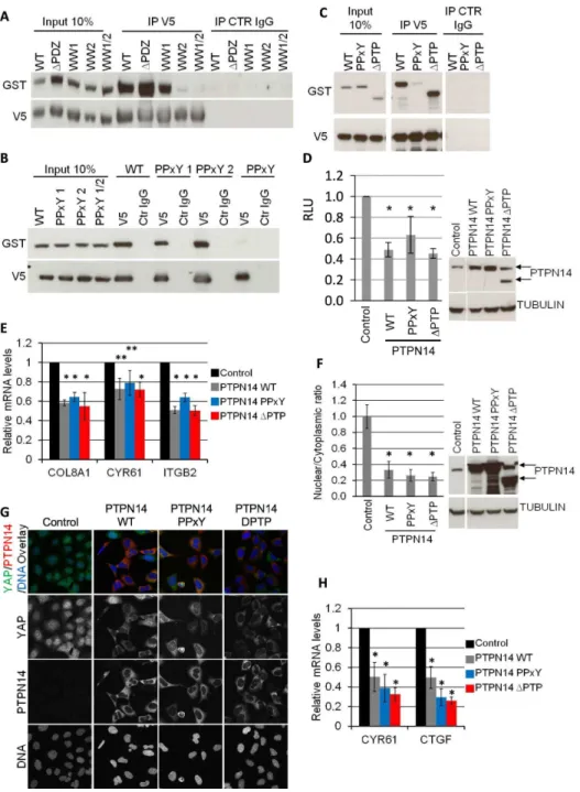
We then aimed at identifying the domains of YAP and PTPN14 that are responsible for the interaction of the two proteins as well as for the regulation of YAP by PTPN14. GST-tagged wild type (WT) PTPN14 and V5-tagged YAP mutants were co-expressed transiently in 293A cells and the ability of the YAP mutants to bind to PTPN14 was tested by a V5 co-IP (Figure 3A). Deletion of the PDZ domain (DPDZ) or mutation of the first WW domain of YAP (WW1) had no effect on the ability of PTPN14 to co-IP with YAP. However, mutation of the second WW domain of YAP (WW2) significantly reduced the binding of the two proteins, indicating that the second WW domain of YAP is necessary for binding to PTPN14. Similarly, co-expression of GST-tagged PTPN14 mutants and V5-tagged WT YAP indicated that mutation of both PPxY motifs of PTPN14 (PPxY) was necessary to abrogate YAP-PTPN14 binding (Figure 3B). Deletion of the phosphatase domain (DPTP) had no effect on PTPN14-YAP binding (Figure 3C). We therefore conclude that, at least in the mechanistic setting of ectopic overexpression, the YAP-PTPN14 interaction is mediated by the second WW domain of YAP and either of the PPxY motifs of PTPN14. The interaction of WW domain of YAP with the PPxY motif of PTPN14 is not unexpected as the binding of the WW domain to the PPxY motif has been identified for a variety of protein interactions [35,36]. In fact, the WW-PPxY interaction is utilized by a variety of binding proteins that associate with YAP [37].
Furthermore, we tested the ability of various PTPN14 mutants to regulate endogenous YAP activity and localization. Interesting-ly, expression of the PPxY mutant or the DPTP mutant downregulated luciferase expression in SF268 MCAT_Luc cells to levels comparable to WT PTPN14 (Figure 3D). In parallel, YAP target genesCOL8A1,CYR61 and ITGB2were also downregulated similarly to PTPN14 WT expression (Figure 3E). Of note, expression of an unrelated protein had no effect on luciferase levels and YAP target gene mRNA levels (data not shown), indicating that the PTPN14 findings are not an artefact of ectopic expression in these cells. In agreement with these results, ectopic expression of the PPxY orDPTP PTPN14 mutants in 293A cells still resulted in the nuclear exclusion of endogenous YAP at low density (Figure 3F and G) and downregulation of YAP target genes CYR61 and CTGF (Figure 3H). We therefore conclude that, at least in the physiological setting with endogenous (rather than ectopic) YAP (over)expression, the PPxY mutant of PTPN14 does not appear to be sufficient to abrogate negative regulation of YAP, and neither is the loss of PTPN14 phosphatase activity.
YAP has been shown to act as an oncogene in MCF-10A cells by protecting from anoikis and inducing EMT and transformation [12]. Interestingly, knockdown of PTPN14 also protected cells from anoikis-mediated cell death to an extent similar to that achieved by YAP overexpression (Figure 4A–B). Knockdown of PTPN14 in YAP-overexpressing cells did not have a significant effect on anoikis-mediated cell death levels. Although this could possibly be due to limitations of the anoikis assay it could also suggest that the effect of PTPN14 in anoikis-mediated cells death Figure 1. PTPN14 is a YAP-binding protein.A) QPCR-based YAP
of MCF-10A cells is YAP-dependent. Finally, we tested the ability of the PTPN14 hairpins to induce transformation of MCF-10A cells. In the control MCF-10A cell line, knockdown of PTPN14 caused an increase in the number of colonies growing in soft agar,
on average 20% of the number achieved by YAP overexpression (Figure 4C–D). However, in the presence of ectopic YAP MCF-10A cells with PTPN14 knockdown formed approximately 3 times more colonies than cells with YAP overexpression alone, Figure 2. PTPN14 is a negative regulator of YAP. A) An SF268 cell line stably expressing the YAP-responsive MCAT_Luc reporter was transfected with two independent siRNAs against YAP and analysed in a Resazurin and luciferase assay 72 h after transfection. mRNA levels of YAP were determined by QRTPCR. Luciferase results are normalized based on the Resazurin results. All results are the average of 5 experiments6STDEV. Statistical analysis was carried out with a 2-tailed paired t-test for each siRNA; * p,0.0001. B) An SF268 cell line stably expressing the YAP-responsive MCAT_Luc reporter was transduced with lentivirus encoding for dox-inducible PTPN14 expression. After pharmacological selection, luciferase expression was measured for both cell lines in the presence and absence of dox (upper panel). A Resazurin assay was carried out in parallel for each sample and used to normalize the luciferase readings. Results are shown as the average of three independent experiments6STDEV. Statistical analysis was carried out with a 2-tailed paired t-test; * p,0.0001. RLU: relative luciferase units. PTPN14 expression achieved by dox and YAP levels in each sample was analyzed by Western blot (lower panel). Tubulin serves as loading control C) 293A cells were transduced with lentivirus encoding dox-inducible PTPN14 expression. YAP localization in control and PTPN14-expressing cell lines was analysed 72 hours post dox induction by IF at low density using confocal miroscopy. D) 293A cells were transduced with lentivirus encoding two independent dox-inducible shRNAs targeting PTPN14. The nuclear/cytoplasmic ratio of YAP was quantified at high density 72 hours post dox induction using a Cellomics automated imager with a conventional microscope, and expressed relative to the control shGFP (left upper panel). Results are shown as the average of 3 independent experiments6STDEV. For each experiment, the average ratio was calculated from three wells per sample (10 images per well). Statistical analysis was carried out with a 2-tailed paired t-test; * p,0.001, ** p,0.05. Western blot analysis of PTPN14 levels, indicating the efficiency of each shRNA (left lower panel). Tubulin serves as a loading control. Confocal microscopy images of cells 72 hours post dox induction (right panel).
indicating that downregulation of PTPN14 can further promote the transforming phenotype of YAP (Figure 4C–D). We conclude that PTPN14 downregulation can phenocopy YAP activation with respect to protection from anoikis and possibly transformation in human cells and it can further promote YAP-mediated transfor-mation.
Discussion
We have demonstrated that PTPN14 is a YAP-binding protein that can inactivate YAP by promoting nuclear exclusion and/or cytoplasmic retention at low density in human cells, resulting in downregulation of YAP target genes. Mechanistically, the interaction between YAP and PTPN14 can be mediated through the WW2 domain of YAP and the PPxY motifs of PTPN14. The nature of the YAP-PTPN14 interaction has also been studied in three previous reports, which also indicate that the YAP-PTPN14 interaction is mediated by WW-PPxY binding [28–30]. However, there is a discrepancy with regards to the contribution of the individual domains to the binding between all four studies. Liuet al. demonstrate that loss of either WW domain can abrogate binding whereas Huanget al.show that loss of both domains is necessary to disrupt binding, Wang et al identify the second WW domain being more important than the first for this interaction
and we show that the second WW domain is primarily responsible for YAP-PTPN14 binding. With regard to the PPxY motifs, Liuet al.and we demonstrate that loss of both is necessary for abrogation of YAP-binding whereas Huanget al.and Wang et al. indicate that deletion of either motif dramatically reduces the interaction of the two proteins. The reason for these differences is not yet clear. The discrepancy might in part be due to the different methods used to create the mutants in each study: for the PPxY motif, mutation of tyrosine residue alone [28,29] or mutation of both prolines and the tyrosine to alanine (this study) vs full deletion of the entire motif [30]; and for the WW domains, mutation of the tryptophan and proline residues to alanine (this study) vs full deletion of the domain [28–30]. Most importantly however, all four studies indicate that the YAP-PTPN14 interaction is mediated by WW domain-PPxY motif binding.
We find that loss of YAP-binding through mutation of the PPxY motifs, as determined by biochemical IP in an ectopic expression setting, does not appear to abrogate regulation of endogenous YAP activity and localization by PTPN14. Opposite to our findings, Liuet al.and Huang et al.show that mutation of both PPxY motifs of PTPN14 can, albeit only partially, abrogate negative regulation of YAP activity using a reporter gene assay (RGA). Notably, these studies rely on the ectopic overexpression of both PTPN14 and YAP (i.e., identical to the mechanistic IP
* p,0.05.
doi:10.1371/journal.pone.0061916.g003
Figure 4. Down-regulation of PTPN14 enhances the oncogenic phenotype of YAP.A) MCF-10A cells were transduced with lentivirus encoding for YAP expression and after pharmacological selection transduced with lentivirus encoding for dox-inducible shRNAs targeting either PTPN14 or GFP (control) (two independent shRNAs targeting PTPN14). All six cell lines were subjected to an anoikis assay in low-attachment plates in the presence of dox for 48 hours. Results are shown as the average of three experiments6STDEV. Statistical analysis was carried out with a 2-tailed paired t-test; * p,0.05, ** p = 0.05. B) Western blot analysis of cells from (A) 48 hours post dox induction. Tubulin serves as a loading control. C) Soft agar assay quantification of cells from (A). Results were obtained two weeks post dox induction and are the average of three experiments6STDEV. D) Images of wells from (C) at the time of quantification. Scale: 1 mm.
setting), to assess the impact of binding mutants in RGA, whereas we explore the functional PTPN14 domain contributions on the activity of endogenous YAP in models which possibly implicate physiological formation of larger complexes (see below). However, and again opposite to our findings, Wanget al. demonstrate that deletion of both PPxY motifs abolishes PTPN14-mediated regulation of endogenous YAP localisation in MCF-10A cells. The difference between our data and of the report of Wanget al. could, at least in part, be caused by the fact that we used alanine substitutions to generate the PTPN14 mutants whereas Wanget al. deleted the entire motif, which could result in a stronger phenotype. Finally, the discrepancies between the four studies could potentially reflect model or cell-line specific differences in PTPN14-mediated regulation of YAP.
Our results indicate that mutation of the two PPxY motifs of PTPN14 can disrupt YAP-binding but cannot abolish PTPN14-mediated regulation of YAP. Importantly, it should be noted that apart from two PPxY motifs, PTPN14 contains an LPxY and a VPxY motif, both of which have been shown to interact with WW domains in vitro, albeit with lower affinity than the PPxY motif [38,39]. It is therefore possible that binding of the WW domains of endogenous YAP to the LPxY and VPxY motifs of (PPxY-mutated), overexpressed PTPN14 would be sufficient to mediate the translocation of endogenous YAP to the cytoplasm as shown in figure 3G and to downregulate endogenous YAP activity in an RGA as shown in figure 3D. Furthermore, it is possible that the interaction between ectopically-expressed WT YAP and PPxY-mutant PTPN14 would not be detectable in an IP experiment as shown in figure 3B, due to the reduced affinity of the remaining LPxY and VPxY motifs for the WW domains of YAP. Further mutating the LPxY and VPxY motifs in PTPN14 should bypass the problem of motif redundancy within the protein and provide further insight as to the role of YAP-binding in the regulation of its activity and localization.
Another possible explanation for the uncoupling of YAP-binding and YAP regulation by PTPN14 as indicated by our experiments is that PTPN14 and YAP are part of a larger complex, possibly formed at adherens junctions, and in line with the findings for the Pez/Kibra interaction inDrosophila(30), with the complex components interacting through multiple domains. Although loss of the PPxY motif abolishes binding to YAP as demonstrated by mechanistic co-IP experiments, it is conceivable that this does not destabilize a potentially larger complex sufficiently to completely abrogate PTPN14-mediated regulation of YAP localization and activity in a physiological setting. Yet another possibility is that PTPN14 can function as a dimer/ multimer. In the presence of the ectopic PPxY mutant, the endogenous wild-type protein could heterodimerize with the ectopic mutant and create dimers that, although not interacting strongly enough with YAP to detect by co-IP experiments, can still regulate YAP localization and activity in a physiological setting.
We demonstrate that the phosphatase activity of PTPN14 is dispensable for YAP regulation. This is shown not only in an RGA, but also by means of expression levels of YAP target genes which are similar to those seen by expression of WT PTPN14, as well as YAP localization which remains cytoplasmic at low density in the presence of the DPTP mutant. Furthermore, and in agreement with our findings, the PTP domain is also dispensable for PTPN14-mediated control of YAP localization in MCF-10A cells as well as for the growth-regulatory role of Pez in the Drosophila midgut and wing development [30,40]. However, in their recent publication, Liu and colleagues claim that YAP is a direct target of PTPN14. Although we cannot formally exclude that YAP is targeted by PTPN14 under certain circumstances or in
certain cell types, we fail to see a role for the PTP domain in YAP-binding and regulation in our setting. As the Y357 phosphoryla-tion which according to Liuet al. is targeted by PTPN14, has been implicated in the regulation of YAP’s tumor suppressor phenotype in response to DNA damage [21], it is possible that tyrosine dephosphorylation of YAP by PTPN14 is more relevant in that setting.
Tyrosine phosphatases are known to stabilize adherens junc-tions by means of counteracting tyrosine kinases. However, deletion of the phosphatase domain does not affect the ability of PTPN14 to regulate YAP activity and localization, indicating that PTPN14 might have both phosphatase-dependent functions at adherens junctions and phosphatase-independent functions in regulating YAP activity. Whether PTPN14-mediated YAP regu-lation is at all linked to the presence of functional adherens junctions remains to be elucidated.
Lastly, we provide evidence that PTPN14 downregulation can phenocopy YAP over-expression in its ability to rescue MCF-10A cells from anoikis. Furthermore, and in agreement with Wanget al. [30], we show that downregulation of PTPN14 can induce transformation of MCF-10A cells and can exacerbate the transforming phenotype of ectopic YAP in MCF-10A cells. While the induction of transformation upon PTPN14 downregulation -which results in prolonged nuclear localization/activity of YAP ((Figure 2D and Figure S2) and other reports [28,30]) - may be attributed to a number of downstream events, a key contribution is likely provided by the increased survival under loss of matrix attachment, as evidenced by the anoikis data (Figure 4A). Together with the finding that PTPN14 loss can induce aberrant acini formation in MCF-10A cells [30], these results indicate a potential tumor suppressive role for PTPN14 in human cells, in agreement with previous reports of PTPN14 loss of heterozygosity (LOH) in colorectal cancer [41].
Supporting Information
Figure S1 Comparison of luciferase reporter activity in SF268 and 293A cells.SF268 and 293A cells were transduced with lentivirus encoding for the YAP/TEAD-responsive MCAT luciferase reporter. Luciferase expression in each stable cell line was analysed in parallel in the absence of exogenous YAP (left panel). The same number of cells was plated for each cell line and a luciferase and resazurin assay was carried out 72 hours after plating. Luciferase expression was normalized based on Resazurin readings. WB analysis of YAP levels in SF268 and 293A cells (right panel). CDK4 serves as loading control. s.e.: short exposure; l.e.: long exposure. All lanes are from a single blot and exposure. The lane after SF268 was left empty.
(TIF)
Figure S2 YAP localization in SF268 cells.SF268 cells were transduced with lentivirus encoding for constitutive PTPN14 knock down. Stable cell lines were plated onto Lab-TekTM 8 chamber glass slides and allowed to reach confluency. Seventy-two hours after seeding, cell lines were analysed by immunofluores-cence for PTPN14 and YAP expression using a confocal microscope. DAPI was used for DNA staining.
(TIF)
Acknowledgments
The authors would like to thank Ken Crawford and Janet Sim for providing the pLenti6.3TM-EF1aplasmid; Frank Stegmeier for the
Xu Wu, Jiayi Yang and Jun Liu for the YAP immunostaining protocol in 293A. We are grateful to NIBR Oncology presidential postdocs and supervisors as well as members of the Christofori lab for helpful discussions.
Author Contributions
Conceived and designed the experiments: CM TS GH WRS FH HV. Performed the experiments: CM WL TM CD LB EB HV. Analyzed the data: CM WL TS MI HV. Contributed reagents/materials/analysis tools: AH MW HN MED CW GC AB. Wrote the paper: CM TS.
References
1. Camargo FD, Gokhale S, Johnnidis JB, Fu D, Bell GW, et al. (2007) YAP1 increases organ size and expands undifferentiated progenitor cells. Curr Biol 17: 2054–2060.
2. Dong J, Feldmann G, Huang J, Wu S, Zhang N, et al. (2007) Elucidation of a universal size-control mechanism in Drosophila and mammals. Cell 130: 1120– 1133.
3. Zhao B, Lei QY and Guan KL (2008) The Hippo-YAP pathway: new connections between regulation of organ size and cancer. Curr Opin Cell Biol 20: 638–646.
4. Huang J, Wu S, Barrera J, Matthews K and Pan D (2005) The Hippo signaling pathway coordinately regulates cell proliferation and apoptosis by inactivating Yorkie, the Drosophila Homolog of YAP. Cell 122: 421–434.
5. Lai ZC, Wei X, Shimizu T, Ramos E, Rohrbaugh M, et al. (2005) Control of cell proliferation and apoptosis by mob as tumor suppressor, mats. Cell 120: 675–685.
6. Tao W, Zhang S, Turenchalk GS, Stewart RA, St John MA, et al. (1999) Human homologue of the Drosophila melanogaster lats tumour suppressor modulates CDC2 activity. Nat Genet 21: 177–181.
7. Wu S, Huang J, Dong J and Pan D (2003) hippo encodes a Ste-20 family protein kinase that restricts cell proliferation and promotes apoptosis in conjunction with salvador and warts. Cell 114: 445–456.
8. Boggiano JC and Fehon RG (2012) Growth control by committee: intercellular junctions, cell polarity, and the cytoskeleton regulate Hippo signaling. Dev Cell 22: 695–702.
9. Genevet A and Tapon N (2011) The Hippo pathway and apico-basal cell polarity. Biochem J 436: 213–224.
10. Zhao B, Tumaneng K and Guan KL (2011) The Hippo pathway in organ size control, tissue regeneration and stem cell self-renewal. Nat Cell Biol 13: 877– 883.
11. Pan D (2010) The hippo signaling pathway in development and cancer. Dev Cell 19: 491–505.
12. Overholtzer M, Zhang J, Smolen GA, Muir B, Li W, et al. (2006) Transforming properties of YAP, a candidate oncogene on the chromosome 11q22 amplicon. Proc Natl Acad Sci U S A 103: 12405–12410.
13. Schlegelmilch K, Mohseni M, Kirak O, Pruszak J, Rodriguez JR, et al. (2011) Yap1 acts downstream of alpha-catenin to control epidermal proliferation. Cell 144: 782–795.
14. Mauviel A, Nallet-Staub F and Varelas X (2012) Integrating developmental signals: a Hippo in the (path)way. Oncogene 31: 1743–1756.
15. Varelas X and Wrana JL (2012) Coordinating developmental signaling: novel roles for the Hippo pathway. Trends Cell Biol 22: 88–96.
16. Ito M, Barys L, O’Reilly T, Young S, Gorbatcheva B, et al. (2011) Comprehensive mapping of p53 pathway alterations reveals an apparent role for both SNP309 and MDM2 amplification in sarcomagenesis. Clin Cancer Res 17: 416–426.
17. Gaffney CJ, Oka T, Mazack V, Hilman D, Gat U, et al. (2012) Identification, basic characterization and evolutionary analysis of differentially spliced mRNA isoforms of human YAP1 gene. Gene 509: 215–222.
18. Wee S, Wiederschain D, Maira SM, Loo A, Miller C, et al. (2008) PTEN-deficient cancers depend on PIK3CB. Proc Natl Acad Sci U S A 105: 13057– 13062.
19. Wiederschain D, Wee S, Chen L, Loo A, Yang G, et al. (2009) Single-vector inducible lentiviral RNAi system for oncology target validation. Cell Cycle 8: 498–504.
20. Basu S, Totty NF, Irwin MS, Sudol M and Downward J (2003) Akt phosphorylates the Yes-associated protein, YAP, to induce interaction with 14-3-3 and attenuation of p73-mediated apoptosis. Mol Cell 11: 11–23. 21. Levy D, Adamovich Y, Reuven N and Shaul Y (2008) Yap1 phosphorylation by
c-Abl is a critical step in selective activation of proapoptotic genes in response to DNA damage. Mol Cell 29: 350–361.
22. Shevchenko A, Wilm M, Vorm O and Mann M (1996) Mass spectrometric sequencing of proteins silver-stained polyacrylamide gels. Anal Chem 68: 850– 858.
23. Chan SW, Lim CJ, Chong YF, Pobbati AV, Huang C, et al. (2011) Hippo pathway-independent restriction of TAZ and YAP by angiomotin. J Biol Chem 286: 7018–7026.
24. Oka T, Schmitt AP and Sudol M (2012) Opposing roles of angiomotin-like-1 and zona occludens-2 on pro-apoptotic function of YAP. Oncogene 31: 128– 134.
25. Paramasivam M, Sarkeshik A, Yates JR, 3rd, Fernandes MJ, and McCollum D (2011) Angiomotin family proteins are novel activators of the LATS2 kinase tumor suppressor. Mol Biol Cell 22: 3725–3733.
26. Wang W, Huang J and Chen J (2011) Angiomotin-like proteins associate with and negatively regulate YAP1. J Biol Chem 286: 4364–4370.
27. Zhao B, Li L, Lu Q, Wang LH, Liu CY, et al. (2011) Angiomotin is a novel Hippo pathway component that inhibits YAP oncoprotein. Genes Dev 25: 51– 63.
28. Liu X, Yang N, Figel SA, Wilson KE, Morrison CD, et al. (2012) PTPN14 interacts with and negatively regulates the oncogenic function of YAP. Oncogene.
29. Huang JM, Nagatomo I, Suzuki E, Mizuno T, Kumagai T, et al. (2012) YAP modifies cancer cell sensitivity to EGFR and survivin inhibitors and is negatively regulated by the non-receptor type protein tyrosine phosphatase 14. Oncogene. 30. Wang W, Huang J, Wang X, Yuan J, Li X, et al. (2012) PTPN14 is required for
the density-dependent control of YAP1. Genes Dev 26: 1959–1971. 31. Wadham C, Gamble JR, Vadas MA and Khew-Goodall Y (2003) The protein
tyrosine phosphatase Pez is a major phosphatase of adherens junctions and dephosphorylates beta-catenin. Mol Biol Cell 14: 2520–2529.
32. Jiang SW, Desai D, Khan S and Eberhardt NL (2000) Cooperative binding of TEF-1 to repeated GGAATG-related consensus elements with restricted spatial separation and orientation. DNA Cell Biol 19: 507–514.
33. Miller E, Yang J, DeRan M, Wu C, Su AI, et al. (2012) Identification of serum-derived sphingosine-1-phosphate as a small molecule regulator of YAP. Chem Biol 19: 955–962.
34. Yu FX, Zhao B, Panupinthu N, Jewell JL, Lian I, et al. (2012) Regulation of the Hippo-YAP pathway by G-protein-coupled receptor signaling. Cell 150: 780– 791.
35. Bork P and Sudol M (1994) The WW domain: a signalling site in dystrophin? Trends Biochem Sci 19: 531–533.
36. Chen HI and Sudol M (1995) The WW domain of Yes-associated protein binds a proline-rich ligand that differs from the consensus established for Src homology 3-binding modules. Proc Natl Acad Sci U S A 92: 7819–7823.
37. Webb C, Upadhyay A, Giuntini F, Eggleston I, Furutani-Seiki M, et al. (2011) Structural features and ligand binding properties of tandem WW domains from YAP and TAZ, nuclear effectors of the Hippo pathway. Biochemistry 50: 3300– 3309.
38. Chen HI, Einbond A, Kwak SJ, Linn H, Koepf E, et al. (1997) Characterization of the WW domain of human yes-associated protein and its polyproline-containing ligands. J Biol Chem 272: 17070–17077.
39. Fotia AB, Ekberg J, Adams DJ, Cook DI, Poronnik P, et al. (2004) Regulation of neuronal voltage-gated sodium channels by the ubiquitin-protein ligases Nedd4 and Nedd4-2. J Biol Chem 279: 28930–28935.
40. Poernbacher I, Baumgartner R, Marada SK, Edwards K and Stocker H (2012) Drosophila Pez acts in Hippo signaling to restrict intestinal stem cell proliferation. Curr Biol 22: 389–396.