Peptide-Recombinant VP6 Protein Based
Enzyme Immunoassay for the Detection of
Group A Rotaviruses in Multiple Host Species
Naveen Kumar1☯¤, Yashpal Singh Malik1☯
*, Satish Kumar2☯, Kuldeep Sharma3☯, Subhankar Sircar1‡, Sharad Saurabh1‡, Baldev R. Gulati4‡, Neeraj Singh1‡, Arvind Kumar Singh2‡, Vinay G. Joshi2‡, Krisztian Banyai5‡, Kuldeep Dhama6‡
1Biological Standardization Division, Indian Veterinary Research Institute (IVRI), Izatnagar, Uttar Pradesh, India,2Division of Animal Biotechnology, Indian Veterinary Research Institute (IVRI), Izatnagar, Uttar Pradesh, India,3National Institute of Research in Tribal Health, Jabalpur, Madhya Pradesh, India, 4National Research Center on Equines (NRCE), Hisar, Haryana, India,5Institute for Veterinary Medical Research, Centre for Agricultural Research, Hungarian Academy of Sciences, Hungáriakrt. 21, Budapest, Hungary,6Division of Veterinary Immunology, Indian Veterinary Research Institute (IVRI), Izatnagar, Uttar Pradesh, India
☯These authors contributed equally to this work.
¤ Current address: National Institute of High Security Animal Diseases (NIHSAD), Bhopal, Madhya Pradesh,
India
‡These authors also contributed equally to this work.
Abstract
We developed a novel enzyme immunoassay for the detection of group A rotavirus (RVA) antigen in fecal samples of multiple host species. The assay is based on the detection of conserved VP6 protein using anti-recombinant VP6 antibodies as capture antibodies and anti-multiple antigenic peptide (identified and constructed from highly immunodominant epi-topes within VP6 protein) antibodies as detector antibodies. The clinical utility of the assay was evaluated using a panel of 914 diarrhoeic fecal samples from four different host species (bovine, porcine, poultry and human) collected from diverse geographical locations in India. Using VP6- based reverse transcription-polymerase chain reaction (RT-PCR) as the gold standard, we found that the diagnostic sensitivity (DSn) and specificity (DSp) of the new assay was high [bovine (DSn = 94.2% & DSp = 100%); porcine (DSn = 94.6% & DSp = 93.3%); poultry (DSn = 74.2% & DSp = 97.7%) and human (DSn = 82.1% & DSp = 98.7%)]. The concordance with RT-PCR was also high [weighted kappa (k) = 0.831–0.956 at 95%
CI = 0.711–1.0] as compared to RNA-polyacrylamide gel electrophoresis (RNA-PAGE).
The performance characteristics of the new immunoassay were comparable to those of the two commercially available ELISA kits. Our results suggest that this peptide-recombinant protein based assay may serve as a preliminary assay for epidemiological surveillance of RVA antigen and for evaluation of vaccine effectiveness especially in low and middle income settings.
a11111
OPEN ACCESS
Citation:Kumar N, Malik YS, Kumar S, Sharma K, Sircar S, Saurabh S, et al. (2016) Peptide-Recombinant VP6 Protein Based Enzyme Immunoassay for the Detection of Group A Rotaviruses in Multiple Host Species. PLoS ONE 11 (7): e0159027. doi:10.1371/journal.pone.0159027
Editor:Sabato D'Auria, CNR, ITALY
Received:April 8, 2016
Accepted:June 24, 2016
Published:July 8, 2016
Copyright:© 2016 Kumar et al. This is an open access article distributed under the terms of the Creative Commons Attribution License, which permits unrestricted use, distribution, and reproduction in any medium, provided the original author and source are credited.
Data Availability Statement:All relevant data are within the paper and its Supporting Information files.
Funding:The financial support from Education Division, Indian Council of Agricultural Research, Ministry of Agriculture & Farmer’s Welfare, Govt. of India, New Delhi in the form of National Fellow project is acknowledged. Dr. Krisztian Banyai was supported by the Hungarian Scientific Research Fund (OTKA, T100727).
Introduction
Rotaviruses (RVs) belong to the familyReoviridaeand are a leading cause of diarrhea in
humans and animals worldwide. For example, RVs account for approximately one fourth of global mortality in Indian children annually [1–3]. The genome consists of 11 dsRNA seg-ments, which encode six structural proteins (VP1–4, VP6 and VP7) and five/six non-structural proteins (NSP1–NSP5/6) [4]. RVs are classified into at least nine distinct serological species or groups (A to I), of which group A rotaviruses (RVAs) are frequently associated with acute diar-rhea [1,2,5]. Commonly used dual classification system for RVAs designate P- and G-geno-types to the genes coding for outer most proteins VP4 and VP7, respectively. At least 27 G and 37 P genotypes of RVAs have been identified in humans and animals, reflecting huge genetic diversity among RVs [2,6].
Rapid and accurate diagnosis of RVAs remains a major hurdle, especially in the low and middle income countries. Currently, the diagnosis of RVAs relies on the detection of either the virus antigen or its genome. Although highly sensitive, the reverse transcription- polymerase chain reaction (RT-PCR) is costly and is not affordable for conducting surveillance studies, especially in poor countries [7–8]. On the other hand, electropherotyping and latex agglutina-tion tests are cost effective, but their low sensitivity and specificity limits their use on a large scale [9–10]. In this context, enzyme-linked immunosorbent assay (ELISA) is considered an ideal diagnostic tool because it is known to be effective and economical for RVA surveillance studies. Unfortunately, the use of polyclonal serum against whole virus antigen makes this test prone to false reactions. Synthetic peptides prepared from antigenic regions of the virus may provide an alternative to the whole virus and has several other advantages, e.g., it is stable at pH 2–9, avoids risk of infection, instant manufacturing on a large scale, and durable storage [11–12].
Despite the huge genetic diversity of RVAs, the sole structural protein of the middle of three capsid layers (VP6) is highly immunogenic and conserved among RVAs [13], which makes it preferable to be used as a universal diagnostic reagent. However, tracing the regions within the VP6 protein that are highly reactive with anti-RVA antibodies is imperative for effective use of peptides in a diagnostic assay. The anti-peptide antibodies (usually targeted against one or two epitopes) could serve as homogenous antibodies comparable to monoclonal antibodies, thereby increasing the specificity as well as reducing the cost and clumsy procedure employed in mono-clonal antibody production [14]. Further, engaging multiple antigenic peptides (MAPs) may overcome the problem of low titer anti-peptide serum [15–16]. MAPs have essentially three parts viz. an amino acid (alanine/cysteine) linked to a solid support or resin, an inner lysine core matrix, and a surface layer of four or eight peptides of the same/different sequences attached to the core matrix. Attaching the four or eight peptides over the lysine core matrix increases total molecular weight and mimics closely the native antigenic sites present on the virus surface. Owing to these advantages, anti-peptide antibodies have been used for serodiag-nosis of hepatitis C virus [17], infectious bronchitis virus [18], infectious bursal disease virus [19–20] and peste-des-petits ruminants virus [21].
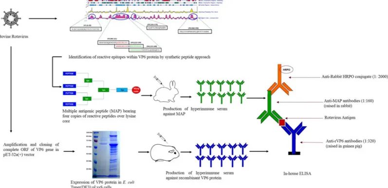
This study was undertaken to develop a novel methodology for RVAs diagnosis utilizing the potential of viral peptides. A schematic representation of the test development is provided in
Materials and Methods
Ethics statement
Approval to conduct this study was obtained from the Institute Animal Ethical Committee (IAEC) [vide approval no. 1-53/2012-13/JD(R)] of Indian Council Agricultural Research-Indian Veterinary Research Institute (ICAR-IVRI), Izatnagar, Bareilly (U.P.), India. The hyper-immune sera were produced in laboratory animals in accordance with the guidelines set by the Committee for the Purpose of Control and Supervision of Experiments on Animals (CPCSEA), Ministry of Environment and Forestry, Government of India.
Chemicals and Reagents
All reagents used for peptide synthesis were mostly dry and of HPLC grade. The Fmoc-amino acids, coupling reagent, and 2-(1H-Benzotriazole-1-yl)-oxo-1,2,3,3-tetramethyluronium hexa-fluorophosphate (HBTU) were obtained from GL Biochem (Shanghai, China); Wang resin was from Nova Biochem, Switzerland; and 1-Hydroxybenzotriazole (HOBt), N,N´-Diisopropylcar-bodiimide (DIPC), 2,2,2-Trifluoroethanol (TFE), and N,N-Diisopropyl ethylamine (DIEA) were from Spectrochem, India.
Cloning vector pET-32a(+) and fourE.coliexpression strains [BL21(DE3)pLysS, Origami
(DE3)pLysS, Rosetta-gami(DE3)pLysS, and Tuner(DE3)pLysS] were from Novagen (Ger-many). African green monkey kidney (MA-104) cell-adapted bovine RVA (positive control in assay development) and anti-RVA polyclonal serum were kindly provided by Dr. Minakshi Prasad, Department of Animal Biotechnology, Lala Lajpat Rai University of Veterinary and Animal Sciences, Hisar, Haryana, India.
Fig 1. Schematic of the development process of peptide-recombinant VP6 protein based enzyme immunoassay for group A rotaviruses.
Synthesis and purification of peptides
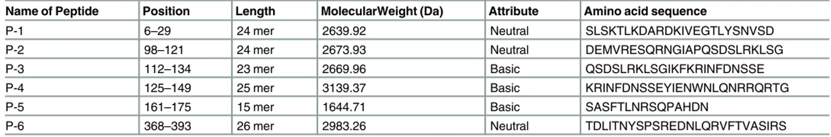
The selection of bovine RVA (UKD-M1/09) strain for this study was based on multiple sequence alignments with public domain retrieved RVAs of different host species. The UKD-M1/09 strain is a multiple-species reassortant isolated in 2009 from a male cross-bred calf with gastroenteritis in the North-West Himalayan city of Mukteswar (Nainital), Uttara-khand, India. We identified a total of six regions within VP6 protein of this strain that showed high Jameson-Wolf antigenic index (Protean software, DNASTAR) as given inTable 1andS1 Fig.
The peptides covering identified regions were manually synthesized on Wang resin (Nova Biochem, Switzerland) using Fmoc-(Nα-9-Fluorenylmethyloxycarbonyl) chemistry and solid phase peptide synthesis as per the methods described earlier [22–24]. Briefly, Fmoc-glycine was activated with N,N´-Diisopropylecarbodiimide (DIPC) and 4-Dimethylaminopyridine (DMAP) followed by its coupling with Dimethylformamide (DMF) pre-swollen Wang resin beads. The capping of free functional groups on Wang resin was done with an acetylation mix-ture [DMF: Acetic anhydride: N,N-Diisopropylethylamine (DIEA), (193: 6: 1, v/v)] to avoid undesired peptide synthesis. Chemical deprotection of N-terminal Fmoc group of glycine was done by 20% piperidine in DMF (v/v). Each successive Fmoc-amino acid to be used in peptides synthesis was activated by HOBt and HBTU, and coupled in the presence of DIEA. Rest steps were performed in a similar manner until the desired length of the peptide was obtained. The last deprotection (removal of side chain protecting groups) and cleavage from resin beads were done by treating the resin bound peptides with a cleavage mixture [Trifluoroacetic acid (TFA): Phenol: Thioanisol: Ether: Water (82.5: 5: 5: 5: 2.5, v/v)]. Finally, the cleaved peptide was pre-cipitated with chilled (dry) diethyl ether, vacuum dried and stored under dry conditions until used. Purification of peptides was done by Reversed Phase- High Performance Liquid Chroma-tography (RP-HPLC) performed on a Shimadzu SCL-10AVP ver. 5.42 liquid chromatograph equipped with semi-preparative, RP C-18 column (Toyasoda, Japan). Linear gradients of aceto-nitrile in aqueous 0.1% TFA (v/v) were used to elute peptides. Peptide elution was monitored at 220nm using a SPDM10AVP photodiode array detector. Chromatograph was analyzed using inbuilt software.
Synthesis and purification of multiple antigenic peptides (MAPs)
The MAPs were constructed on peptidyl core of three radially branched lysine residues onto which desired peptide sequences were built using step-wise Fmoc solid-phase peptide synthesis [22–23]. Cysteine was attached to Wang resin after activation in the presence of DIPC and DMAP. The capping of free functional groups on Wang resin and chemical deprotection of N-terminal Fmoc group of cysteine was done as described above. The peptide branching was achieved to form four arm structure using di-Fmoc-Lys-OH to provide lysine core over which chosen peptide sequence was synthesized (S2 Fig). Fmoc-amino acids coupling was done as
Table 1. Peptides spanning the major immunodominant epitopes within the bovine RVA VP6 protein.
Name of Peptide Position Length MolecularWeight (Da) Attribute Amino acid sequence
P-1 6–29 24 mer 2639.92 Neutral SLSKTLKDARDKIVEGTLYSNVSD
P-2 98–121 24 mer 2673.93 Neutral DEMVRESQRNGIAPQSDSLRKLSG
P-3 112–134 23 mer 2669.96 Basic QSDSLRKLSGIKFKRINFDNSSE
P-4 125–149 25 mer 3139.37 Basic KRINFDNSSEYIENWNLQNRRQRTG
P-5 161–175 15 mer 1644.71 Basic SASFTLNRSQPAHDN
P-6 368–393 26 mer 2983.26 Neutral TDLITNYSPSREDNLQRVFTVASIRS
described above, until the desired length of the multimeric peptide was obtained. Final chemi-cal deprotection and cleavage were done by treating resin bound peptides with the cleavage mixture. Lastly, cleaved MAP was precipitated with chilled (dry) diethyl ether, vacuum dried and stored under dry conditions until used. The MAPs were purified by RP-HPLC.
Circular Dichroism (CD) Spectroscopy
The CD spectra were recorded at room temperature (~20°C) in a quartz cuvette of 0.1 cm path length on a JASCO-J810 spectropolarimeter calibrated with d-camphor-10-sulfonate. The experiments were performed in polar (HPLC water) and non-polar (Trifluoroethanol) solvents to investigate the effect on conformation of both peptides and multiple antigenic peptides. The RP-HPLC purified peptides were used at a concentration of 100μg/ml. Each spectrum was
recorded in a continuous scanning mode (at an average of four repeated scans with 100nm/sec scanning speed), and response time of 1 sec. The contribution of buffer/solvent was duly sub-tracted from each spectrum. Respective intensities were expressed in mean residue molar ellip-ticity [ϑ], calculated from the following equation:
½y ¼ 100c
CL
Where [ϑ] is molar ellipticity in deg.cm2.decimole-1,Cis observed ellipticity in degree, C is concentration in mole Lit-1, L is the path length in cm.
The line shapes of recorded spectra were analyzed using a least-square fitting routine in comparison to poly-lysine standards representing 100%α-helix,β-turn or random coil, respec-tively. The secondary structure estimation by Spectra Manager software provided an assess-ment of secondary structural eleassess-ments of each peptide in solution [25].
Peptide- ELISA (P- ELISA)
The six peptides covering the major immunodominant epitopes of bovine RVA VP6 protein were screened by P- ELISA. In brief, each peptide was coated in duplicate wells (10μg/well) of
high binding 96-well polystyrene Maxisorp ELISA plate (Nunc, Denmark) in carbonate-bicar-bonate buffer, pH 9.6. The plate was incubated overnight at 4°C. The unoccupied sites on the well surfaces were blocked with 200μl of 2% bovine serum albumin (BSA) after washing three
times with phosphate buffered saline (PBS, pH 7.4) and incubated at 37°C for 2 hours. The peptides bound to polystyrene surface were made to react with 50μl of rabbit anti-RVA
poly-clonal serum (1:100 dilutions in 2% BSA) for 1 hour at 37°C. After washing three times with PBS, the plate was incubated with 50μl of anti-rabbit HRP conjugate (1:1000 in 2% BSA)
(Sigma-Aldrich, USA) for 1 hour at 37°C. The plate was washed again three times with PBS and then left for colour development at 37°C for 15 minutes after addition of 50μl of OPD
(o-phenylenediamine dihydrochloride) substrate (Sigma-Aldrich, USA). The reaction was stopped using 1M sulfuric acid. Optical density was recorded at a 492nm wavelength in an ELISA reader (Biorad, Model 680).
Multiple antigenic peptide- ELISA (MAP- ELISA)
All steps in MAP-ELISA were similar to those of P-ELISA as described above except that MAPs were used as the coating antigen instead of peptides. Each of the MAPs was coated on to duplicate wells of ELISA plate at 1μg/well concentration prepared in carbonate-bicarbonate
Construction of VP6 recombinant plasmid and it’s over expression
RNA from bovine rotavirus strain (UKD-M1/09) was extracted using Quick-RNATMMini Prep Kit (Prolab, India) followed by reverse transcription. The complete VP6 ORF was ampli-fied using sense primer having an EcoRI site (sequence underlined); VP6_EcoRI_ F [+] (
aac-GAATTCATGGATGTCCTATACTCTTTGT) and anti-sense primer containing XhoI site (sequence underlined); VP6_XhoI_R [–] (tcagCTCGAGCTCATTTGACAAGCATGC). The PCR reaction mixture consisted of 2μL of cDNA (2μg), 25μL of 2X DreamTaq PCR Mater
Mix (Thermo Scientific), 1μL of each primer (10 pmol) and 21μL of NFW. The PCR was
per-formed at initial denaturation step of 95°C for 5 min followed by 35 cycles of 94°C for 1 min, 54°C for 1 min, 72°C for 1 min 15 sec and final extension at 72°C for 10 min. The PCR prod-ucts were purified using a GeneJETTMGel Extraction kit (Thermo Scientific) and further sub-jected to nucleotide sequencing (SciGenom Labs Pvt. Ltd., India).
To construct VP6 recombinant plasmid, doubly (EcoRI and XhoI) digested VP6 amplicon and pET-32a(+) vector were ligated in the presence of T4 DNA ligase followed by transforma-tion inE.coliTOP 10 F’competent cells. The recombinant clones were confirmed by colony
PCR and nucleotide sequencing. The pET-32a(+)-VP6 recombinant plasmid was transformed into four different expression hosts, e.g., BL21(DE3)pLysS, Origami(DE3)pLysS, Rosetta-gami (DE3)pLysS, and Tuner(DE3)pLysS. These transformed cells were grown in Luria Bertani (LB) broth (HiMedia, India) supplemented with 1% glucose and antibiotics [ampicillin (50μg/ml)
and chloramphenicol (35μg/ml)] on shaker-cum-incubator at 37°C (180 rpm). When the
opti-cal density of the culture at 600nm reached 0.8, isopropyl beta-D-thiogalactoside (IPTG) was added at to a final concentration of 1 mM followed by incubation at different temperatures and times to optimize the induction conditions. The size and yield of 6xHis-tagged recombinant VP6 (rVP6) protein were confirmed by Sodium Dodecyl Sulphate–Polyacrylamide Gel Electro-phoresis (SDS-PAGE) analysis.
Western blot analysis and rVP6 purification under denaturing conditions
Further confirmation of rVP6 protein was done by Western blot analysis utilizing two approaches viz. polyHistidine based recombinant protein detection, and anti-RVA polyclonal serum based specific detection of recombinant proteins using a semi-dry blot system [26–27]. Briefly, rVP6 protein was subjected to SDS-PAGE and protein bands were transferred over Polyvinylidene difluoride (PVDF) membrane (MDI, India) using constant voltage of 6–8 V (0.8–1 mA/cm2of the gel) for 60 min. The protein bands were allowed to react with monoclo-nal anti-polyHistidine peroxidase conjugate (1:1000 in 3% BSA) and rabbit anti-RVA poly-clonal serum (1:500 in 3% BSA) separately, after blocking the unoccupied sites by 3% BSA at 4°C overnight. The anti-RVA polyclonal serum reacted protein bands were further incubated with anti-rabbit HRPO conjugate (1:1000 in 3% BSA) after washing with PBST (0.1% Tween-20 in PBS). The protein bands were visualized using 3,30-Diaminobenzidine (DAB) (Sigma-Aldrich, USA).
The over-expressed 6xHis-tagged rVP6 protein formed inclusion bodies as revealed by solu-bility analysis. Therefore, rVP6 protein was purified by Nickel- Nitrilotriacetic acid (Ni-NTA) affinity chromatography after solubilizing the inclusion bodies in 6M guanidium hydrochlo-ride. Briefly, an equal volume of Lysis-Equilibration (LE) buffer (50mM NaH2PO4, 300mM
NaCl, 8M Urea, pH-8.0) with guanidium hydrochloride treated cell lysate was allowed to pass through Ni-NTA agarose column after equilibration. The column was washed with five bed volumes of eight wash buffers (50mM NaH2PO4, 300mM NaCl, 30mM Imidazole, 7 to 0 M
(50mM NaH2PO4, 300mM NaCl, 250mM Imidazole, pH-8.0). The flow through collected at
each step were analyzed by SDS-PAGE. After purification, rVP6 protein was dialyzed overnight against PBS (pH, 7.4) at 4°C by using cellulose membrane (12 kDa) (Sigma-Aldrich, USA) and quantified spectrophotometrically at 280 nm using Nanodrop (ND-1000, Thermo-Scientific, USA).
Generation of hyperimmune serum in rabbits and guinea pigs
Four New Zealand white rabbits (females aged 4 months and weighing 2.5–3.0 kg) and six guinea pigs (females aged 6 months and weighing 600–700 gm) were procured from Labora-tory Animal Resources (LAR), ICAR-IVRI, Bareilly, Uttar Pradesh, India. All the guidelines of CPCSEA were strictly followed to minimize stress and discomfort and the animals were moni-tored daily for any disease condition. Pre-immunization serum was collected aseptically from the marginal ear vein of rabbits and saphenous vein of guinea pigs. Emulsion preparations con-sisting of an equal volume of antigens (250μg of antigen/rabbit, and 100μg of antigen/guinea
pig) and Freund’s complete adjuvant (FCA) were formulated. Two rabbits each were immu-nized with an emulsion prepared from MAP and rVP6 protein by subcutaneous route at 4–6 sites (0.2 ml/site). Three guinea pigs each were inoculated with an emulsion prepared from MAP and rVP6 protein by subcutaneous route at 3–5 sites (0.2 ml/site). Subsequent boosters at 21th, 28thand 35thdays post-first immunization were administered using an emulsion made of an equal volume of antigens and Freund’s incomplete adjuvant (FIA). Finally, exsanguination was performed on 42ndday post-first immunization through cardiac puncture after adminis-tration of general anesthesia in rabbit [Xylazine (5 mg/kg) + ketamine (30 mg/kg)] and guinea pig [Xylazine (2 mg/kg) + ketamine (80 mg/kg)] by the intramuscular route followed by pento-barbital overdose in accordance with CPCSEA. The sera after extraction from blood were stored at -20°C till further use.
Development and validation of sandwich ELISA for RVA antigen
detection
Hyperimmune sera generated against rVP6 and MAP were used as capture and detector antibodies, respectively and alsovice-versa. Optimal dilutions of the detector and capture
antibodies from both animal species were determined by standard checkerboard titrations. Sandwich ELISA was optimized as follows: 50μl of guinea pig anti-rVP6 antibodies (1:320
in carbonate-bicarbonate buffer, pH 9.6) were coated onto high binding 96 well polystyrene Maxisorp ELISA plates (Nunc, Denmark) and kept at 4°C overnight. Next day, the wells were incubated with 200μl blocking buffer (5% Bovine serum albumin and 5% Skimmed
milk powder in the ratio of 1:1) at 37°C for 2 hrs, after three washes with 0.1% Tween-20 in PBS (PBST). The wells were incubated with 50μl of processed fecal samples (10%
suspen-sions made in PBS) in duplicate at 37°C for 1 hr after one time wash with PBST. Then, the wells were incubated with rabbit anti-MAP antibodies (1:160 in blocking buffer) at 37°C for 1 hr, after three washes with PBST. Finally, 50μl of anti-rabbit HRP conjugate
(Sigma-Aldrich) diluted to 1:2000 in blocking buffer was transferred in each well and incubated at 37°C for 45 min. The optical density (OD492) values were recorded in an ELISA reader after
the addition of OPD (o-phenylenediamine dihydrochloride) (50μl/well) for 10 min at 37°C
followed by stopping the reaction with 1M H2SO4. The OD492was recorded for each of the
samples in duplicate.
formula:
Percent positivityðPPÞvalue¼ ðOD
S ODCCÞ=ðODP ODCCÞx100
Where ODSis the OD of test samples, ODCCis the OD of conjugate control, ODPis the OD of
positive control.
For determination of cut-off values, 40 each of RVA positive and negative samples from each of the species were tested in triplicate in two separate experiments. The end-point cut off (in terms of PP value) was estimated by Receiver Operating Characteristic (ROC) curve analy-sis so as to achieve maximum diagnostic sensitivity (DSn) and diagnostic specificity (DSp). In addition, the specificity of the optimized in-house ELISA was checked with other enteric (rota-virus group B, rota(rota-virus group C, picobirna(rota-virus) and non-enteric (bluetongue (rota-virus) (rota-viruses.
For estimating the repeatability of the assay, we tested eight each of RVA positive and nega-tive fecal samples having different PP values. For intra-assay (within-plate) repeatability, three replicates of the same fecal sample were tested in the same ELISA plate. The inter-assay (between-run) repeatability was performed by two technicians in two different labs by testing each sample in triplicate.
Comparison of three diagnostic assays (RNA-PAGE, diagnostic
RT-PCR and in-house ELISA) and commercially available ELISA
diagnostic kits
A total of 914 diarrhoeic fecal samples collected from different species [bovine (n = 368); por-cine (n = 317); human (n = 111) and poultry (n = 118)] from different geographical regions of India were screened for the presence of RVA antigen by three diagnostic assays (i.e. RNA--PAGE, diagnostic RT-PCR and in-house ELISA) (S1 Table). Briefly, RNA-PAGE was per-formed in 10% polyacrylamide gel using the discontinuous buffer system [28] and genome segments were visualized by silver staining [29–30]. The diagnostic RT-PCR was performed using self-designed primers targeting the conserved region of VP6 gene; VP6_F (885–902) [+] (5´-ACGWCCACCRAATATGAC-3´) and VP6-R (979–1000) [–] (5´-GATTCACAAACTG CAGATTCAA-3´). The PCR reaction mixture contained 2μL of cDNA (2μg), 25μL of 2X
DreamTaq PCR Mater Mix (Thermo Scientific), 1μL of each primer (10 pmol) and 21μL of
NFW. Reaction conditions were: 35 cycles of 5 min at 95°C, 30 sec at 94°C, 10 sec at 46°C, 10 sec at 72°C and final extension at 72°C for 5 min. The in-house sandwich ELISA was per-formed as described elsewhere in the manuscript.
Further, we compared in-house ELISA with commercially available diagnostic kits (IDEXX Rotavirus ELISA kit and Bio-X Diagnostics ELISA kit). Randomly chosen bovine (n = 39), and small ruminants (sheep and goat) (n = 25) fecal samples were screened for the detection of RVA antigen and compared with the IDEXX Rotavirus ELISA kit and Bio-X Diagnostics ELISA kit, respectively.
Data analysis
ROC curve analysis was performed by MedCalc1statistical software 13.1.1.0 [31] where true RVA positive and negative samples were assigned the values of 1 and 0, respectively. The area under the ROC curve (AUC) was then calculated for samples from each of the species. The AUC values close to 1 indicated a highly precise test [31]. The cut-off points were also deter-mined using ROC curve analysis for each species.
Prism 6 (GraphPad Software, San Diego, California, USA). Further, the concordance of the three assays was estimated by inter-rater reliability analysis using Kappa (κ) statistic [32]. Val-ues ofκ<0 indicate poor agreement; 0.00 to 0.20, slight agreement; 0.21 to 0.40, fair agree-ment; 0.41 to 0.60, moderate agreeagree-ment; 0.61 to 0.80, substantial agreement and 0.81 to 1.00, perfect agreement [32].
Results
We identified six regions of high antigenic index within bovine RVA VP6 protein and corre-spondingly synthesized peptides by solid phase peptide synthesis over Wang resin. Purity of all peptides was greater than 85% as determined in semi-preparative RP-HPLC.
Characterization of peptides by CD spectroscopy
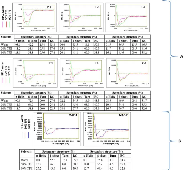
The behavior of peptides and MAPs were examined in both polar and non-polar solvents. The CD spectra analysis revealed that all six peptides in polar solvent had a negative minimum below 200 nm in a far UV range. Changing the environment from polar to non-polar [HPLC water to Trifluoroethanol (TFE)] resulted in a decline in ellipticity at 197nm followed by increase in negative ellipticity around 220nm for all the peptides, which is generally credited to the formation of more ordered conformation [33]. We further analyzed secondary structure compositions of all peptides in different environments. In polar solvent, the disordered confor-mation predominated in all six peptides (Fig 2A). Addition of non-polar solvent (50% TFE) led to induction of ordered structures. Further increase in non-polar solvent concentration to 90% TFE led to additional improvement in the ordered structures of all six peptides. We observed a peculiar secondary structure composition of P4 peptide i.e. total ordered conformations of P4 in polar and non-polar solvents were 72.4% and 77.5%, respectively (Table 2).
On analyzing the CD spectra of MAPs, interestingly,β-sheet composition dominated in both MAPs in the polar solvent i.e. 52.8%β-sheet in MAP-1 (four arm copy of P-3) and 75.6%
β-sheet in MAP-2 (four arm copy of P-4) (Fig 2B). Similar to peptides, both MAPs displayed ordered structures in non-polar environment. Total ordered conformations of MAP-2 in both polar (75.6%) and non-polar (77.1%) solvents were higher than those of MAP-1 (Table 2).
Reactivity of peptides with rabbit anti- RVA polyclonal serum
Reactivity of all peptides was evaluated in an indirect ELISA. Two peptides synthesized from an overlapping region, P-3 (QSDSLRKLSGIKFKRINFDNSSE-Gly; 112-134aa) and P-4 (KRINFDNSSEYIENWNLQNRRQRTG-Gly; 125-149aa) were reactive with anti- RVA polyclonal serum (Fig 3A). Since peptides are less immunogenic because of low molecular weight, four arms copy of these peptide sequences (P-3 and P-4) were synthesized separately on lysine mosaic prepared on Wang resin. Reactivity levels of MAP-2 [(KRINFDNSSEYIENW NLQNRRQRTG)4-Lys2-Lys-Cys] was higher in comparison to that of MAP-1 [(QSDSLRKLS
GIKFKRINFDNSSE)4-Lys2-Lys-Cys] in an indirect ELISA (Fig 3B), indicating that the region
spanning 125-149aa within VP6 protein of bovine RVA was highly reactive with rabbit anti-RVA polyclonal serum. Hence, MAP-2 was used for generation of hyperimmune serum in lab-oratory animals.
VP6 protein: Over-expression, purification and characterization
analysis. Expression levels in other two hosts were very low. Solubility analysis revealed the for-mation of inclusion bodies by over-expressed rVP6 protein. The inclusion bodies were first sol-ubilized in 6M guanidium hydrochloride followed by allowing the folding of denatured rVP6 protein at a constant rate by decreasing the concentration of urea (8M to 0M) from the washing buffer step-by-step by Immobilized-Metal Affinity Chromatography (IMAC). Thus, we allowed two processes (washing to get rid of undesired proteins and refolding of denatured
protein) to occur at the same time in Ni-NTA resin column. A total yield of ~16 mg/liter was obtained after purification under denaturing conditions.
The presence of purified rVP6 protein resolved in SDS-PAGE was confirmed by both rabbit anti- RVA polyclonal serum as well as monoclonal anti-polyHistidine peroxidase conjugate, both of which produced an intense brown color reaction with the protein size corresponding to ~62 kDa for VP6 protein (Fig 4B). Further, competence of anti-rVP6 antibodies in detecting RVA antigen in different bovine fecal samples was evaluated in dot-blot assay (Fig 4C).
Development and validation of sandwich ELISA
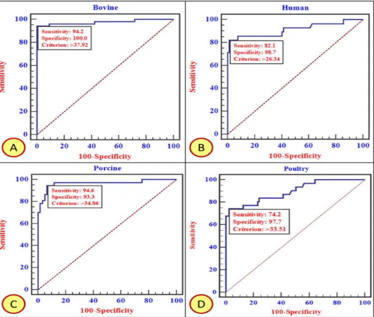
An initial set of experiments were performed to determine the optimal concentration of the coating and detector sera. The DSn and DSp of in-house ELISA at a particular cut off values (PP values) were estimated using ROC curve analysis (Fig 5andTable 3).
We observed no cross-reactivity with other enteric and non-enteric viruses, ascertaining its specificity for RVA only. The intra-assay coefficient of variation (CV) of eight well-defined
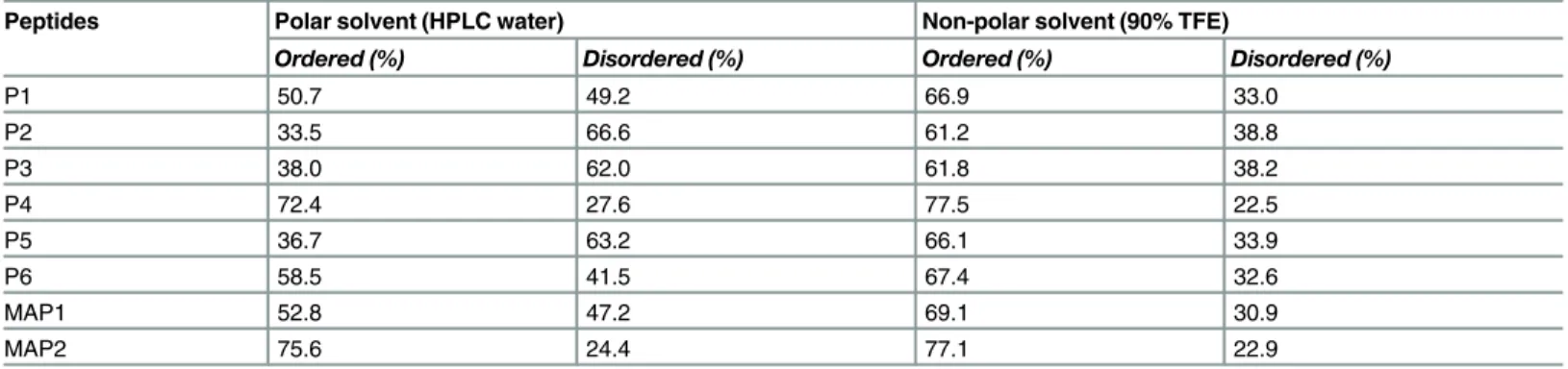
Table 2. Behavior of peptides in polar and non-polar solvents as determined by CD spectroscopy.
Peptides Polar solvent (HPLC water) Non-polar solvent (90% TFE)
Ordered (%) Disordered (%) Ordered (%) Disordered (%)
P1 50.7 49.2 66.9 33.0
P2 33.5 66.6 61.2 38.8
P3 38.0 62.0 61.8 38.2
P4 72.4 27.6 77.5 22.5
P5 36.7 63.2 66.1 33.9
P6 58.5 41.5 67.4 32.6
MAP1 52.8 47.2 69.1 30.9
MAP2 75.6 24.4 77.1 22.9
doi:10.1371/journal.pone.0159027.t002
RVA positive samples ranged from 1.02% to 3.33%, while those of eight RVA negative samples ranged from 0.02% to 4.91%. The inter-assay CV for RVA positive samples (n = 8) was between 0.36% and 41.79%, whereas the CV for RVA negative samples (n = 8) was between 0.49% and 36.01%. It is evident that this assay is repeatable and yielded a low and acceptable variation.
Further, we evaluated the performance characteristics of three diagnostic tests (RNA-PAGE, in-house ELISA, and RT-PCR) for detection of RVA antigen in the fecal samples of four spe-cies, bovine (n = 368), porcine (n = 317), poultry (n = 118) and human (n = 111) (Table 4).
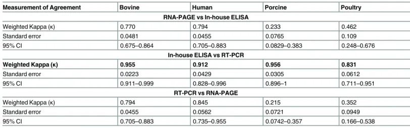
Results of Chi-square test suggested that the frequency of RVA positive samples by either of the diagnostic assay combinations were different at a highly significant level (p<0.001). We observed severe deviation in terms of RVA positivity shown by RNA-PAGE in comparison to in-house ELISA and RT-PCR, especially in porcine and poultry samples. The agreement among these three diagnostic tests was estimated by inter-rater reliability analysis based on Kappa values (κ) and results are presented inTable 5. The performance of in-house ELISA with respect to RT-PCR disclosed a perfect agreement (κvalues>0.81) for all the species tested. However, other combinations of tests showed fair to perfect agreements in different host species.
Furthermore, comparative performance of in-house ELISA revealed three and one RVA positive samples in bovine, and sheep and goat species, respectively, which were in complete concordance with the two commercial kits evaluated (Table 6).
Discussion
The development of a high sensitive and specific sandwich ELISA is described for the detection of RVA antigen in multiple host species using a novel peptide-recombinant protein approach. The commonly used commercially available diagnostic kits for RVA detection use either anti-RVA polyclonal serum or a combination of anti- VP6 monoclonal antibodies and anti- anti-RVA polyclonal serum. The unique combination of anti-rVP6 antibodies as coating and anti-MAP
Fig 4. (A) Coomassie-blue stained SDS–PAGE of cell lysates with (indicated by red arrow) and without IPTG-induction in expression host [E.coliTuner (DE3)pLysS cells], (B) Western blot analysis of recombinant hexahistidine-tagged VP6 fusion protein of bovine RVA on polyvinyl difluoride (PVDF) membrane (indicated by red arrow), (C) Dot blot assay for detection of RVA antigen in bovine fecal samples using anti-rVP6 antibodies.
antibodies as detector antibodies is novel; diagnostic use of peptides and their multimeric form has also not been documented so far in RVAs.
Fig 5. ROC curve analysis based on in-house ELISA test results obtained on screening of fecal samples from Bovine (A), Human (B), Porcine (C), and Poultry (D). Every species specific curve is provided with DSn, DSp and cut-off threshold. The area enclosed under the curve with diagonal base represents Area Under Curve (AUC).
doi:10.1371/journal.pone.0159027.g005
Table 3. Diagnostic specificity (DSp) and sensitivity (DSn) of in-house ELISA for RVA antigen detec-tion in multiple host species.
S.No. Species PP cut off value (%) DSn (%) DSp (%)
1. Bovine 37.92 94.2 100
2. Porcine 34.86 94.6 93.3
3. Poultry 33.51 74.2 97.7
4. Human 26.34 82.1 98.7
Previously, rVP6 protein was used either for sero-diagnosis of RVA infection in humans [34–35] or for detection of human RVA antigen using anti-rVP6 antibodies in latex agglutina-tion test format [36]. In latex-agglutination test, antibodies against the conserved N-terminal portion of the VP6 (1–245aa) displayed high sensitivity (98.5%) and specificity (100%), which were calculated by comparing with less sensitive RNA-PAGE [36]. Recently, another study uti-lized anti-rVP6 antibodies in differentiation of porcine rotaviruses from other porcine viruses, but lacked validation part of the assay [37]. The major limitation of all these assays was spe-cific-specific usage only.
To overcome these limitations, we exploited the peptide-recombinant protein approach. We identified a region spanning 125-149aa within the conserved VP6 protein of bovine RVA as highly reactive with rabbit anti- RVA polyclonal serum. Previous studies on protective epi-topes mapping of RVA VP6 protein were limited to c-terminal. For examples, the region of the VP6 protein spanning from amino acids 197 to 397 has been identified to contain multiple pro-tective epitopes (227–244aa, 232–261aa, 249–277aa, 283–307aa, 368–397aa) using a library of 11 overlapping peptides in H-2d BALB/c mice [38–39]. However, a 14 mer peptide covering 289-302aa region provided almost complete protection against EDIM challenge in BALB/c mice [39]. In another study, a VP6 epitope (amino acids, 242–259) was identified as immuno-dominant CD4+T cell epitope following intranasal immunization of BALB/c mice [40]. Though, 368–397aa region provided protection against challenge in a mouse model in the ear-lier study, this region did not react well with rabbit anti- RVA polyclonal serum in our study.
Further, we tried to explain as why a region spanning 125-149aa of VP6 protein was highly immune-reactive by studying the behavior of peptides of this region (P-4 and MAP-2) in polar and non-polar solvents. It has been suggested that antibodies prefer binding to ordered region of antigen and that more avidity is seen with an increase in ordered conformation [41]. Hence, high immune-reactivity of P-4 peptide with anti-RVA polyclonal serum might be triggered by
Table 4. Comparison of RNA-PAGE, in-house ELISA and RT-PCR for RVA antigen detection in multiple host species.
Species tested RNA-PAGE In-house ELISA RT-PCR
Bovine (n = 368) 115/386 (31.25%) 143/386 (38.85%) 154/368 (41.84%)
Porcine (n = 317) 25/317 (7.88%) 120/317 (37.85%) 125/317 (39.43%)
Poultry (n = 118) 8/118 (6.80%) 23/118 (19.50%) 30/118 (25.42%)
Human (n = 111) 29/111 (26.12%) 30/111 (27.02%) 34/111 (30.63%)
doi:10.1371/journal.pone.0159027.t004
Table 5. Inter-rater reliability analysis among three diagnostic tests for RVA detection in multiple host species.
Measurement of Agreement Bovine Human Porcine Poultry
RNA-PAGE vs In-house ELISA
Weighted Kappa (κ) 0.770 0.794 0.233 0.462
Standard error 0.0481 0.0455 0.0765 0.109
95% CI 0.675–0.864 0.705–0.883 0.0829–0.383 0.248–0.676
In-house ELISA vs RT-PCR
Weighted Kappa (κ) 0.955 0.912 0.956 0.831
Standard error 0.0223 0.0429 0.0305 0.0612
95% CI 0.911–0.999 0.828–0.996 0.896–1 0.711–0.951
RT-PCR vs RNA-PAGE
Weighted Kappa (κ) 0.794 0.845 0.215 0.352
Standard error 0.0455 0.0562 0.0721 0.0949
95% CI 0.705–0.883 0.735–0.955 0.0742–0.357 0.166–0.538
the following possible reasons: (i) more uniform change in the ordered structures with an increase in non-polar solvent concentration; (ii) high ordered conformation in both polar (72.4%) and non-polar (77.5%) solvents; (iii) corresponding decrease in %β-sheet structure on increasing the non-polar solvent concentration; or (iv) lower negative molar ellipticity value (<–8000 deg.cm2.decimole-1) compared to other peptides. Comparatively high immune-reac-tivity of MAP-2 with anti-RVA polyclonal serum may be explained by high ordered secondary structure in both polar (75.6%) and non-polar (77.1%) solvents or by, lower negative molar ellipticity value for MAP-2 (<–8000 deg.cm2.decimole-1) compared to MAP-1.
We expressed full length VP6 recombinant protein in prokaryotic expression system. Our goal was to produce mono-specific antibodies that react with diverse RVA strains. The eukary-otic expression system is usually preferred for full expression of VP6 mainly because of the inhibitory effect of recombinant VP6 onE.coligrowth leading to low yield [42–44]. Even,
some researchers have used codon optimized VP6 protein to achieve high yield [45]. However, we were successful in achieving a significantly high yield of VP6 protein inE.coliTuner(DE3)
pLysS cells under optimized induction conditions without codon optimization.
Peptide-recombinant protein approach exploited capture antibodies against rVP6 protein (thus avoiding the use of whole virus antigen which is more prone to yield false reactions) and detector antibodies against MAP-2 (more or less equivalent to monoclonal antibodies) to achieve high DSn and DSp. However, comparatively lower DSn especially in poultry samples might be due to low RVA excreted from their faeces. It is notable that concordance between in-house ELISA and RT-PCR tests performance (κvalues close to 1) was almost in perfect agree-ment in all the species evaluated in comparison to other combinations of tests. The RT-PCR, no doubt is a highly sensitive test, still ELISA is preferred for RVA antigen detection because results of ELISA correlate well with the clinical disease [46–47]. Also, use of enzyme immuno-assay has been suggested for vaccine efficacy evaluations in patients with acute gastroenteritis [46].
Recently, quantitative real time RT-PCR has been developed to distinguish symptomatic infections from asymptomatic based on the threshold cycle cut-off values [47], but again the cost involved might restrict its usage on a large scale especially in the low income settings. The performance of most of the RVA diagnostic tests is concentrated from well-resourced settings [48–49]. However, the pattern of RVA-associated gastroenteritis in developing countries differs from that in developed countries in terms of high levels of infection, early disease onset, and delayed acquisition of immunity, which may impact accurate and prompt diagnosis [50–51]. Based on the comparable performance of our test with the two commercial ELISA kits together with the results of inter-rater reliability assay, our test might be more suitable as a preliminary assay in RVA surveillance studies, especially in resource poor countries.
In conclusion, peptide-recombinant protein approach using anti-rVP6 antibodies as coating and anti-MAP2 antibodies as detector antibodies in sandwich ELISA format provides a tial diagnostic assay for RVA antigen detection in multiple host species. This assay has a poten-tial use in surveillance studies and in monitoring vaccine effectiveness in low and middle income settings. Adaptation of the newly developed reagents to a rapid-antigen test format would be ideal since no additional equipment would be needed.
Table 6. Comparative performance of in-house ELISA with commercially available diagnostic kits for RVA antigen detection.
Species No. of samples In-house ELISA IDEXX Rotavirus ELISA Bio-X Diagnostic ELISA
Bovines 39 3 3
-Sheep & Goat 25 1 - 1
Supporting Information
S1 Fig. Identification of regions within bovine RVA VP6 protein showing high antigenic index by bioinformatics tool.
(TIF)
S2 Fig. Schematic representation of Multiple Antigenic Peptide (MAP) design.
(TIF)
S1 Table. Details of fecal samples collected from different species in diverse geographical locations of India.
(DOCX)
Acknowledgments
We thank the Indian Council of Agricultural Research (ICAR)-Indian Veterinary Research Institute, Izatnagar, for infrastructural support. The financial support from Education Division, Indian Council of Agricultural Research, Ministry of Agriculture & Farmer’s Welfare, Govt. of India, New Delhi in the form of National Fellow project is acknowledged. Dr. Krisztian Banyai was supported by the Hungarian Scientific Research Fund (OTKA, T100727). We thank Dr. Sagar Goyal, Professor University of Minnesota, USA for critical reviewing the manuscript.
Author Contributions
Conceived and designed the experiments: NK YSM SK. Performed the experiments: NK KS AKS SBS NS SS. Analyzed the data: NK YSM SK. Contributed reagents/materials/analysis tools: YSM SK BRG NK. Wrote the paper: NK YSM KB KD VGJ.
References
1. Matthijnssens J, Otto PH, Ciarlet M, Desselberger U, Van Ranst M, Johne R. VP6-sequence-based cut-off values as a criterion for rotavirus species demarcation. Arch Virol. 2012; 157(6):1177–1182. doi:
10.1007/s00705-012-1273-3PMID:22430951
2. Papp H, László B, Jakab F, Ganesh B, De Grazia S, Matthijnssens J, et al. Review of group A rotavirus strains reported in swine and cattle. Vet Microbiol. 2013; 165(3–4):190–199. doi:10.1016/j.vetmic.
2013.03.020PMID:23642647
3. Tate JE, Burton AH, Boschi-Pinto C, Steele AD, Duque J, Parashar UD. Estimate of worldwide rotavi-rus-associated mortality in children younger than 5 years before the introduction of universal rotavirus vaccination programmes: a systematic review and meta-analysis. Lancet Infect Dis. 2012; 12(2):136– 141. doi:10.1016/S1473-3099(11)70253-5PMID:22030330
4. Greenberg HB, Estes MK.Rotaviruses: from pathogenesis to vaccination. Gastroenterology. 2009; 136:1939–1951. doi:10.1053/j.gastro.2009.02.076PMID:19457420
5. Mihalov-Kovács E, Gellért Á, Marton S, Farkas SL, Fehér E, Oldal M, et al. Candidate New Rotavirus Species in Sheltered Dogs, Hungary.Emerg Infect Dis. 2015; 21(4):660–663. doi:10.3201/eid2104.
141370PMID:25811414
6. Matthijnssens J, Ciarlet M, McDonald SM, Attoui H, Bányai K, Brister JR, et al. Uniformity of rotavirus strain nomenclature proposed by the Rotavirus Classification Working Group (RCWG). Arch Virol. 2011; 156(8):1397–1413. doi:10.1007/s00705-011-1006-zPMID:21597953
7. Gouvea V, Glass RI, Woods P, Taniguchi K, Clark HF, Forrester B, et al. Polymerase chain reaction amplification and typing of rotavirus nucleic acid from stool specimens. J ClinMicrobiol. 1990; 28 (2):276–282.
8. Taniguchi K, Wakasugi F, Pongsuwanna Y, Urasawa T, Ukae S, Chiba S, et al. Identification of human and bovine rotavirus serotypes by polymerase chain reaction. Epidemiol. Infect. 1992; 109:303–312. PMID:1327857
10. Sanekata T, Yoshida Y, Okada H. Detection of rotavirus in faeces by latex agglutination. J Immun Meth-ods. 1981; 41:377–385.
11. Langeveld JPM, Casal JI, Oster Haus ADME, Cortes E, Swart RD, Vela C, et al. First peptide vaccine providing protection against viral infection in the target animal: studies on canine parvo virus in dogs. J Virol. 1994; 68:4506–4513. PMID:8207825
12. Zamorano PI, Wigdorovitz A, Filguirea DMP, Escribano JM, Sadir AM, Borca MV. Induction of anti-foot and mouth disease virus T and B cell responses in cattle immunized with a peptide representing ten amino acids of VP1. Vaccine. 1998; 16(6):558–563. PMID:9569465
13. Tang B, Gilbert JM, Matsui SM. Comparison of the rotavirus gene 6 from different species by sequence analysis and localization of subgroup-specific epitopes using site-directed mutagenesis. Virology. 1997; 237:89–96. PMID:9344910
14. Dyson HJ, Lerner RA, Wright PE. The physical basis for induction of protein-reactive antipeptide anti-bodies. Annu Rev Biophys Biophys Chem. 1988; 17:305–324. PMID:2456075
15. Tam JP. Synthetic peptide vaccine design: synthesis and properties of a high-density multiple antigenic peptide system. Proc Natl Acad Sci. 1988; 85(15):5409–5413. PMID:3399498
16. Posnett DN, McGrath H, Tam JP. A novel method for producing anti-peptide antibodies. Production of site-specific antibodies to the T cell antigen receptor beta-chain. J Biol Chem. 1988; 263:1719–1725. PMID:3276675
17. Simmonds P, Rose KA, Graham S. Mapping of serotype-specific, immuno-dominant epitopes in the NS-4 region of hepatitis C virus (HCV): use of type specific peptides to serologically differentiate infec-tions with HCV types 1, 2, and 3. J Clin Microbiol. 1993; 31:1493–1503. PMID:7686182
18. Jackwood MW, Hilt DA. Production and immunogenicity of multiple antigenic peptide (MAP) constructs derived from the S1glycoprotein of infectious bronchitis virus (IBV). Adv Exp Med Biol. 1995; 380:213– 219. PMID:8830482
19. Saravanan P, Kumar S, Kataria JM. Detection of infectious bursal disease virus by ELISA using an anti-peptide antibody raised against VP3 region. Acta Virol. 2004a; 48:39–45.
20. Saravanan P, Kumar S, Kataria JM. Use of multiple antigenic peptides related to antigenic determi-nants of infectious bursal disease virus (IBDV) for detection of anti- IBDV-specific antibody in ELISA-quantitative comparison with native antigen for their use in serodiagnosis. J Immunol Methods. 2004b; 293:61–70.
21. Dechamma HJ, Dighe V, Kumar CA, Singh RP, Jagadish M, Kumar S. Identification of T-helper and lin-ear B epitope in the hyper variable region of nucleocapsid protein of PPRV and its use in the develop-ment of specific antibodies to detect viral antigen. Vet Microbiol. 2006; 118:201–211. PMID:16962260
22. Merrifield B.Solid phase peptide synthesis. I. The synthesis of a tetrapeptide. J Am Chem Soc.1963; 85(14):2149–2154.
23. Merrifield B. The chemical synthesis of proteins. Prot Sci. 1996; 5(9):1947–1951.
24. Nilsson BL, Soellner MB, Raines RT. Chemical synthesis of proteins. Annu Rev Biophys Biomol Struct. 2005; 34:91–118. PMID:15869385
25. Yang JT, Wu CS, Martinez HM. Calculation of protein conformation from circular dichroism. Meth Enzy-mol. 1986; 130:208–269. PMID:3773734
26. Towbin H, Staehelin T, Gordon J. Electrophoretic transfer of proteins from polyacrylamide gels to nitro-cellulose sheets: procedure and some applications. Proc Natl Acad Sci. 1979; 76:4350–4354. PMID:
388439
27. Sambrook J, Russel D. Molecular Cloning: A Laboratory Manual, 3rded, Cold Spring Harbor Labora-tory Press, Cold Spring Harbor, NY; 2001.
28. Laemmli UK. Cleavage of structural proteins during the assembly of the head of bacteriophage T4. Nature. 1970; 227(5259):680–685. PMID:5432063
29. Malik YPS, Sharma K, Vaid N, Chakravarti S, Chandrashekar KM, Basera SS, et al. Frequency of group A rotavirus with mixed G and P genotypes in bovines: predominance of G3 genotype and its emergence in combination with G8/G10 types. J Vet Sci. 2012; 13(3):271–278. PMID:23006956
30. Svensson L, Uhnoo I, Grandien M, Wadell G. Molecular epidemiology of rotavirus infections in Uppsala, Sweden, 1981: disappearance of a predominant electropherotype. J Med Virol. 1986; 18:101–111. PMID:3005484
31. Kawada T.Receiver operating characteristic curve analysis, sensitivity comparison and individual differ-ence. Clin Radiol. 2012; 67(9):940. doi:10.1016/j.crad.2012.04.002PMID:22608247
33. Greenfield NJ. Methods to estimate the conformation of proteins and polypeptides from circular dichro-ism data. Anal Biochem. 1996; 235:1–10. PMID:8850540
34. Mukhopadhya I, Anbu D, Iturriza-Gomara M, Gray JJ, Brown DW, et al. Anti-VP6 IgG antibodies against group A and group C rotaviruses in South India. Epidemiol Infect. 2010; 138:442–447. doi:10.1017/ S0950268809990732PMID:19723364
35. Seo JH, Kim SY, Park JS, Lim JY, Park CH, Woo HO, et al. Usefulness of Escherichia coli-expressed recombinant VP6 proteins of Group A Rotavirus in serodiagosis of Rotavirus infection. Korean J Pediatr Gastroenterol Nutr. 2010; 13(2):134–145.
36. de Góes AC, de Moraes MT, de Castro Silveira W, Araújo IT, de H'alluin JC, et al. Development of a rapid and sensitive latex agglutination-based method for detection of group A rotavirus. J Virol Methods. 2008; 148:211–217. doi:10.1016/j.jviromet.2007.11.013PMID:18241934
37. Zhu J, Yang Q, Cao L, Dou X, Zhao J, Zhu W, et al. Development of porcine rotavirus vp6 protein based ELISA for differentiation of this virus and other viruses. Virol J. 2013; 10:91. doi: 10.1186/1743-422X-10-91PMID:23517810
38. Choi AH, Basu M, McNeal MM, Flint J, VanCott JL, Clements JD, et al. Functional mapping of protective domains and epitopes in the rotavirus VP6 protein. J Virol. 2000; 74:11574–11578. PMID:11090155
39. Choi AH, McNeal MM, Basu M, Bean JA, VanCott JL, Clements JD, et al. Functional mapping of protec-tive epitopes within the rotavirus VP6 protein in mice belonging to different haplotypes. Vaccine. 2003; 21(7–8):761–767. PMID:12531356
40. McNeal MM, Basu M, Bean JA, Clements JD, Choi AH, Ward RL. Identification of an immunodominant CD4+ T cell epitope in the VP6 protein of rotavirus following intranasal immunization of BALB/c mice. Virology. 2007; 363(2):410–418. PMID:17337285
41. Lang E, Szendrei GI, Lee VM, Otvos L Jr. Spectroscopic evidence that monoclonal antibodies recog-nize the dominant conformation of medium-sized synthetic peptides. J Immunol Methods. 1994; 170:103–115. PMID:7512605
42. Ernst H, Stroup D. Synthesis of the major inner capsid protein VP6 of the human rotavirus Wa in Escherichia coli. Gene. 1988; 68(2):345–356. PMID:2851499
43. Erk I, Huet JC, Duarte M, Duquerroy S, Rey F, Cohen J, et al. A zinc ion controls assembly and stability of the major capsid protein of rotavirus. J Virol. 2003; 77:3595–3601. PMID:12610135
44. Mathieu M, Petitpas I, Navaza J, Lepault J, Kohli E, Pothier P, et al. Atomic structure of the major capsid protein of rotavirus: implications for the architecture of the virion. EMBO J. 2001; 20:1485–1497. PMID:
11285213
45. Choi AH, Basu M, McNeal MM, Bean JA, Clements JD, Ward RL. Intranasal administration of an Escherichia coli-expressed codon-optimized rotavirus VP6 protein induces protection in mice. Protein Expr Purif. 2004; 38(2):205–216. PMID:15555936
46. Tate JE, Mijatovic-Rustempasic S, Tam KI, Lyde FC, Payne DC, Szilagyi P, et al. Comparison of 2 assays for diagnosing rotavirus and evaluating vaccine effectiveness in children with gastroenteritis. Emerging Infect Dis. 2013; 19(8):1245–1252. doi:10.3201/eid1908.130461PMID:23876518 47. Bennett A, Bar-Zeev N, Jere KC, Tate JE, Parashar UD, Nakagomi O, et al. Determination of a Viral
Load Threshold To Distinguish Symptomatic versus Asymptomatic Rotavirus Infection in a High-Dis-ease-Burden African Population. J ClinMicrobiol. 2015; 53(6):1951–1954.
48. Phillips G, Lopman B, Tam CC, Iturriza-Gomara M, Brown D, Gray J. Diagnosing rotavirus A associated IID: using ELISA to identify a cut-off for real time RT-PCR. J Clin Virol. 2009; 44(3):242–245. doi:10. 1016/j.jcv.2008.12.001PMID:19179107
49. Iturriza-Gomara M, Elliot AJ, Dockery C, Fleming DM, Gray JJ. Structured surveillance of infectious intestinal disease in pre-school children in the community:‘The Nappy Study’. Epidemiol Infect. 2009; 137(7):922–931. doi:10.1017/S0950268808001556PMID:19017426
50. Gladstone BP, Ramani S, Mukhopadhya I, Muliyil J, Sarkar R, Rehman AM, et al. Protective effect of natural rotavirus infection in an Indian birth cohort. N Engl J Med. 2011; 365(4):337–346. doi:10.1056/ NEJMoa1006261PMID:21793745