Vimentin and Anti Vimentin Antibodies in Chagas’ Disease
Marilda Savoia Nascimento, Anna Maria Simonsen Stolf, Heitor Franco de Andrade Junior, Ramendra Pati Pandey,
Eufrosina Setsu Umezawa
Universidade de São Paulo (USP), São Paulo, SP – Brazil
Mailing Address: Marilda Savoia Nascimento •
Av. Senador Vergueiro, 608. Postal Code 09750-000, Centro, São Bernardo do Campo, São Paulo, SP – Brazil
E-mail: [email protected]
Manuscript received on June 13, 2017; revised manuscript on September 05, 2017; accepted on October 06, 2017
DOI: 10.5935/abc.20180038
Abstract
Background: Vimentin is a main structural protein of the cell, a component of intermediate cell filaments and immersed in cytoplasm. Vimentin is mimicked by some bacterial proteins and anti-vimentin antibodies occur in autoimmune cardiac disease, as rheumatic fever. In this work we studied vimentin distribution on LLC-MK2 cells infected with T. cruzi and anti-vimentin antibodies in sera from several clinical pictures of Chagas’ disease or American Trypanosomiasis, in order to elucidate any vimentin involvement in the humoral response of this pathology.
Objective: We standardized an indirect immunofluorescence assay (IFI) to determine sub cellular expression in either parasites and host cells, and ELISA to evaluate anti-vimentin antibodies in sera fron chagasic patients.
Methods: We analyzed the distribution of vimentin in culture cells using indirect fluorescent assays, using as external controls anti-T. cruzi sera, derived from chronic infected patients for identification of the parasites in the same model. After infection and growth of T.cruzi amastigotes, those cells express larger amounts of vimentin, with heavy staining of cytoplasm outside the parasitophorous vacuole and some particle shadowing patterns, suggesting that vimentin are associated with cell cytoplasm. Anti-vimentin antibodies were present in most American trypanosomiasis samples, but notably, they are much more present in acute (76, 9%) or clinical defined syndromes, especially cardiac disease (87, 9%). Paradoxically, they were relatively infrequent in asymptomatic (25%) infected patients, which had a clearly positive serological reaction to parasite antigens, but had low frequency of anti-vimentin antibodies, similar to controls (2,5%).
Conclusion: Our current data revealed that anti-vimentin antibodies induced during T. cruzi infection could be a marker of active disease in the host and its levels could also justify drug therapy in American Trypanosomiasis chronic infection, as a large group of asymptomatic patients would be submitted to treatment with frequent adverse reactions of the available drugs. Anti-vimentin antibodies could be a marker of cardiac muscle cell damage, appearing in American Trypanosomiasis patients during active muscle cell damage. (Arq Bras Cardiol. 2018; 110(4):348-353)
Keywords: Chagas Disease; Heart Diseases; Trypanossoma Cruzi; Rheumatic Fever; Vimentin; Antibodies, Monoclonal.
Introduction
Chagas’ disease or American Trypanosomiasis is a
peculiar parasitic infection as Trypanosoma cruzi is a
unique intracellular parasite which resulted in cytoplasmic presence of amastigotes forms, a rare cellular event in nature, as cytoplasm is usually free from parasites in almost
all intracellular infections1. After its reproduction, the
parasite had a set of enzymes, as sialidases, that transfers host cell molecules to their surface, allowing cell evasion
without disruption2. All those processes could alter cell
cytoskeleton and its proteins, probably generating in the host cell signals that alters the protein synthesis of structural proteins. Vimentin is a main structural protein of the cell, a
component of intermediate cell filaments and immersed in
cytoplasm3. Vimentin is expressed in normal cardiac muscle
and their tumors, and autoantibodies against a vimentin re
found in allograft rejection4. or cardiac models of allograft
rejection5.Vimentin is mimicked by some bacterial proteins
and anti-vimentin antibodies occur in autoimmune cardiac
disease, as rheumatic fever6. In this work we studied
vimentin distribution on LLC-MK2 cells infected with T.cruzi
and anti-vimentin antibodies in sera from several clinical pictures of American Trypanosomiasis, in order to elucidate any vimentin involvement in the humoral response of this pathology.
Methods
Parasites and serum samples
Trypanosoma cruzi epimastigotes were grown from Y strain routinely maintained in our lab on Liver Infusion Tryptose (LIT) culture media supplemented with 10% fetal
calf serum. T. cruzi trypomastigotes were obtained from cell
vimentin from bovine lens was obtained commercially (Sigma Aldrich, Saint Louis, Missouri, USA). A serum from patient with cardiac chronic American Trypanosomiasis was
used as anti T.cruzi antibody. Human sera from American
Trypanosomiasis patients and controls were used from the
biorepository of T.cruzi patients samples from E.S.Umezawa,
Lab.Protozoology, IMTSP, serologically characterized in TESA specific serology tests and published previously in several articles, were recovered and comprising 26 sera from acute disease, 33 from isolated cardiac disease, 17 from isolated digestive disease, 20 without clinical disease (asymptomatic disease) and 40 sera from patients outside endemic area. All clinical data were maintained by the attendant physician and not available for this study.
Antigen expression and morphology
All morphological assays were performed in a Zeiss Axioplan epifluorescent microscope with fluorescein filters. For antigen detection, we fixed LLC-MK2 control cells,
T.cruzi infected LLC-MK2 cells and T.cruzi epimastigotes
and permeated cell surface with Triton X-1007 with either
anti-Vimentin mAb or anti-T.cruzi antibodies as elsewhere
described. After this step, bound antibodies were revealed with adequate fluorescein conjugate, carefully washed and mounted in glycerin for observation. Representative Fields were digitalized at high power field using a Canon camera.
TESA and vimentin ELISA
T.cruzi trypomastigotes excreted secreted antigen was
obtained as elsewhere described8. TESA (1/80) and Vimentin
(0.06ug/ml) in carbonate 0.05 M pH9.6 were adsorbed overnight to wells of 96 wells high binding ELISA plates (Corning Inc. New York, USA). After washing and blocking with PBS Tween 20, 0,05% plus 5% milk or BSA 0.5%, adequate dilution of sera (1/50 vimentin and 1/200 TESA) were incubated for one hour. After new washings, adequate dilution of peroxidase conjugate were added for another hour, washed and bound conjugate revealed by 1 h with orto-phenylenediamine and hydrogen peroxide. After 30 min in 37°C, reaction was stopped with 4N HCl and 492 nm absorbance determined in a microplate reader (Multiskan-Titertek II).
Statistical analysis
All quantitative data, such as O.D. ELISA, were analyzed using ANOVA after the Levene test for variance check, with intragroup comparisons by the Bonferroni's test, if there are uniformity of variances. In the absence of this homogeneity, data were analyzed by Kruskal-Wallis tests with Dunns post-tests. We opt for graphical representation of individual data in dot plot with association of mean and SEM for comparison. Qualitative analysis, as frequency of positive sera in the group, was analyzed by Fisher exact tests in two group analysis. We also included 95% confidence interval of estimated proportion. Significant difference was considered when the probability of equality (H1 = H0) was less than
0.05(p≤0.05), using two-tailed analysis and power greater
than 90%. We used the statistical package GraphPad Prism 7.0 for all statistical analysis and plotting.
Results
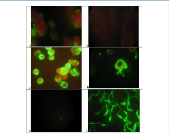
We analyzed the distribution of Vimentin in culture cells using indirect fluorescent assays as described in Methods,
using as external controls anti-T.cruzi sera, derived from
chronic infected patients for identification of the parasites in the same model, as could be seen in figure 1. LLC-MK2 cells,
the host cell used for intracellular growth of T.cruzi, showed
a discrete and uniform cytoplasmic staining, uniform in most
cells (Figure 1A). Those cells are no reactive to anti-T-cruzi
antibodies, without any staining (Figure 1B). After infection
and growth of T.cruzi amastigotes, those cells express larger
amounts of vimentin, with heavy staining of cytoplasm outside the parasitophorous vacuole and some particle shadowing patterns, suggesting that vimentin are associated with cell cytoplasm (Figure 1C). Vimentin could involve unstained cytoplasmic parasites, but no specific staining of parasites was seen. Those parasites were easily identified
by anti-T.cruzi antibodies showing a typical morular pattern
in the cytoplasm of infected cells (Figure 1D). No staining of those parasites was observed with anti-vimentin mAbs, which demonstrate the absence of antigen mimicry, both for amastigotes (Figure 1C) or extracellular parasites (Figure 1E).
Those extracellular parasites are heavily stained by anti T.cruzi
antibodies as well as intracellular amastigotes (Figure 1F).
Anti-vimentin auto antibodies
Figure 1 – Distribution of Vimentin or Trypanosoma cruzi antigen on control or infected cells and parasites. A and B) Uninfected LLC MK2 cells reacted to antivimentin abs(A) or anti-T.cruzi abs(B). C and D) T.cruzi infected LLC MK2 cells reacted to antivimentin abs(C) or anti-T.cruzi abs(D). E and F) T.cruzi promastigote forms from in vitro culture reacted to antivimentin abs (E) or anti-T.cruzi abs(F). Cells, infected cells or parasites forms were reacted with Anti Vimentin mAb or chronic infection chagasic serum and revealed with adequate conjugate (x1000) (see Methods).
sera from patients with clinical active disease for any origin was in higher frequency than in patients without active disease or non-infected controls. Data were compared mainly with active or undetermined without clinical forms of Chagas' disease shows greater difference as expected with high statistical difference (p < 0.01) and also demonstrated by 95% confidence interval of the proportion
The Table 1 Summarizes the data obtained in Figure 2 and provides ELISA positivity indexes with commercial Vimentin, showing that the percentage of positive sera from the groups of chronic patients with clinical manifestations of Chagas’ disease and the group of patients from the acute phase was higher than that observed in the indeterminate group of chagasic patients.
The positivity index of sera from patients in the acute phase was 76.9% with 20 positive sera from the 26 analyzed. In the group of chronic digestive tract positive percentage was 70.5% with 12 positive in the 17 analyzed, the cardiac patients had a positive percentage of 87.9% with 29 positive
sera from the 23 analyzed and in the group of indeterminate patients, the Index was 25% with 5 positive of the 20 analyzed. The positivity of the non-chagasic sera was 2.5% or only a positive serum in 40 analyzed.
Discussion
This intracytoplasmic infection resulted in altered expression of cell fibrillary proteins as vimentin, as we clearly show in immunofluorescence of infected cells. This altered vimentin production is devoid of association with the parasite, which has no reactivity with antivimentin antibodies in any form. Vimentin is important for specific virus entry, another possible cytoplasmic pathogen and are used by Foot-and-mouth disease virus (FMDV) for virus
mounting inside the cells.9
Figure 2 – Sera reactivity profile of patients with different clinical forms of Chagas disease by ELISA with T. cruzi TESA antigens (A) and commercial vimentin (B). Groups were compare with ANOVA with Bonferronipost tests.
3.0
2.5
2.0
1.5
1.0
0.5
0.0
2.5
2.0
1.5
1.0
0.5
0.0
TESA
ELISA
(492nm)
V
imentin ELISA
(492nm)
A
B p < 0.001
p < 0.001 p < 0.001
p > 0.05
acute asymptomatic digestive cardiac controls
acute asymptomatic digestive cardiac controls
Table 1 – Percentage of positivity of sera with different clinical forms of Chagas disease for Vimentin antigen in the ELISA reaction
Clinical form Samples (n) Positives (n) Negatives (n) Positivity (%) 95% C.I.
(p vs w/o Chagas) p VS undetermined
Acute 26 20 6 76.9 53-87 (p < 0.001) p < 0.001
Cardiac 33 29 4 87.8 67-93 (p < 0.001) p < 0.001
Digestive 17 12 5 25 42-84 (p < 0.001) p < 0.01
Symptomatic 76 61 15 80,2 70-88 (p < 0.001) p < 0.001
Undetermined 20 5 15 70,5 5-44 (p < 0.05)
Total Chagas 96 66 30 68,5 52-75 (p < 0.001)
Without Chagas 40 1 39 2,5 1-4%
1. Calvet CM, Melo TG, Garzoni LR, Oliveira FO Jr, Neto DT, N S L M, et al. Current understanding of the Trypanosoma cruzi-cardiomyocyte interaction. Front Immunol. 2012 Oct 30;3:327. doi: 10.3389/fimmu.2012.00327.
2. Freire-de-Lima L, Fonseca LM, Oeltmann T, Mendonça-Previato L, Previato JO. The trans-sialidase, the major Trypanosoma cruzi virulence factor: three decades of studies. Glycobiology. 2015;25(11):1142-9. doi: 10.1093/ glycob/cwv057.
3. Lowery J, Kuczmarski ER, Herrmann H, Goldman RD. 2015. Intermediate filaments play a pivotal role in regulating cell architecture and function. J Biol Chem. 2015;290(28):17145-53. doi: 10.1074/jbc.R115.640359.
4. Mahesh B. Leong HS, McCormack A, Sarathchandra P, Holder A, Rose ML. Autoantibodies to vimentin cause accelerated rejection of cardiac allografts. Am J Pathol. 2007;170(4):1415-27. doi: 10.2353/ajpath.2007.060728.
5. Azimzadeh AM, Pfeiffer S, Wu GS, Schröder C, Zhou H, Zorn GL 3rd,
et al. Humoral immunity to vimentin is associated with cardiac allograft injury in nonhuman primates. Am J Transplant. 2005;5(10):2349-59. doi: 10.1111/j.1600-6143.2005.01022.x.
6. Delunardo F, Scalzi V, Capozzi A, Camerini S, Misasi R, Pierdominici M, et al. Streptococcal-vimentincross-reactive antibodies induce microvascular cardiac endotelial pro inflammatory phenotype in rheumatic heart disease. Clin Exp Immunol. 2013;173(3):419-29. doi: 10.1111/cei.12135.
7. Kaverina I, Rottner K, Small JV. Targeting, capture, and stabilization of microtubules at early focal adhesions. J Cell Biol. 1998;142(1):181-90. PMID: 9660872.
8. Umezawa ES, Nascimento MS, Stolf AM. Enzyme-linked immunosorbent assay with Trypanosoma cruzi excreted-secreted antigens (TESA-ELISA) for serodiagnosis of acute and chronic Chagas disease. Diagn Microbiol Infect Dis. 2001;39(3):169-76. PMID: 11337184.
References
vimentin production is devoid of association with the parasite, which has no reactivity with anti-vimentin antibodies in any form. Viral infection alters host cell architecture similarly, as
parvovirus in mice10 but other pathogens also affects vimentin
distribution in infected cells, with similar perivacuolar
distribution, as in Salmonella infections.11 Proteomics studies
in experimental models of T.cruzi infection had shown higher
plasma levels of vimentin related to disease severity,12 which
can offer to the immune response intracellular filaments for antibody production. Those data were expected as vimentin autoantibodies could be related to antigen exposure during active infection, as proposed in experimental models of
T.cruzi infection.12 Several other immune diseases that
interact with cardiac muscle cells also presented anti-vimentin antibodies. Murine models of viral myocarditis presented those
antibodies13 and as well as post-streptococcal rheumatic fever
patients14. Noninfectious myocarditis, as in coronary artery
disease patients15 and kidney or heart transplants recipients16
also showed those antibodies resulted from any exposure of antigen, unregard the origin of cardiac muscle cell damage. Our data were similar to those findings and anti-vimentin
antibodies induced during T.cruzi infection could be a marker
of active disease in the host and its levels could also justify drug therapy in American Trypanosomiasis chronic infection, as a large group of asymptomatic or indeterminate patients would be submitted to treatment with frequent adverse reactions of the available drugs. Anti-vimentin antibodies could be a marker of cardiac muscle cell damage, appearing in American Trypanosomiasis patients during active muscle cell damage.
Conclusions
Our data revealed that anti-vimentin antibodies induced
during activity of T. cruzi infection could be a marker of
active disease in the host, despite absence of evident clinical involvement. This assay could be also a non-invasive follow-up test during drug therapy in Chagas’ disease or American Trypanosomiasis. This test could allow the selection of possible active patients for therapy and also to supply a marker of disease activity after therapy, avoiding that a large group of asymptomatic patients without active disease were
submitted to treatment with frequent adverse reactions. Anti-vimentin antibodies could be a marker of cardiac muscle cell inflammatory involvement, showed by American Trypanosomiasis patients with active muscle cell damage and must be tested in other cardiac muscle inflammatory conditions as viral myocarditis.
Author contributions
Conception and design of the research: Nascimento MS, Stolf AMS, Umezawa ES; Acquisition of data: Nascimento MS, Stolf MAS; Analysis and interpretation of the data: Nascimento MS, Stolf AMS, Andrade Junior HF, Pandey RP, Umezawa ES; Statistical analysis: Nascimento MS, Andrade Junior HF; Obtaining financing: Nascimento MS, Umezawa ES; Writing of the manuscript: Nascimento MS, Stolf AMS, Pandey RP; Critical revision of the manuscript for intellectual content: Nascimento MS, Andrade Junior HF, Pandey RP.
Potential Conflict of Interest
No potential conflict of interest relevant to this article was reported.
Sources of Funding
This study was funded by FMUSP.
Study Association
This article is part of the thesis of master submitted by Marilda Savoia Nascimento, from Universidade de São Paulo.
Ethics approval and consent to participate
This is an open-access article distributed under the terms of the Creative Commons Attribution License
9. Gladue DP, O’Donnell V, Baker-Branstetter R, Holinka LG, Pacheco JM, Fernández Sainz I, et al. Foot-and-mouth disease virus modulates cellular vimentin for virus survival. J Virol. 2013;87(12):6794-803. doi: 10.1128/JVI.00448-13
10. Nüesch JP, Lachmann S, Rommelaere J. Selective alterations of the host cell architecture upon infection with parvovirus minute virus of mice.Virology. 2005;331(1):159-74. doi: 10.1016/j.virol.2004.10.019.
11. Finlay BB, Ruschkowski S, Dedhar S. Cytoskeletal rearrangements accompanying salmonella entry into epithelial cells. J Cell Sci. 1991;99(Pt 2):283-96. PMID: 1909337.
12. Wen JJ, Garg NJ. Proteome expression and carbonylation changes during Trypanosoma cruzi infection and Chagas disease in rats. Mol Cell Proteomics. 2012;11(4):M111.010918. doi: 10.1074/mcp.M111.010918.
13. Sato Y, Matsumori A, Sasayama S. Autoantibodies against vimentin in a murine model of myocarditis. Autoimmunity. 1994; 18(2):145-8. PMID: 7742476.
14. Guilherme L, Kalil J. Rheumatic fever and rheumatic heart disease: cellular mechanisms leading autoimmune reactivity and disease. J Clin Immunol. 2010;30(1):17-23. doi: 10.1007/s10875-009-9332-6.
15. Nikkari ST, Solakivi T, Sisto T, Jaakkola O. Antibodies to cytoskeletal proteins in sera of patients with angiographically assessed coronary artery disease. Atherosclerosis. 1993;98(1):11-6. PMID: 8457245.