Histological response to platelet-rich plasma added to
polypropylene mesh implemented in rabbits
_______________________________________________
Oscar Rubini Ávila
1, Natália Gomes Parizzi
2, Ana Paula Mayumi Souza
2, Dayane Silvestre Botini
2, João
Ytimura Alves
2, Silvio Henrique Maia Almeida
21 Departamento de Cirurgia, Universidade do Oeste Paulista, Presidente Prudente, São Paulo, Brasil; 2 Departamento de Cirurgia, Universidade Estadual de Londrina, Londrina, Paraná, Brasil
ABSTRACT
ARTICLE
INFO
______________________________________________________________ ______________________
Introduction: The platelet-rich plasma (PRP) is part of a set of biotechnologies, provid-ing some growth factors that promote repair of different tissues. The polypropylene meshes (PPM) are applied in the correction of abdominal defects, pelvic floor and uri-nary incontinence, however, they induce many significant complications, as a result of an inappropriate inflammatory response.
Purpose: To investigate the changes caused by PRP associated with the implantation of PPM in the abdomen of female rabbits, in the production of collagen I and III and the inflammatory infiltrate (ININ).
Materials and Methods: We performed implant meshes with and without PRP in adult rabbits (n=30) and euthanasia at 7, 30 and 90 days. Two plates were prepared from each animal and analyzed in five different fields. The ININ was evaluated by quanti-fication of inflammatory cells using hematoxylin-eosin and the collagen by Sirius red method. The results were analyzed applying the Wilcoxon, Kruskal-Wallis, Junckheere and Friedmann tests.
Results: There was a significant difference in the number of inflammatory cells be-tween the groups with and without PRP (p=0.01) at 90 days. There was increased pro-duction of collagen I, III and total with the use of PRP, at seven days.
Conclusion: The PPM coating with PRP was associated with increased ININ at the im-plant area, and an increasing trend during the process of tissue repair. The PPM coated with PRP was related to increased concentration of collagen I, collagen III and the concentration of total collagen increased after seven days of implantation.
Keywords:
Histological Techniques; Rats; Blood Platelets
Int Braz J Urol. 2016; 42: 993-8
_____________________
Submitted for publication: June 14, 2015
_____________________
Accepted after revision: October 26, 2015
INTRODUCTION
Synthetic mesh, of varying compositions, has been widely used for several years in the repair of hernias and in Urogynecology. Howe-ver, its diverse properties and the inflammatory reaction caused by synthetic materials are still being studied (1-3).
Using synthetic mesh to repair abdominal hernias and urinary incontinence represents the
largest group of implants in modern medicine, with about 1 million surgeries performed worl-dwide (4). Despite this fact, few randomized, con-trolled trials have evaluated the effects of the co-ated mesh during host incorporation.
genital prolapse repair (6). The host’s reaction to the implant is related to the type of material used; for example, an autologous fascia promotes lower in-flammation and produces less collagen (7). Various protocols tested bioactive substances that act on inflammation and accelerate the repair process (8, 9). However, the complexity of the implant-host in-teraction impedes their use and causes controversy. The platelet-rich plasma (PRP) definition is a plasma sample with a higher concentration of platelets, on average 2-3 times more, and is asso-ciated with clotting factors when compared to the peripheral blood (10). The potential therapeutic effect of PRP is the ability to promote tissue rege-neration by releasing growth factors (CF) present in platelet alpha granules. These granules release growth factors soon after clot formation (11), and the proteins contained in it have a strong influence on reparative phenomena of wounds. The proteins include PDGF (“platelet derived growth factor”), TGF-Beta (“transforming growth factor”), and IGF (“insulin-like growth factor”), which exerts chemotactic, mitogenic, and angiogenic activity, which influences the inflammatory process. Some proteins released by platelets are absent in chronic wounds, emphasizing the role of these substances in tissue repair (12).
The PRP was chosen to study the accelera-tion of the repair process because it is a source of many growth factors (GF). It is already widely used in other medical fields (dermatology, orthopedics, dentistry and plastic surgery) and is widely availa-ble at a low cost (12, 13). The objective of this study was to examine the inflammatory infiltrate (ININ) response and collagen production induced by the implantation of polypropylene mesh in two groups, with or without PRP, using an animal model.
MATERIALS AND METHODS
The Institutional Animal Care Utilization Committee approved the study. Thirty white New Zealand adult rabbits were submitted to sub apo-neurotic implant of polypropylene mesh 1.0 x 1.0cm fragments (with pores of the 1500 microns).
The specimens were anesthetized with in-tramuscular Ketamine and Xylazine. The PRP gel was obtained after removal of 10mL of blood by
cardiac puncture, and immediately taken to the laboratory for preparation using the protocol des-cribed by Anitua et al. (14).
Platelet counts in plasma of 25% of the samples chosen randomly after the process of pre-paration of PRP gel were performed to confirm the increase in the number of platelets. The gel count presented on average three times the plate-let count than the peripheral blood (Figure-1).
A one-centimeter abdominal incision was performed, and the implant was accomplished without fixation to prevent tissue reactions. The mesh was implanted in a standardized manner, between the hypodermis and the fascia of the
ab-dominal muscles. The animals were divided into 2 groups (15 each): mesh or PRP covering the mesh. Benzathine Penicillin was the antibiotic of choice.
The animals were euthanized after 7, 30 and 90 days after the implant 95 animals each from each group, with and without PRP.
All animals were anesthetized before the lethal injection. It was then removed from the block comprising the implant site interesting the skin, subcutaneous, mesh and muscle apo-neurosis. At each time point, the wounds were harvested, and their histologic features were as-sessed in paraffin-embedded sections using
he-Figure 1 - Mean platelet concentration in the blood compared to the centrifuged platelet-rich plasma (PRP).
300.000
250.000
200.000
150.000
100.000
50.000
0
Platelets
Blood
matoxylin & eosin and picro-sirius stains at a
magnification of 10 X 40.
One pathologist was blinded to the mesh type and time from wounding evaluated all speci-mens. The slides were scanned under an Olympus microscope and a 3CCD pro-series digital camera; the capture and analysis program used was Image Pro-Plus (Media Cybernetics, Silver Spring, MD). The collagen type I, III, and total were assessed using polarized light (Sirius red), by computer morphometry (14) (density per micra²), while in-flammatory infiltrate (H & E) were counted in
di-fferent fields (400x). Four fragments of the mate-rial were placed on each slide.
For statistical studies were performed, in-cluding the Wilcoxon test (differences between the PRP groups and collagen types), the Kruskal--Wallis test (difference between point times), the Jonckheere trend test, and the Friedman test (di-fferences inside groups). All statistical analysis was carried out using the SPSS version 13.0 data analysis system. P–values <0.05 were considered to be statistically significant for all comparisons.
RESULTS
Figure-2 shows the average sum of ININ per microscopic field to the rabbits euthanized at 7, 30, and 90 days comparing the group with
Figure 2 - Curves of inflammatory infiltrate by time, accordingly with or without PRP.
with PRP without PRP
700
600
500
400
300
200
100
0
7 30 90
Days Inflammatory
cells
or without PRP. The Jonckheere Test showed an increasing trend in the number of inflammatory cells into the euthanasia (p=0.027) in both groups.
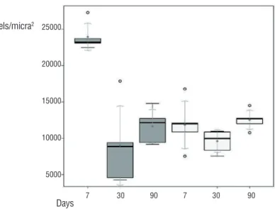
Figure-3 shows a significant increase of to-tal collagen to rabbits euthanized after seven days for the group with PRP (Wilcoxon test, p=0.008). At seven days, both type I collagen and the type III specimens in the PRP group are shown enlarged (Figure-4 (Wilcoxon test, p=0.001)).
DISCUSSION
25000
20000
15000
10000
5000
7 30 90 7 30 90
Pixels/micra2
Days
Figure 3 - Total collagen boxplots for rabbits sacrificed at 7, 30, and 90 days per group with and without PRP.
Days
7 3 0 9 0 16000,00
14000,00
12000,00
10000,00
8000,00
6000,00
4000,00
2000,00
0,00
Pixels/micra2
Type III with PRP Type III without PRP Type I with PRP Type I without PRP
Figure 4 - Curve of the average of collagens I and III for the rabbits sacrificed at 7, 30, and 90 days per group with and without PRP.
is independent of the implantation site and cannot be influenced by cover plasma.
The data evaluation shows no significant change in inflammatory cells in an initial phase (7 days). This result demonstrates that the PRP does not alter the initial inflammatory response and, therefore, results in no impact at this stage.
platelets. So it is assumed that the continued mi-gration of inflammatory cells favors the end repair probably with macrophages and lymphocytes, which are mainly responsible for maintaining the healing process in the late phase (19).
Our experiment shows an increase main-ly of type I collagen when comparing the groups with and without PRP for rabbits sacrificed at se-ven days, reaching a difference of about 4.5 times. This finding is relevant since this type of collagen is the most common late in the tissue repair pro-cess. Therefore, an increase early in the process suggests a possible acceleration in the repair by the action of PRP. However, there was no differen-ce at the late point (90 days), which kept the same level of concentration in individuals who did not undergo PRP use.
The type III collagen also presents an in-creased production in rabbits euthanized at seven days, but ordinarily, at this early stage, the collagen type III is more active and responsible for tissue re-pair. Then, the addition of PRP response to the ini-tial increase collagen production can create a better relationship between the mesh and the host. These reflections are evident when integrating the two variable’s results: the inflammatory infiltration and collagen production. In an initial process when the intense inflammatory reaction can cause a rejection of the implanted material (20, 21), the PRP did not affect this response. However, when the concen-tration of inflammatory cells is necessary for the maintenance of the chronic process, providing an adequate material integration, the PRP provides a positive response. Also, the total collagen produc-tion increases in the early process, showing a pos-sible acceleration of tissue repair. So it is pospos-sible to suggest that a PRP action occurs in the initial phase and the late stage of the process. The PRP acts in different ways but synergistically and probably by the action of growth factors provided by plasma alpha granules.
This study used rabbits in reproductive adulthood with abdominal implants, but the mesh for gynecological use is employed in menopausal women with vaginal implants. Despite the same pa-thophysiology of healing, abdominal implants oc-cur in sterile conditions differing from the vagina, a potentially contaminated environment.
Since the majority of pelvic reconstructive surgeries are being performed in postmenopausal women, it is essential to understand how estrogen deficiency affects this process.
Another issue is the study times. In a study of animals, Gerullis et al. found that the use of plasma coated mesh did not influence the early inflammatory reaction but did influence the in-flammation in the medium and long terms (21). Unfortunately, this experiment is not contempla-ted in later times to euthanasia, so it is impossible to compare results.
An immunohistochemical evaluation and analysis of the presence of PDGF in the various sta-ges of the process with their comparison to groups with and without PRP at different times can con-tribute to a better understanding of the subject. Also, the PRP’s presence should be evaluated by tests of pro-inflammatory and anti-inflammatory markers in the various stages of the repair process. This strategy can answer questions of the intensity of the inflammatory response necessary for proper mesh integration and if the changes are caused by the PRP’s action.
Thus, further studies should be conducted to improve understanding of PRP action in host tissue and its actual validity for use in the clinic.
CONCLUSIONS
The PPM coating with PRP was associa-ted with increased ININ at the implant area, and an increasing trend during the process of tissue repair. The PPM coated with PRP was related to increased concentration of collagen I, collagen III and the concentration of total collagen increased after seven days of implantation.
CONFLICT OF INTEREST
None declared.
REFERENCES
2. Afonso JS, Martins PA, Girao MJ, Natal Jorge RM, Ferreira AJ, Mascarenhas T, et al. Mechanical properties of polypropylene mesh used in pelvic floor repair. Int Urogynecol J Pelvic Floor Dysfunct. 2008;19:375-80. Erratum in: Int Urogynecol J Pelvic Floor Dysfunct. 2008;19:381.
3. Gerullis H, Georgas E, Eimer C, Arndt C, Barski D, Lammers B, et al. Coating with autologous plasma improves biocompatibility of mesh grafts in vitro: development stage of a surgical innovation. Biomed Res Int. 2013;2013:536814. 4. Schumpelick V, Klinge U, Welty G, Klosterhalfen B. [Meshes
within the abdominal wall]. Chirurg. 1999;70:876-87. 5. Klinge U, Schumpelick V, Klosterhalfen B. Functional
assessment and tissue response of short- and long-term absorbable surgical meshes. Biomaterials. 2001;22:1415-24.
6. Baessler K, Maher CF. Mesh augmentation during pelvic-floor reconstructive surgery: risks and benefits. Curr Opin Obstet Gynecol. 2006;18:560-6.
7. de Almeida SH, Rodrigues MA, Gregório E, Crespígio J, Moreira HA. Influence of sling material on inflammation and collagen deposit in an animal model. Int J Urol. 2007;14:1040-3.
8. Dias FG, Prudente A, Siniscalchi RT, de Vidal BC, Riccetto CL. Can highly purified collagen coating modulate polypropylene mesh immune-inflammatory and fibroblastic reactions? Immunohistochemical analysis in a rat model. Int Urogynecol J. 2015;26:569-76.
9. Prudente A, Riccetto CL, Simões MM, Pires BM, de Oliveira MG. Impregnation of implantable polypropylene mesh with S-nitrosoglutathione-loaded poly(vinyl alcohol). Colloids Surf B Biointerfaces. 2013;108:178-84.
10. Andrew JG, Hoyland JA, Freemont AJ, Marsh DR. Platelet-derived growth fator expression in normally healing human fractures. Bone. 1995;16:455-60.
11. Anitua E. Plasma rich in growth factors: preliminary results of use in the preparation of future sites for implants. Int J Oral Maxillofac Implants. 1999;14:529-35.
12. Taylor DW, Petrera M, Hendry M, Theodoropoulos JS. A systematic review of the use of platelet-rich plasma in sports medicine as a new treatment for tendon and ligament injuries. Clin J Sport Med. 2011;21:344-52.
13. Everts PA, Overdevest EP, Jakimowicz JJ, Oosterbos CJ, Schönberger JP, Knape JT, et al. The use of autologous platelet-leukocyte gels to enhance the healing process in surgery, a review. Surg Endosc. 2007;21:2063-8.
14. Anitua E, Andía I, Sanchez M, Azofra J, del Mar Zalduendo M, de la Fuente M, et al. Autologous preparations rich in growth factors promote proliferation and induce VEGF and HGF production by human tendon cells in culture. J Orthop Res. 2005;23:281-6.
15. Fanning J, Murrain L, Flora R, Hutchings T, Johnson JM, Fenton BW. Phase I/II prospective trial of autologous platelet tissue graft in gynecologic surgery. J Minim Invasive Gynecol. 2007;14:633-7.
16. Gorlero F, Glorio M, Lorenzi P, Bruno-Franco M, Mazzei C. New approach in vaginal prolapse repair: mini-invasive surgery associated with application of platelet-rich fibrin. Int Urogynecol J. 2012;23:715-22.
17. Einarsson JI, Jonsdottir K, Mandle R. Use of autologous platelet gel in female pelvic organ prolapse surgery: a feasibility study. J Minim Invasive Gynecol. 2009;16:204-7. 18. Barski D, Gerullis H, Georgas E, Bär A, Lammers B,
Ramon A, et al. Coating of mesh grafts for prolapse and urinary incontinence repair with autologous plasma: exploration stage of a surgical innovation. Biomed Res Int. 2014;2014:296498.
19. Klosterhalfen B, Junge K, Hermanns B, Klinge U. Influence of implantation interval on the long-term biocompatibility of surgical mesh. Br J Surg. 2002;89:1043-8.
20. Culligan P, Heit M, Blackwell L, Murphy M, Graham CA, Snyder J. Bacterial colony counts during vaginal surgery. Infect Dis Obstet Gynecol. 2003;11:161-5.
21. Gerullis H, Georgas E, Borós M, Klosterhalfen B, Eimer C, Arndt C, et al. Inflammatory reaction as determinant of foreign body reaction is an early and susceptible event after mesh implantation. Biomed Res Int. 2014;2014:510807.