* Study carried out at the Post-Graduation Program in Respiratory Sciences of the Universidade Federal do Rio Grande do Sul – UFRGS, Federal University of Rio Grande do Sul – Porto Alegre, Brazil.
1. PhD in Medicine. Consultant Radiologist at the Cardiothoracic Centre – Liverpool NHS Trust, Liverpool, UK. 2. Resident Physician in Radiology. Santa Casa de Porto Alegre, Porto Alegre, Brazil.
3. PhD in Medicine. Universidade Federal Fluminense – UFF, Fluminense Federal University – Rio de Janeiro, Brazil.
4. PhD in Medicine. Pereira Filho Pavillion of the Santa Casa Hospital Complex and Mãe de Deus Hospital, Porto Alegre, Brazil. 5. PhD in Medicine. Universidade Federal de Santa Maria – UFSM, Federal University of Santa Maria – Santa Maria, Brazil. 6. Physician. Irion Radiologia, Porto Alegre, Brazil.
Correspondence to: Bruno Hochhegger. Rua João Alfredo, 558/301, Cidade Baixa, CEP 90050-230, Porto Alegre, RS, Brasil. Tel 55 51 3286-4230. Fax 55 51 3214-8000. E-mail: [email protected]
Submitted: 13 July 2007. Accepted, after review: 15 August 2007.
Klaus Loureiro Irion1, Bruno Hochhegger2, Edson Marchiori3,
Nelson da Silva Porto4, Sérgio de Vasconcellos Baldisserotto5, Pablo Rydz Santana6
Abstract
Emphysema is a condition of the lung, characterized by the abnormal increase in the size of the airspace distal to the terminal bronchioles. Currently, emphysema is the fourth leading cause of death in the USA, affecting 14 million people. The present article describes the principal tools in the imaging diagnosis of emphysema, from the early days until the present. We describe traditional techniques, such as chest X-ray, together with the evolution of computed tomography (CT) to more advanced forms, such as high resolution CT, as well as three-dimensional CT densitometry and volumetric assessment.
Lesions identified by pathologists were not always accompanied by alterations in the clinical examina-tion or pulmonary funcexamina-tion tests, and not even in radiological studies. The author quotes a definition of emphysema: “elle consist dans la simple dilata-tion des vesicles ou cellules dont elle compose”, which can be translated as: “it consists in the simple dilatation of the air spaces (alveoli) it comprises”.(7)
Further in her book, the author quotes two articles in which this concept is expanded upon. The altera-tion is no longer restricted to the alveoli, and can compromise the whole respiratory portion of the lungs, including any portion of air spaces distal to the terminal bronchioles, that is, in the lobes.(8,9) In
this same text, the author discusses the definition of emphysema adopted by the American Thoracic Society in 1962, according to which there should necessarily be destruction of the alveolar walls, recalling that not all of the causes of emphysema would be included in this criterion, especially the forms associated with hypoplasia or atrophy. The following concept is proposed: “Emphysema is a condition of the lung, characterized by the abnormal enlargement of air spaces distal to the terminal bronchioles, that is, in the lobes”. Reduction of pulmonary vasculature, especially of capillary bed, is emphasized as common finding in the various forms of emphysema, either by joint destruction of capillaries and alveolar walls, or by the loss of fibers caused by the elongation resulting from hyperdis-tention of air spaces that surround these capillaries, reminding that the nature and quantity of altered blood vessels depend on the type of emphysematous lesion. This critical analysis proposed by the author had not been properly valued, since the definition currently accepted is described as “a condition of the lung characterized by abnormal and permanent increase of air spaces distal to the terminal bron-chioles, accompanied by destruction of its walls (air spaces) without obvious fibrosis. The ordered aspect of the lobes and their content is compromised and can be lost”,(10) and the destruction of alveolar walls
remains within the concept.
Clinical diagnosis is usually difficult, since patients with small volumes of emphysema are typically asymptomatic.(10) Disseminated
emphy-sema, however, provokes nonproductive cough and progressive dyspnea upon exertion. Except for paraseptal emphysema, the degree of functional impairment is related to the extent of the
emphy-Introduction
The concern with the study of lung alterations caused by emphysema is quite ancient in medicine and has become even more important over time, especially due to the great increase in smoking. Currently, emphysema is the fourth leading cause of death in the United States of America, affecting 14 million people.(1)
Although pulmonary emphysema is a disease of universal distribution, it is more common in polluted and industrialized cities. In general, it is quite common, principally in its mild forms. Some degree of emphysema is reported in 50% of autop-sies at various centers worldwide.(2) Its prevalence
peaks in individuals near 70 years of age, and it is twice as common in men.(2)
Although the pathogenesis of emphysema is complex, two mechanisms are quite important: first, the structural frailty caused by elastolysis, which can be secondary to a constitutional disturbance or to the increase of proteolysis; and second, the obstruction of the airways that results from the loss of airway support (loss of elastic traction), or due to inflammatory alterations in the airway walls.
The most important etiological factor of the disease is the smoking habit, the effects of which are expressed in various ways. In addition to smoking, other inhaled pollutants have also been identified, principally cadmium chloride, nitrogen oxides and phosphagen. It has been reported that the intra-venous injection of methylphenidate tablets results in emphysema with distribution in the caudal portions of the lungs.(3) The pathogenesis of early
emphysema is related to this as well as to other recreational intravenous drugs. However, it has not been fully clarified.(4) Various genetic disturbances
associated with emphysema have been described, including alpha-1 antitrypsin deficiency, as well as hereditary connective tissue diseases such as cutis laxa,(5) osteogenesis imperfecta, Marfan syndrome
and familial emphysema.
One of the milestones in the study of this disease was presented by a book,(6) in which the definition,
and progresses to situations of hypoxia, initially during sleep or exercise. There is no hypercapnia, since response capacity to levels of arterial oxygen remains intact.
Anatomopathological classification
of emphysema
There are four principal causal mechanisms that can participate, together or in isolation, in the development of emphysema: hypoplasia (Figure 1), atrophy, hyperdistention, and partial or total destruc-tion of the alveoli. Hypoplasia occurs due to failure in the development of the alveoli. Atrophy results from atrophy of alveolar walls, of former normal development. Hyperdistention represents disten-tion of the alveoli beyond its normal capacity at maximum inspiration. Destruction is represented by loss of substance of alveolar walls, anatomopatho-logically distinct from atrophy.
In 1958,(13) a very important article was published,
which pointed out errors in the preparation of pulmonary specimens in order to understand emphysematous lesions, describing better disten-tion and fixadisten-tion techniques. In a review of previous studies,(6) one author thus defined
anatomopatho-logical parameters, according to their distribution in relation to the lobes, (Figure 2): centrilobular, paraseptal or perilobular, panlobular and irregular. sema, more than to the type.(10) Emphysema tends
to be associated with the pink-puffer phenotype, characterized by dyspnea, nonproductive cough and relatively normal blood gas, at the expense of tachypnea.(11) Nonetheless, there is a considerable
overlap with extreme chronic bronchitis of the blue-bloater phenotype of the spectrum.
Respiratory function tests in the diagnosis of emphysema reflect three important alterations(1):
obstruction of small airways results in loss of support and inflammatory alteration of their walls,(2) loss of elasticity or pulmonary shrinkage,
and(3) loss of alveolar surface.(12) Airway obstruction
reduces peak expiratory flow and forced expira-tory volume in one second (FEV1). The loss of lung elastic recoil is compensated by the expansion of the chest wall; when the former is reduced, the chest wall expands and thereby the various static pulmonary volumes increase (residual volume, func-tional residual capacity, and total lung capacity). Decreased gas exchange surfaces resulting from the destruction of alveolar walls reflect on the reduc-tion of the diffusing capacity of the lung for carbon monoxide. In addition, respiratory effort increases
a
Figure 1 - a) X-ray of a case of bronchial atresia with mucocele; and b) computed tomography of bronchial atresia in the same case.
Centrilobular emphysema is strongly associated with smoking and chronic bronchitis, with predominance of males. Inflammatory alterations in small airways are common, with obstruction, mural infiltrate and fibrosis, leading to stenosis, airflow obstruction, in addition to distortion and destruction of the anatomy of the center of the lobes.
The centrilobular type, as the name suggests, affects the central portion of the lobes, next to respiratory bronchioles. There is selective dilata-tion, with confluence of the central elements in the lobes, principally respiratory bronchioles and their alveoli. The process tends to be more pronounced in the upper thirds of upper and lower lobes.
a
13 mm
b
c
d
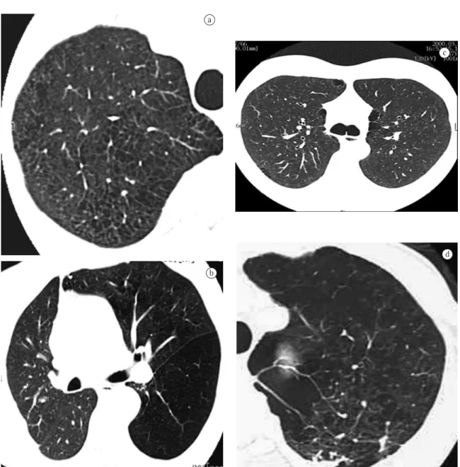
Figure 2 - Computed tomography (CT) and high-resolution CT (HRCT) images: a) representation of centrilobular emphysema - CT reveals predominant degree III and IV lesions; b) representation of panlobular emphysema obtained by HRCT of patient with right lung transplant by panlobular emphysema – predominant degree IV lesions; c) HRCT scan of patient with emphysema, with predominant paraseptal lesions; and d) photo obtained by paraseptal HRCT of a patient with irregular emphysema, with some areas of ‘scar-related’ emphysema - degree III and IV lesions
volume in relation to the amount of pulmonary tissues and blood that the X-rays come across in their path. This can occur when the lung increases its volume by compensatory expansion, as long as there is no significant increase in blood flow, accom-panying the increased air volume. However, the reduction of blood flow in a certain area of the lung can be the principal cause of reduced attenuation. Therefore, bronchitis and bronchiolitis obliterans can be responsible for the increased transparency seen on X-rays, even before the disease causes the destruction of the alveolar walls.
The yield of conventional chest X-ray in the eval-uation of emphysema is quite limited. When there is no significant air trapping, the principal alteration is reduction of vasculature, which is only perceived too late in the natural history of the disease, and is an extremely subjective criterion. When there is air trapping (Figures 3 and 4), the criteria are safer, and can be divided into 3 basic groups of alterations,(6,8)
presented in Chart 1. When all criteria are present, the diagnosis is definitively confirmed. It should be noted that the increased right chambers of the heart, with reduction of the intrasegmental vascu-lature, can also be identified in pulmonary arterial hypertension, without emphysema. It should also be noted that bullae are only present in approximately one-third of the cases.
The objectives of the radiological study of the chest in the evaluation of emphysema are diag-nosis, identification of lesion type, and evaluation of the extent of the disease. In this sense, there have been studies(6,8) that called attention to some quite
important considerations:
• In centrilobular or paraseptal emphysema, there
is typically no clinical symptomatology, and there can be increase in air spaces, without air trapping or clear radiological alterations; and
• Although panlobular emphysema is typi -cally more relevant in clinical terms, it can be present in the lungs of elderly individuals without causing air trapping.
Some authors(15) reported that the length of the
right lung and the height of the arch of the hemidi-aphragm correlated well with FEV1 and FEV1/vital capacity ratio. In this study, a right lung length of 30 cm or more identified 70% of the patients with respiratory obstruction.(16) However, one author(17)
reported that all of these studies presented errors, since they comprise excessive number of patients The paraseptal type only occurs in those lobes
delimitated by conjunctive tissue, peripheral conjunctive septa, pleura and conjunctive tissue cuffs, peribronchial or perivascular. It tends to develop on regular pulmonary margins. Air spaces in paraseptal emphysema frequently become confluent and develop into bullae, which can be large. It is believed that paraseptal emphysema is the basic lesion in pulmonary bullous disease.(14)
Airway obstruction and respiratory dysfunction are frequently smaller in paraseptal emphysema, despite the large formation of bullae.
Emphysema of panlobular type affects the entirety of the lobes, with dilatation and destruc-tion of their alveoli. Characteristics that usually set apart the alveoli of the alveolar ducts are lost, pores of Kohn increase, and fenestrae appear among the alveoli. This process has been compared to a diffuse simplification of lung architecture. With progressive destruction, all that remains are fine tissue bands surrounding the blood vessels. Panlobular emphysema is the most disseminated and severe type of emphysema and, consequently, the one that will mostly result in clinically signifi-cant disease. Although pathological alterations are seen throughout the lungs, the distribution is frequently predominant in the lower thirds. The type of emphysema that occurs in alpha-1 antit-rypsin deficiency, in Swyer-James syndrome and in cases of familial emphysema is predominantly of the panlobular type. Although it is considered the emphysema of nonsmokers, panlobular emphysema also occurs when induced by smoking, in combina-tion with centrilobular emphysema.
When emphysema is located at the margins of a scar in the lungs, it can be denominated scar-related or irregular emphysema. The classification as irreg-ular emphysema is reserved for cases that cannot possibly be classified in the other three types.
In addition to the classification according to distribution in relation to the lobes, it is also neces-sary to grade emphysematous lesions, so that we can have a notion of the severity of the disease from the anatomopathological point of view. This graduation is necessary, especially in panlobular and centrilobular emphysema.
tion but preserve excellent mobility during expiration; and
• in individuals with asthma, especially older
children and adolescents, in whom the diaphragm can present some of the character-istics similar to those of the diaphragm of the individual with emphysema. However, cardio-vascular alterations are not present.
In the three situations, diaphragmatic excursion should be greater than that of the individual with emphysema.
The use of diaphragmatic excursion also has limitations, especially in: patients with heart failure, where portions of the diaphragm that are not in contact with the heart present greater cranial dislo-cation; and patients with pulmonary edema, since the rigidity caused by the edema can determine less mobility of the diaphragm.
One author comments that the emphysematous pattern defined by the conventional radiological study is only present when the emphysema is so pronounced from the anatomopathological point of view that, in general, pulmonary reserves have already been depleted or pushed to the limit.(6,8) A more
updated review(22) confirms these propositions: • “If the lungs are mildly affected by emphy
-sema, the X-ray is frequently normal”;
• “Emphysema can be diagnosed by X-ray when
the disease is advanced”; and with chronic respiratory obstruction and,
there-fore, radiological characteristics of air obstruction receive “disproportionate value in the recognition of emphysema”. It was also demonstrated that the sensitivity to chest X-ray is not good, ranging from 24%(18) to 80%.(19) In addition there is considerable
intra- and inter-observer variation regarding clas-sical radiological signs.(20) Radiological signs related
to vascularization are subject to even greater intra- and inter-observer variation than are signs related to hyperinflation.(19)
Considering that the hemidiaphragm has a surface of approximately 250 cm2, we can calculate
that each 4 cm dislocation of the diaphragm deter-mines a dislocation equivalent to approximately 1 L of volume in each lung (2 L, if we consider both lungs). When there is air trapping, the mobility of the diaphragm is limited during expiration. End-expiratory pulmonary volume is significant in the recognition of emphysema. In general, the dislocation of the diaphragm between maximum inspiration and expiration ranges from 3 to 10 cm, corresponding to volumes ranging from 1500 mL to 5000 mL. Cranial dislocation <2 or 3 cm during expiration indicates limited diaphragmatic excur-sion in the individual with emphysema.(21)
The use of the height of the diaphragm as a diagnostic indicator is limited in various situations:
• in patients with kyphoscoliosis;
• in some individuals, such as athletes, who
present a low diaphragm during
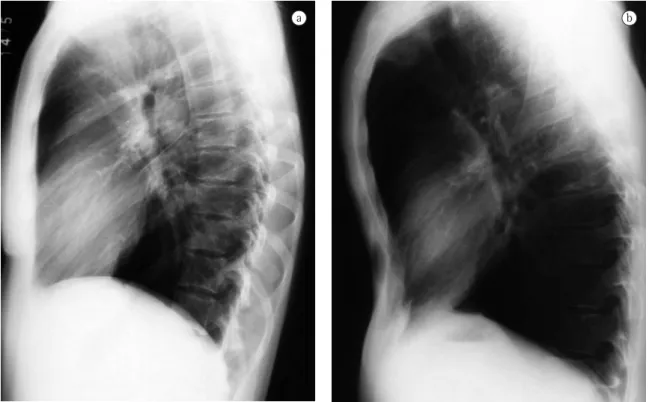
inspira-Figure 3 - Frontal chest X-ray: a) normal patient: and b) patient with chronic obstructive pulmonary disease, with excessive air in the lungs and cardiovascular alterations.
presence of low attenuation areas, with ill-defined margins, generally without visible walls.(44) Lesions
present a ‘moth-eaten’ appearance. When emphy-sematous lesions reach diameters greater than those of the lobes, part of their diameters can become well-defined, mimicking cysts, due to the presence of interstitial septa or major vessels surrounding the lesions. In a study that evaluated various signs of centrilobular emphysema, low attenuation areas in the medulla portion of the lungs, revealed on CT scans, correlated significantly with emphysema evaluated post mortem.(45) Other signs of
emphy-sema include bullae (pseudocysts that contain air, with thin well-defined walls), rarefaction of vessels and distortion of the vasculature. Complementary signs of hyperinflation of the lungs that, when pronounced, are promptly identified on chest X-ray, might not be so obvious on CT. However, the ante-rior joint line of the lungs, measuring 3 cm in the anteroposterior portion, suggests hyperexpansion in the patient with emphysema.(46)
• “Only half of the patients with emphysema of
moderate extent can be diagnosed by chest X-ray.”
Both authors suggest that conventional chest X-ray is not a reliable method for the diagnosis or quantification of emphysema.(22) This limitation is so
significant that some authors(21) demonstrated that,
when conventional radiology managed to diagnose chronic obstructive pulmonary disease, 53% of the patients died within 5 years, and 70% died within 10 years.
Evolution of imaging in the
investigation and quantification
of emphysema
In contrast to conventional chest X-ray, computed tomography (CT) has proven very sensitive and specific in the evaluation of emphysema.(14,23-43) Since
findings were first described in 1982,(14) CT has been
used to detect, characterize and quantify the disease. Centrilobular emphysema is characterized by the
a b
Various studies have been dedicated to the evaluation of detection capacity, anatomopatho-logical correlations and pulmonary function tests, as well as the quantification of the extent of the disease on CT scans.(23-48) It is the opinion of various
authors that the quantification of emphysema by conventional X-ray is limited.(7,23,49) Therefore, CT
has become the imaging method of choice for the quantification in vivo of this disease.
Some authors(39) studied quantification by visual
score, with scale from 1 to 5, according to involve-ment of 0, 25, 50, 75% or 100% of the lungs. The correlation of this technique with pathological anatomy presents correlation of r = 0.91 in vitro (with cadaver pulmonary specimens). In vivo, this correla-tion is r = 0.81.(24-39) We should consider, however,
that there are natural limitations when subjective analysis is used in the quantification of emphysema, whether the pathological anatomy analysis involves radiology, macroscopy or microscopy.
In an attempt to reduce the inherent subjec-tivity to the technique used by one author, other authors(31) proposed the visual stratification in
classes, through overlapped grids on CT images, which presents good anatomopathological correla-tion. However, it is much too complicated and quite subjective. The level and opening of the window in Hounsfield units (HU) selected for CT imaging analysis can influence significantly in the identifi-cation of emphysema areas.(29-34) The best window
selection for the visual identification of emphysema is the one using opening including 1100 HU, with
level or fixed center at −750 HU. Another limita -tion common to the two techniques is the lack of The high-resolution CT (HRCT) can differentiate
among the various types of emphysema, in patients with mild or moderate disease,(47) depicting with
great similarity the findings of pathological anatomy described above, according to which centrilobular emphysema presents: preference for upper thirds,(45)
and it can be confined to these regions; low density multifocal areas resulting from the destruction of the alveoli, predominantly in the medullar portion, distant from the pleura(45); lesions that can be
similar to small cystic air spaces, typically without an obvious wall; and lung surrounding lesions, which can be totally normal. With the progression of the disease, centrilobular emphysematous lesions become confluent.
HRCT, in panlobular emphysema, reveal a more uniform pulmonary destruction, with extensive low density areas, accompanied by vascular distortion and rarefaction. In classical panlobular emphy-sema, small focal low density areas, characteristic of centrilobular emphysema, are not found. In contrast to the latter, distribution is preferential in the lower thirds of the lungs.
Paraseptal emphysema is easily detected on HRCT scans, and presents as low density areas, with well-defined, hair-thin walls.(48) These pseudocysts
are distributed in the subpleural regions or adja-cent regions to larger bronchovascular cuffs. This emphysema pattern curiously have a ‘saw-tooth’ appearance and, occasionally, interstitial septa can seem particularly prominent; sometimes, they can mimic lymphangitic carcinomatosis at chest X-rays, which is promptly revealed on HRCT scans.(48)
Chart 1 - Air trapping criteria for conventional chest X-rays.
i) excessive air in the lungs Lowered or rectified diaphragm, below and anterior to the sixth intercostal space, at maximum inspiration.
Increased retrosternal clear space (>3 cm).
Persistence of increased retrosternal clear space in expiration.
Reduction of diaphragmatic mobility (<3 cm) between deep inspiration and expira-tion. Usual dislocation ranges from 3 to 10 cm.
ii) cardiovascular alterations Elongated heart in a more vertical position, with a transverse diameter of <11.5 cm at its widest point, remaining pliable even with increased right ventricle.
Pulmonary artery can be dilatated, with abnormal subaortic prominence. Hilar branches of the pulmonary artery can be enlarged.
Diversion of blood perfusion toward less affected areas can occur.
structure integral to or contained within the lungs can be achieved in 15-20 s. In multislice CT equip-ment, this time can be further reduced.
Tomographic slices obtained with fine collima-tion and processed with edge-preserving smoothing filters give the visual impression of being better than larger collimation slices (7-10 mm) without post-processing, in the subjective evaluation of emphysema. This occurs because our visual discrim-ination power for densities is lower than that of computers.
Although tomographic slices with fine collima-tion and the use of edge-preserving smoothing filters seem to be more sensitive in the detection of emphysema, CT slices with larger collimation (7-10 mm), without edge-preserving smoothing filters, are more reliable in relation to original data measured by detectors; this is true in acquisitions and measurements carried out based on densi-ties measured. In addition, the finer the slices, the more absorption artifacts occur and the greater is the filament current needed to minimize these arti-facts. Edge-preserving smoothing filters manipulate original data. Consequently, those intermediary densities resulting from the partial volume between the margins of the vessels and airways, with the surrounding air or with the air they contain, are transformed into densities close to air density or to densities equivalent to vascular or bronchial structures.
Therefore, mean density of the lungs is altered, and proportions of low density areas can vary significantly. During the First World Congress of Thoracic Imaging (Florence, Italy, 2005), some authors presented a measured volume variation from 131 mL to 860 mL, when measured in post-processed data with smoothing filters, in the same collimation. Collimation was less influential, and the volume of emphysema measured in data obtained with 1-mm collimation decreased from 131 mL to 60 mL when collimation was selected at 5 mm, both without post-processing filters.
Various authors have used collimation between 7 and 10 mm for volumetric quantification of emphysema by spiral CT. However, it is of note that the use of 7 or 10 mm-spiral scan is not recom-mended in the investigation of emphysema by subjective visual analysis.(39)
The acquisition of data volume in one single breath-hold allows us to calculate total volumes consensus on the minimum number of necessary
tomographic slices for a representative quantifica-tion of the whole of the lungs. In addiquantifica-tion to these problems, it is reported that the tube current also influences significantly in the visual subjective iden-tification of emphysema on HRCT scans.(39)
Predictably, the comparison between quantifica-tion methods by visual scores and those performed automatically by graphic computing demonstrated significant difference in favor of automation.(27)
One study(50) demonstrated that regardless of the
experience of the observer, there is a tendency to overestimate emphysema at visual analysis, and that densitometry correlates better with morphometric data.
In this evolution toward automation, it is not possible to overlook the important step represented by the research conducted in another study,(28) in
which it was demonstrated that patients with emphysema present more areas with density from
−900 HU to −1000 HU than do those without emphy -sema. Later, other studies confirmed the proposal that CT can objectively discriminate patients with and without emphysema.(37,41) These techniques
were studied in detail by one author who demon-strated the advantages of the use of density masks, which present correlation r = 0.89 with pathological anatomy.(43) The density mask is one of the most
important techniques in the semi-automatic evalua-tion of emphysema. Other authors(21) studied the use
of the technique of projections of minimum intensi-ties, which subtracts all high density structures from the image, highlighting the areas of emphysema. The studies of some authors(34) represented a new
milestone in the investigation of this disease,
estab-lishing the threshold value of −950 HU as adequate
to set apart lung with normal density and lung with density too close to air or with emphysema. This threshold has, since then, been one of the most frequently used in the quantification of emphysema by CT.
sarily volume. Other authors have studied CT-guided pulmonary densitometry and volumetric assessment and have demonstrated the applicability of the method, when compared to traditional methods of detection and quantification of emphysema, such as spirometry, quantification by visual score and analysis by histograms.(40,51)
The acquisition of data volume in a single breath-hold allows us to reconstruct data in three-dimensional format, using three-three-dimensional CT. This technological resource has been more frequently used in the investigation of chest altera-tions.(51-56) One of the pioneers in the dissemination
of the technique(56) demonstrated its efficiency in
the quantification of emphysema. The denomina-tion three-dimensional CT-guided densitometry and volumetric assessment for emphysema(55) has
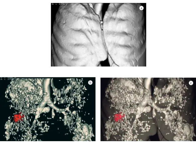
been currently suggested for this quantification process of the volume of emphysema, based on data obtained through spiral CT scan with three-di-mensional representation of emphysematous lesions (Figures 5 and 6). The test measures the whole pulmonary volume with abnormal density and, also, the whole lung volume with normal density,(41,56-61)
setting them apart based on the separation threshold
generally selected at −950 HU, as per the suggestion
of one author. Among the practical applications of the technique, one study demonstrated that normal CT-guided pulmonary densitometry and volumetric of normal lung and emphysematous areas, or
emphysema-like, based on the density measured using Hounsfield scale. This technique is generally described as quantitative CT and could be better denominated as CT-guided pulmonary densitom-etry and volumetric assessment, since quantitative CT can be performed to measure area, not
neces-Figure 5 - Normal pulmonary densitometry and volumetric assessment, with full lungs. The numbers in green
correspond to lung volume with density between −250 HU and −950 HU (normal lung) and lung volume with density below −950 HU (volume of emphysema-like areas).
a b
neoplastic lesion (in red) is found, by unfortunate and common chance, in the lobe that was less affected by emphysema. In this case, lower right lobectomy will be almost equivalent to pneumon-ectomy, since the right upper lobe, which would be the remaining lobe, is almost completely compro-mised by emphysema.
With the establishment of normality reference values,(55) it is already possible, as well, to use
three-dimensional CT-guided densitometry and volumetric assessment in the early diagnosis and graduation of the severity of the disease, and this early detection and demonstration of three-dimensional imaging to the patient has been suggested as potentially useful in techniques of smoking cessation.(60) In addition,
it also seems promising in the evaluation of patients whose occupational activities can induce the devel-opment of emphysema.
assessment excels HRCT, and should be included in the preoperative evaluation of patients for whom surgery for volumetric reduction of the lungs has been indicated.(58) This clinical applicability of
three-dimensional CT-guided densitometry and volumetric assessment used for the same purpose has been known in Brazil for some time.(39) In a
recent consensus, the use of three-dimensional CT-guided densitometry and volumetric assessment was suggested for the evaluation of therapeutic efficacy in the treatment of emphysema, replacing FEV1.(59)
The evaluation of advantages of three-dimen-sional CT-guided densitometry and volumetric assessment for therapeutic decision in patients with operable pulmonary carcinoma from the point of view of tumor-node-metastasis staging is under study. However, with borderline pulmonary func-tion. Figure 7 exemplifies a case in which the
a
b c
17. Thurlbeck WN. Chronic airflow obstruction in lung disease. In Bennington JL. Major problems in pathology, v. 5. Philadelphia: Saunders; 1976. p. 221-302.
18. Thurlbeck WM, Simon G. Radiographic appearance of the chest in emphysema. AJR Am J Roentgenol. 1978;130(3):429-40. 19. Sanders C. The radiographic diagnosis of emphysema. Radiol
Clin North Am. 1991;29(5):1019-30.
20. Nicklaus TM, Stowell DW, Christiansen WR, Renzetti AD Jr. The accuracy of the roentgenologic diagnosis of chronic pulmonary emphysema. Am Rev Respir Dis. 1966;93(6):889-99.
21. Simon G, Medvei VC. Chronic bronchitis: radiological aspects of a five-year follow-up. Thorax. 1962;17:5-8.
22. McLoud TC. Chronic obstructive pulmonary disease. In: McLoud TC, editor, Thoracic radiology: the requisites. 1998. St. Louis: Mosby Inc; 1998. p. 287-300.
23. Gould GA, MacNee W, McLean A, Warren PM, Redpath A, Best JJ, et al. CT measurements of lung density in life can quantitate distal airspace enlargement--an essential defining feature of human emphysema. Am Rev Respir Dis. 1988;137(2):380-92.
24. Miller RR, Müller NL, Vedal S, Morrison NJ, Staples CA. Limitations of computed tomography in the assessment of emphysema. Am Rev Respir Dis. 1989;139(4):980-3. 25. Kuwano K, Matsuba K, Ikeda T, Murakami J, Araki A, Nishitani
H, et al. The diagnosis of mild emphysema. Correlation of computed tomography and pathology scores. Am Rev Respir Dis. 1990;141(1):169-78.
26. Morgan MD. Detection and quantification of pulmonary emphysema by computed tomography: a window of opportunity. Thorax. 1992 Dec;47(12):1001-4.
27. Spouge D, Mayo JR, Cardoso W, Müller NL. Panacinar emphysema: CT and pathologic findings. J Comput Assist Tomogr. 1993;17(5):710-3.
28. Hayhurst MD, MacNee W, Flenley DC, Wright D, McLean A, Lamb D, et al. Diagnosis of pulmonary emphysema by computerised tomography. Lancet. 1984;2(8398):320-2. 29. Stern EJ, Frank MS. CT of the lung in patients with pulmonary
emphysema: diagnosis, quantification, and correlation with pathologic and physiologic findings. AJR Am J Roentgenol. 1994;162(4):791-8.
30. Stern EJ, Webb WR, Gamsu G.Dynamic quantitative computed tomography. A predictor of pulmonary function in obstructive lung diseases. Invest Radiol. 1994;29(5):564-9. 31. Thurlbeck WM, Müller NL. Emphysema: definition,
imaging, and quantification. AJR Am J Roentgenol. 1994;163(5):1017-25.
32. Bhalla M, Naidich DP, McGuinness G, Gruden JF, Leitman BS, McCauley DI. Diffuse lung disease: assessment with helical CT--preliminary observations of the role of maximum and minimum intensity projection images. Radiology. 1996;200(2):341-7.
33. Weeb WR, Muller NL. Principles and techniques of thoracic Computed Tomography and Magnetic Resonance. Naidich DP, Muller NL. Computed tomography and magnetic resonance of the thorax. Philadelphia: Lippincott-Raven; 1999: p. 1-37.
34. Gevenois PA, Scillia P, de Maertelaer V, Michils A, De Vuyst P, Yernault JC. The effects of age, sex, lung size, and hyperinflation on CT lung densitometry. AJR Am J Roentgenol. 1996;167(5):1169-73.
35. Remy-Jardin M, Remy J, Gosselin B, Copin MC, Wurtz A, Duhamel A. Sliding thin slab, minimum intensity projection
Final considerations
The use of CT, including HRCT and three-di-mensional CT-guided densitometry and volumetric assessment, represents an important advance in the investigation of patients with chronic obstructive pulmonary disease, allowing better discrimination between the predominance of bronchitis or emphy-sema, as well as the identification of lesions in an earlier phase and the quantification of the extent of the disease. These imaging techniques have been widely used in clinical practice, and the new appli-cation possibilities are expanding.
References
1. Gurney JW. Pathophysiology of obstructive airways disease. Radiol Clin North Am. 1998;36(1):15-27.
2. Sobonya RE, Burrows B. The epidemiology of emphysema. Clin Chest Med. 1983;4(3):351-8.
3. Stern EJ, Frank MS, Schmutz JF, Glenny RW, Schmidt RA, Godwin JD. Panlobular pulmonary emphysema caused by i.v. injection of methylphenidate (Ritalin): findings on chest radiographs and CT scans. AJR Am J Roentgenol. 1994;162(3):555-60.
4. Weisbrod GL, Rahman M, Chamberlain D, Herman SJ. Precocious emphysema in intravenous drug abusers. J Thorac Imaging. 1993;8(3):233-40.
5. Corbett E, Glaisyer H, Chan C, Madden B, Khaghani A, Yacoub M. Congenital cutis laxa with a dominant inheritance and early onset emphysema. Thorax. 1994;49(8):836-7. 6. Reid L. The pathology of emphysema. London: Lloyd-Luke;
1967. p. 384.
7. Laennec RTH. Traité de lauscultation médiate et des maladies des pulmouns et du cour. Paris: J S Chaudé; 1826. 8. Reid L, Simon G. III. Pathological findings and radiological
changes in chronic bronchitis and emphysema. Br J Radiol. 1959;32(377):291-305.
9. Thurlbeck WM, Simon G. Radiographic Appearance of the Chest in Emphysema. AJR Am J Roentgenol. 1978;130(3):429-440.
10. The definition of emphysema. Report of a National Heart, Lung, and Blood Institute, Division of Lung Diseases workshop. Am Rev Respir Dis. 1985;132(1):182-5.
11. Dornhorst AC. Respiratory insufficiency. Lancet. 1955;268(6876):1185-7.
12. Robins AG. Pathophysiology of emphysema. Clin Chest Med. 1983;4(3):413-20.
13. Heard BE. A pathological study of emphysema of the lungs with chronic bronchitis. Thorax. 1958;13(2):136-49. 14. Snider GL. A perspective on emphysema. Clin Chest Med.
1983;4(3):329-36.
15. Gavelli G, Zompatori M, Bernasconi A, Canini R. Diagnostic imaging of pulmonary emphysema. From radiography to high-resolution computerized tomography. Radiol Med (Torino). 1990;80(5):679-87.
48. Miller RR, Müller NL, Vedal S, Morrison NJ, Staples CA. Limitations of computed tomography in the assessment of emphysema. Am Rev Respir Dis. 1989;139(4):980-3. 49. Takasugi JE, Godwin JD. Radiology of chronic
obstructive pulmonary disease. Radiol Clin North Am. 1998;36(1):29-55.
50. Bankier AA, De Maertelaer V, Keyzer C, Gevenois PA. Pulmonary emphysema: subjective visual grading versus objective quantification with macroscopic morphometry and thin-section CT densitometry. Radiology. 1999;211(3):851-8. 51. Johnson PT, Fishman EK, Duckwall JR, Calhoun PS, Heath
DG. Interactive three-dimensional volume rendering of spiral CT data: current applications in the thorax. Radiographics. 1998;18(1):165-87.
52. Aberle DR. Society of Thoracic Radiology. Future directions of research in thoracic imaging. Radiology. 1998;206(1):11-3. 53. Henschke CI, Yankelevitz DF. Supercomputers help in the
medical diagnostic process, Cornell 1998.
54. Gevenois PA, De Vuyst P, Sy M, Scillia P, Chaminade L, de Maertelaer V, et al. Pulmonary emphysema: quantitative CT during expiration. Radiology. 1996;199(3):825-9.
55. Irion KL. Valores Referenciais de Normalidade em Densito Volumetria Pulmonar por Tomografia Computadorizada Helicoidal [thesis]. Porto Alegre: Universidade Federal do Rio Grande do Sul, 2000.
56. Park KJ, Bergin CJ, Clausen JL. Quantitation of emphysema with three-dimensional CT densitometry: comparison with two-dimensional analysis, visual emphysema scores, and pulmonary function test results. Radiology. 1999;211(2):541-7.
57. Mergo PJ, Williams WF, Gonzalez-Rothi R, Gibson R, Ros PR, Staab EV, et al. Three-dimensional volumetric assessment of abnormally low attenuation of the lung from routine helical CT: inspiratory and expiratory quantification. AJR Am J Roentgenol. 1998;170(5):1355-60.
58. Cederlund K, Bergstrand L, Högberg S, Rasmussen E, Svane B, Tylén U, et al. Visual classification of emphysema heterogeneity compared with objective measurements: HRCT vs spiral CT in candidates for lung volume reduction surgery. Eur Radiol. 2002;12(5):1045-51.
59. Newell JD Jr, Hogg JC, Snider GL. Report of a workshop: quantitative computed tomography scanning in longitudinal studies of emphysema. Eur Respir J. 2004;23(5):769-75. 60. Madani A, Keyzer C, Gevenois PA. Quantitative computed
tomography assessment of lung structure and function in pulmonary emphysema. Eur Respir J. 2001;18(4):720-30. technique in the diagnosis of emphysema: histopathologic-CT
correlation. Radiology. 1996;200(3):665-71.
36. Kemerink GJ, Kruize HH, Lamers RJ, van Engelshoven JM. CT lung densitometry: dependence of CT number histograms on sample volume and consequences for scan protocol comparability. J Comput Assist Tomogr. 1997;21(6):948-54. 37. Uppaluri R., McLennan G., Sonka M., Hoffman EA. Computer-based objective quantitative assessment of pulmonary parenchyma via X-ray CT. SPIE. 1998;3337:377-83. 38. Arakawa H, Webb WR, McCowin M, Katsou G, Lee KN,
Seitz RF. Inhomogeneous lung attenuation at thin-section CT: diagnostic value of expiratory scans. Radiology. 1998;206(1):89-94.
39. Webb WR, Muller NL. Diseases characterized primarily by cysts and emphysema. In: Webb WR, Muller NL, Naidich DP, editors. High-Resolution CT of the lung. Philadelphia: Lippincott Williams and Wilkins; 2001. p. 421-60.
40. Kauczor HU, Heussel CP, Fischer B, Klamm R, Mildenberger P, Thelen M. Assessment of lung volumes using helical CT at inspiration and expiration: comparison with pulmonary function tests. AJR Am J Roentgenol. 1998;171(4):1091-5. 41. Uppaluri R, Mitsa T, Sonka M, Hoffman EA, McLennan
G. Quantification of pulmonary emphysema from lung computed tomography images. Am J Respir Crit Care Med. 1997;156(1):248-54.
42. King GG, Müller NL, Paré PD. Evaluation of airways in obstructive pulmonary disease using high-resolution computed tomography. Am J Respir Crit Care Med. 1999;159(3):992-1004.
43. Müller NL, Staples CA, Miller RR, Abboud RT. “Density mask”. An objective method to quantitate emphysema using computed tomography. Chest. 1988;94(4):782-7.
44. Austin JH, Müller NL, Friedman PJ, Hansell DM, Naidich DP, Remy-Jardin M, et al. Glossary of terms for CT of the lungs: recommendations of the Nomenclature Committee of the Fleischner Society. Radiology. 1996;200(2):327-31. 45. Foster WL Jr, Pratt PC, Roggli VL, Godwin JD, Halvorsen
RA Jr, Putman CE. Centrilobular emphysema: CT-pathologic correlation. Radiology. 1986;159(1):27-32.
46. Zompatori M, Rimondi MR, Gavelli G, Canini R. Paraseptal emphysema mimicking unilateral lymphangitic carcinomatosis: CT findings. J Comput Assist Tomogr. 1993;17(5):810-2.