Idiopathic bilateral diaphragmatic paresis*
MÔNICA CORSO PEREIRA1, RODRIGO FRANGE MIZIARA MUSSI2, REINALDO ALEXANDRE DE CARVALHO MASSUCIO2, ANA MARIA CAMINO3,
ARISTÓTELES DE SOUZA BARBEIRO4, WANDER DE OLIVEIRA VILLALBA5, ILMA APARECIDA PASCHOAL6
*Study carried out in the Pulmonology Department of the Universidade Estadual de Campinas (UNICAMP, State University at Campinas) School of Medical Sciences, Campinas, Brazil.
1. Attending Physician and PhD in Pulmonology from the Universidade Estadual de Campinas (UNICAMP, State University at Campinas) School of Medical Sciences, Campinas, Brazil
2. Resident Physician in Pulmonology at the Universidade Estadual de Campinas (UNICAMP, State University at Campinas) School of Medical Sciences, Campinas, Brazil
3. Attending physician in the Department of Pulmonology of the Universidade Estadual de Campinas (UNICAMP, State University at Campinas) School of Medical Sciences, Campinas, Brazil
4. Attending physician and Masters in Pulmonology from the Universidade Estadual de Campinas (UNICAMP, State University at Campinas) School of Medical Sciences, Campinas, Brazil
5. Masters in Physiotherapy from the Universidade Estadual de Campinas (UNICAMP, State University at Campinas) School of Medical Sciences, Campinas, Brazil
6. Associate professor in the Department of Pulmonology of the Universidade Estadual de Campinas (UNICAMP, State University at Campinas) School of Medical Sciences, Campinas, Brazil
Correspondence to: Mônica Corso Pereira. Rua Edilberto Luis Pereira da Silva, 954, Cidade Universitária - CEP: 13083-190, Campinas, SP, Brasil. Tel: 55 19 3287-5655. Email: [email protected].
Submitted: 6 October 2005. Accepted, after review: 29 November 2005.
ABSTRACT
We report the case of a patient with severe dyspnea upon reclining. Lung disease, neuromuscular disorders and heart disease were ruled out. However, during the course of the investigation, bilateral diaphragmatic paresis was discovered. A key sign leading to the diagnosis was evidence of paradoxical respiration in the dorsal decubitus position. When the patient was moved from the orthostatic position to the dorsal decubitus position, oxygenation and forced vital capacity worsened. The orthostatic fluoroscopy was normal. Maximal inspiratory pressure was severely reduced. The responses to transcutaneous electric stimulation of the diaphragm were normal. However, electric stimulation of the phrenic nerve produced no response, leading to the diagnosis of bilateral diaphragmatic paresis.
Keywords: Paresis; Respiratory insufficiency; Diaphragm; Respiration
INTRODUCTION
We report the case of a patient with idiopathic bilateral diaphragmatic paresis, which is a rare and not always easily diagnosed disease.
CASE REPORT
insidious onset, seven months prior, of dyspnea, even at rest, with a pronounced worsening of the symptoms when in the dorsal decubitus position (orthopnea). He reported no cough, expectoration, wheezing, consumptive symptoms or symptoms affecting other organs. He did not report any type of trauma or thoracic surgery.
Physical examination revealed normal respiration at rest, becoming severe dyspnea when the patient assumed the supine position. Upon lying down, the patient immediately presented rapid, shallow respiration with paradoxical movement of the thorax and abdomen. There was no cyanosis or digital clubbing. Upon auscultation, it was noted that the lungs were clean with reduced breath sounds in the bases. The rest of the physical examination, including the neurological evaluation, revealed no alterations.
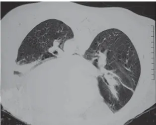
The chest X-ray revealed that the heart area was normal, the hemidiaphragm was elevated, and there were areas of bi-basal hypotransparency consistent with laminar atelectasis. The spirometry showed that forced vital capacity was 55% of predicted, forced expiratory volume in one second was 59% of predicted, and the mean ratio between forced expiratory volume in one second and forced vital capacity was 88%, none of which were altered after bronchodilator use. The chest X-ray revealed only areas of collapse in the lung bases, which were probably secondary to hypoventilation (Figure 1),
and the angiotomography results were normal. The echocardiogram and electrocardiogram results were both normal.
The laboratory testing (complete blood workup, leukocyte count, thyroid function test, renal function test, hepatic function test, evaluation of inflammatory activity and complement testing) did not show significant alterations. Testing for auto-antibodies, including c-ANCA and p-ANCA, was negative.
Fluoroscopy in the orthostatic position, used for evaluation of diaphragm movement, was normal.
Magnetic resonance imaging of the cervical spine was also normal. Testing for neuromuscular diseases (clinical and laboratory exams, as well as electroneuromyographic studies of all four limbs) was negative.
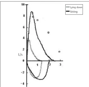
Oxyhemoglobin saturation was 96% (on room air) when it was sitting, decreasing to 87% when the patient was in the decubitus position. Arterial blood gas analysis revealed the following values: pH: 7.436; arterial oxygen tension: 62.8 mmHg; arterial carbon dioxide tension: 34.4 mmHg; bicarbonate: 22.8 mmol/L; oxygen saturation by pulse oximetry: 94.2%. The spirometry was carried out in two positions (sitting and lying down), and the results are presented in Table 1 and Figure 2.
The maximal inspiratory and expiratory pressures were measured by means of a mechanical vacuum pump, which detected a maximal inspiratory pressure of 22 cmH2O and a maximal expiratory pressure of 90 cmH2O.
The six-minute walk test results were normal (the patient covered 420 m without any drop in oxygen saturation by pulse oximetry or any changes in the Borg dyspnea scale score).
Polysomnography revealed alterations in sleep architecture, with a decrease in the percentage of
Figure 1 - Computed tomography scan of the chest in the ventral decubitus position. Note the homogeneous opacities in the lung bases, which are consistent with areas of pulmonary collapse, probably due to hypoventilation
TABLE 1
Spirometry in two positions
Patient sitting Patient lying down
Observed % predicted Observed % predicted % alteration FVC 2,57 66 1,35 35% -47 FEV1 2,12 66 1,02 31% -52 FEV1/FVC 83% 75%
FEF 25%-75% 2,22 64
Figure 2 - Spirometry in two positions. Flow-volume curve in two positions, sitting and lying down. The values are shown in Table 1
Lying down
Sitting
L/s
phase 3 and 4 sleep (14.3%) as well as in that of REM sleep (16.7%), and the apnea/hypopnea index was 5.5/h (acceptable).
To specifically evaluate diaphragmatic function, two additional tests were carried out. External electrostimulation of the diaphragm, a test in which electrodes are positioned in the seventh intercostal space, which then receives direct electrical stimulation, was conducted, revealing visible and palpable bilateral contraction of the diaphragm in response to the electrical charge. External cervical electrostimulation of the phrenic nerves showed severe axonal and myelinic involvement of both phrenic nerves, which is consistent with a clinical diagnosis of severe bilateral mononeuropathy.
DISCUSSION
What called attention in the clinical profile of the patient was the immediate worsening of dyspnea upon reclining, as well as the practically instantaneous improvement upon standing. This indicated that the supine position, in which the abdominal viscera push the diaphragm upwards, thereby hindering its contraction and reducing its effectiveness, triggered the worsening of the symptomatology.
Upon physical examination, the observation of “paradoxical respiration", in which the abdominal
content is "sucked" into the abdominal cavity during inspiration (when normally it is "pushed" outwards), was highly suggestive some type of diaphragmatic dysfunction.
Given this hypothesis, several tests can be useful. Spirometry can either present restriction (decreased vital capacity or forced vital capacity) or be normal (if carried out in the orthostatic position). To make the spirometry test more precise, especially when diaphragmatic weakening is suspected, it can be conducted twice: once with the patient standing and once with the patient sitting or lying down. A 40% or greater reduction in the forced vital capacity measured in the supine position when compared with the sitting/orthostatic position strongly suggests involvement of the diaphragm.(1-2) Patients
with amyotrophic lateral sclerosis(3) and other
neuromuscular diseases present a significant decrease in vital capacity in the supine position when compared with the sitting position.(4)
The measurements of maximal inspiratory pressure and maximal expiratory pressure are frequently used in the overall evaluation of respiratory muscle force.(5-6) Normal values for adults
and children are well established.(5) A test that
results in a highly negative maximal inspiratory pressure (approximately 80 cmH2O) or in a positive maximal expiratory pressure (90 cmH2O) rules out clinically significant inspiratory or expiratory muscular weakness.(7) In this patient, the maximal
inspiratory pressure measured was 25 cmH2O (38% of predicted) and the maximal expiratory pressure was 90 cmH2O (normal).(8)
The principal alteration found in the polysomnography was a decrease in the percentage of phase 3 sleep, phase 4 sleep and REM sleep. In the REM phase, most skeletal muscles lose much of their tone, the exceptions being the diaphragm and extra-ocular muscles. Normally there is a decrease in the tidal volume, minute ventilation and inspiratory flow. In this phase, respiration depends principally on the activity of the diaphragm. Therefore, individuals presenting weakening of the respiratory muscles are more susceptible to hypoventilation in the REM phase and in the transition from the NREM phase to the REM phase. Hypoventilation episodes provoke a drop in arterial oxygen tension and an increase in arterial carbon dioxide tension.(9)
volume in the lung bases, which can be observed in the X-ray and computed tomography of the chest (Figure 1). When the patient changes the decubitus position (from supine to prone), the hypoventilated area is also altered, which shows that these areas of hypoventilation are perfectly expandable.
Although fluoroscopy is a useful test for the evaluation of diaphragmatic excursion, it can, depending on the position in which it is carried out, generate false-negative results.(10)That is what
occurred in this case, in which the results of the test were normal, probably because it was carried out with the patient standing, since he did not tolerate the dorsal decubitus position.
More sophisticated tests, such as electrostimulation of the phrenic nerve, electromyogram of the diaphragm and measurement of transdiaphragmatic pressure, can demonstrate unequivocally the involvement of the phrenic nerve-diaphragm axis.
Direct stimulation of the diaphragm (which was carried out in this case) demonstrates the contractile response of the muscle, whereas the electromyogram provides a qualitative evaluation of muscular activity, which is useful in some neuropathic and myopathic conditions.
The phrenic nerve can be stimulated directly or externally (transcutaneously). The total absence of any diaphragmatic response to external stimulation should raise the suspicion that the phrenic nerve is dislocated.(11) This test is quite accurate and has the
advantage of being noninvasive
Transdiaphragmatic pressure is measured by calculating the difference between gastric pressure and esophageal pressure, reflecting diaphragmatic tension. It is an invasive test that is sometimes poorly tolerated by patients, since the placement of two balloons (one in the mid-esophagus and another in the stomach) is required in order to measure maximal inspiratory pressure.
Bilateral diaphragmatic mononeuropathy is a rare condition that is difficult to identify, and the mean time to diagnosis varies from six weeks to ten years.(2)
The delay in diagnosis leaves the patient chronically exposed to periods (generally nocturnal) of hypoventilation, which occasionally produces serious consequences such as cor pulmonale.
Various situations can be associated with paresis or diaphragmatic paralysis. Such situations include the following: muscular or neuromuscular
diseases (myopathies, motor neuron diseases); traumas leading to lesions in the bone marrow; direct surgical traumas or traumas secondary to hypothermia during cardioplegia (heart surgery); polyneuropathies (infectious, noninfectious and idiopathic); lupus erythematosus; and paraneoplasic syndrome. In many cases, however, it is not possible to establish the etiology.(2,10,14)
The principal objective of the treatment is to maintain adequate ventilation, thereby avoiding complications associated with chronic hypoventilation. In some cases of idiopathic paresis or paresis associated with viral diseases, there can be spontaneous regression of neural or muscular involvement.(14)
Many patients have already been treated with invasive mechanical ventilation, either through tracheostomy or through negative pressure ventilators, such as the so-called “cuirass" or abdominal ventilators (Pneumobelt).(2)
Currently, however, the best options of ventilatory support are offered by ventilators involving masks (noninvasive ventilation), such as those with two levels of positive pressure (BiPAP® type).
After the diagnosis, this patient remained at home, using noninvasive positive pressure ventilation at an inspiratory pressure of 12 cmH2O and an expiratory pressure of 6 cmH2O, pressure levels to which he adapted well. This type of home ventilatory support has some advantages, such as its relatively low cost (when compared with invasive measures) and the portability of the equipment, which permits the individual to lead a practically normal life with nearly no limitations to his daily activities.
The use of a diaphragmatic pacemaker is yet another therapeutic option, albeit temporary, since its prolonged use is not possible due to provoking fatigue of the diaphragm muscle.
REFERENCES
1. Sociedade Brasileira de Pneumologia e Tisiologia. III Consenso Brasileiro no Manejo da Asma. J Pneumol. 2002;28(Suppl 1):S1-S238.
2. Chan CK, Loke J, Virgulto JA, Mohsenin V, Ferranti R, Lammertse T. Bilateral diaphragmatic paralysis: clinical spectrum, prognosis, and diagnostic approach. Arch Phys Med Rehabil. 1988;69(11):976-9.
4. Fromageot C, Lofaso F, Annane D, Falize L, Lejaille M, Clair B, et al. Supine fall in lung volumes in the assessment of diaphragmatic weakness in neuromuscular disorders. Arch Phys Med Rehabil. 2001;82(1):123-8.
5. Black LF, Hyatt RE. Maximal respiratory pressures: normal values and relationship to age and sex. Am Rev Respir Dis. 1969;99(5):696-702.
6. Perrin CMC, Fanfulla F, Fiorentini M, Casali L. Reference values of maximal inspiratory mouth pressures: a population based study. Am Rev Respir Dis. 1992;146(3):790-3. 7. DePalo VA, McCool FD. Respiratory muscle evaluation
of the patient with neuromuscular disease. Semin Respir Crit Care Med. 2002;23(3):201-9.
8. Polkey MI, Green M, Moxham J. Measurement of respiratory muscle strength. Thorax. 1995;50(11):1131-5.
9. Black LF, H, Unterborn JN, Ambrosio CD, Hill NS. Pulmonary complications of chronic neuromuscular diseases and their management. Muscle Nerve. 2004;29(1):5-27.
10. Lin MC, Liaw MY, Huang CC, Chuang ML, Tsai YH. Bilateral diaphragmatic paralysis - a rare cause of acute respiratory failure managed with nasal mask bilevel positive airway pressure (BiPAP) ventilation. Eur Respir J. 1997;10(8):1922-4.
11. Loh L, Goldman M, Davis JN. The assessment of diaphragm function. Medicine (Baltimore). 1977;56(2):165-9. 12. Similowski T, Mehiri S, Duguet A, Attali V, Straus C,
Derenne JP. Comparison of magnetic and electrical phrenic nerve stimulation in assessment of phrenic nerve conduction time. J Appl Physiol. 1997;82(4):1190-9. 13. Sociedade Brasileira de Pneumologia e Tisiologia.
Consenso Brasileiro sobre Espirometria. J Pneumol. 1996;22(3):105-64.
14. Murray JF, Broaddus VC, Nadel JA. In: Mason RJ, Broaddus VC, Murray JF, Nadel JA, editors. Textbook of respiratory medicine. 4th ed. Philadelphia, Pennsylvania, Elsevier; 2005.