C A SE R EPORT
356 J Vasc Bras. 2015 Oct.-Dec.; 14(4):356-359 http://dx.doi.org/10.1590/1677-5449.07514
Endovascular treatment of delayed bleeding after tonsillectomy
Tratamento endovascular de sangramento tardio pós tonsilectomia
Gustavo Henrique Dumont Kleinsorge1
*
, André Mourão de Sousa1, Lucas Ferreira Botelho1, Marina Barros Mourão1,
Roberio Rodrigo Hora Melo2, Rodrigo Di Vita do Lago1
Abstract
Tonsillectomy is one of the most common procedures that otorhinolaryngologists perform. Hemorrhages are the principal complication. Some cases of hemorrhage can have delayed onset and these are related to formation of pseudoaneurysms. While this complication is rare, it is serious and can be fatal if it is not treated correctly. In cases with signiicant bleeding, surgical reintervention is needed and the 3 most common methods are suture, cauterization and endovascular treatment. he objective of this article is to report on the case of a 28-year-old female patient with massive post-tonsillectomy bleeding 31 days after the operation. An endovascular approach was taken and an injury to a left facial artery was diagnosed. Deinitive treatment was achieved by selective embolization of the facial artery with microcoils and 500 μm particles of polyvinyl alcohol (P.V.A.), in that sequence, in order to avoid distal embolization. he endovascular method proved to be safe, selective and deinitive.
Keywords: tonsillectomy; pseudoaneurysm; endovascular procedures; embolization.
Resumo
A tonsilectomia é um dos procedimentos mais realizados por otorrinolaringologistas e possui como principal complicação a hemorragia. Alguns casos podem se manifestar tardiamente e se relacionam com a formação de pseudoaneurismas. Apesar de rara, essa é uma complicação grave e pode levar ao óbito se não tratada devidamente. Em casos de sangramento signiicativo, as reintervenções cirúrgicas são necessárias, sendo as 3 formas mais comuns: sutura, cauterização ou por tratamento endovascular. O nosso estudo tem por objetivo apresentar o histórico de uma paciente de 28 anos, sexo feminino, com sangramento maciço pós tonsilectomia no 31º dia de pós-operatório. Foi realizada abordagem endovascular e diagnosticada lesão em artéria facial esquerda. O tratamento deinitivo foi por embolização seletiva de artéria facial com micromola e partículas de polivinil álcool (P.V.A.) 500 μm, nessa ordem de utilização, a im de evitar-se embolização distal. O método endovascular mostrou-se seguro, deinitivo e seletivo.
Palavras-chave: tonsilectomia; pseudoaneurisma; procedimentos endovasculares; embolização.
1 Fundação Hospitalar do Estado de Minas Gerais – FHEMIG, Hospital João XXIII, Belo Horizonte, MG, Brazil. 2 Hospital Municipal São José, Joinville, SC, Brazil.
Financial support: None.
Conlicts of interest: No conlicts of interest declared concerning the publication of this article. Submitted: October 30, 2014. Accepted: July 01, 2015.
357 J Vasc Bras. 2015 Oct.-Dec.; 14(4):356-359 Gustavo Henrique Dumont Kleinsorge, André Mourão de Sousa et al.
INTRODUCTION
Surgical interventions involving craniomaxillofacial structures, such as, for example, tonsillectomy, are common procedures. However, even when performed by experienced surgeons, there are not free from immediate or delayed complications.1 The most
common complication during the postoperative period following tonsillectomy is hemorrhage, with incidence rates ranging from 3 to 3.9%.2,3 The most
serious causes of hemorrhage are associated with arterial dissections and pseudoaneurysms, which most often occur in patients aged 30 to 34 years and more than 24h after surgery.2
Intraoperative hemorrhage (within 24h of the procedure) is associated with operating technique and coagulopathy, but delayed hemorrhages (more than 24h after the procedure), which are most common 5 to 7 days after surgery, are associated with detachment of the blood clot from the granulating tonsillar fossa.2
There are three treatment options accepted in the literature: local maneuvers, surgical ligature and endovascular treatment.3
Cases of endovascular treatment of post-tonsillectomy pseudoaneurysms have been reported. The most commonly described treatment options involve use of microcoils and particles of polyvinyl alcohol (P.V.A.).2,3
In this article we report on a case of a 28-year-old female patient in which we employed this treatment option, embolization of a pseudoaneurysm of the facial artery, because of massive post-tonsillectomy hemorrhage (31 days after surgery).
CASE DESCRIPTION
Patient C.C.D., a 28-year-old female underwent a tonsillectomy in March of 2014 because of recurrent tonsillitis. She suffered episodes of bleeding 7, 12 and 23 days after surgery, which were treated with simple stitches at the surgery site. Thirty-1 days after the original operation, the patient suffered massive hemorrhage and was referred to the emergency service in her town for treatment. On clinical examination she was lethargic and high volume bleeding from the left tonsillar region was observed. Her airway was safeguarded by orotracheal intubation and the bleeding tissue was clamped with curved hemostatic forceps. Finding that her hemoglobin level was 4g/dl, she was given four bags of packed red blood cells. After clinical stabilization, the patient was transported in a mobile intensive care ambulance to a tertiary hospital in the city of Belo Horizonte, MG, Brazil.
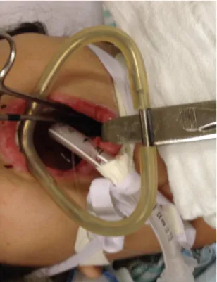
On admission the patient was hemodynamically stable (Hemoglobin 12.7; HR = 65bpm; BP = 101×74),
on mechanical ventilation via an orotracheal tube,
itted with a McIvor mouth retractor and with the
curved hemostatic forceps clamped to the left tonsillar region (Figure 1). The examining otorhinolaryngologist found that there was no active bleeding, but that further simple suture was not possible because of tissue friability cause by the previous manipulations.
Since the patient was stable hemodynamically and because of the adverse local conditions for surgery, including the friability of tissues, it was decided to take an endovascular approach.
Deinitive treatment was achieved via a right
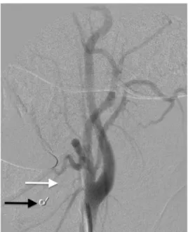
femoral artery access (with a 6F Prelude Meritmedical sheath) and catheterization of the left common carotid artery with a 5F Performa Meritmedical vertebral catheter and a 0.35 mm, 260 cm Cook Roadrunner guide wire. Arteriography was then conducted with non-ionic contrast (Ultravist 300) to diagnose the origin of the bleeding, revealing an injury to the patient’s facial artery adjacent to the hemostatic forceps (Figures 2 and 3).
Selective catheterization of the facial artery was achieved using a Cook Cantata microcatheter and a 0.014”, 300 cm Zinger Medium Medtronic guide wire. Embolization was achieved with a 3 mm Cook
Nester microcoil, placed distally, and 500 μm Cook
358 J Vasc Bras. 2015 Oct.-Dec.; 14(4):356-359 Treatment of delayed bleeding after tonsillectomy
polyvinyl acid microparticles, used in the order described. Control arteriography revealed treatment success, with an absence of contrast leakage and the left facial artery occluded (Figure 4).
There were no complications during the procedure and the patient was taken to the anesthesia recovery room and extubated.
The postoperative period was free of incidents and the patient was allowed to return to a normal diet 1 day after surgery. As there was no further bleeding,
the patient was discharged 4 days after the operation. She is currently in outpatients follow-up, free from intercurrent conditions of any kind.
DISCUSSION
Hemorrhage is the most common post-tonsillectomy complication, with an estimated incidence ranging from 3 to 3.9%. Later hemorrhage, which has onset more than 24h after surgery, has peak incidence from 5 to 10 days after surgery. The age of the patient is a risk factor for this complication; with no preference for either sex.2
Among the possible causes of late bleeding,
pseudoaneurysm rupture is rare, but can signiicantly
compromise the clinical status of patients.3-5
Manzato et al. conducted a review of the literature, reporting 23 cases of post-tonsillectomy pseudoaneurysm formation, two of which involved the facial artery.
The facial artery follows a variable path close to the submandibular gland, passing close behind and below the tonsillar region. In this region both the facial and lingual arteries are more vulnerable to traumatic lesions during the surgical procedure.2,6,7
There are three treatment options accepted in the literature for late onset bleeding: local maneuvers, surgical ligature and endovascular treatment.3
Ipsilateral ligature of the external carotid artery is one treatment option for massive hemorrhage after tonsillectomy. However, the procedure involves
signiicant risks, such as: (1) injury to the superior
Figure 2. Arteriography of the left common carotid artery and
branches (A.C.I.: Internal Carotid Artery; A.C.E.: External Carotid Artery). White arrow – probable site of pseudoaneurysm rupture, at the tip of the hemostatic forceps.
Figure 3. White arrow – Selective arteriography of left facial artery.
Black arrow – Contrast escaping into oropharynx. Asterisk (*) – Tip of the hemostatic forceps at the site of the injured facial artery.
Figure 4. Post-embolization control arteriography. White arrow
359 J Vasc Bras. 2015 Oct.-Dec.; 14(4):356-359 Gustavo Henrique Dumont Kleinsorge, André Mourão de Sousa et al.
laryngeal nerve and/or vagus nerve, (2) stroke and (3) reduction of the vascular reserve in distribution of the arterial supply to the ligated vessels. Furthermore, even proximal ligature of the external carotid artery or its branches may fail to control very severe hemorrhages.7
Endovascular embolization of pseudoaneurysms is
a treatment option that was irst described in 1975.8
In stable patients its use should be encouraged because it offers a series of advantages over other procedures: it is selective, it is less mutilating and it poses fewer risks to neighboring structures, such as injuries to the vagus and accessory nerves.2,3,9 Another advantage
is that diagnostic angiography can be followed by therapeutic embolization during the same surgical intervention.7
Occlusion of the affected vessel can be accomplished with coils or microcoils, since using P.V.A. particles in isolation is undesirable because it is associated with additional risks such as ischemic necrosis of the tip of the tongue due to embolization of branches terminating in the area.3
In the case described here, the decision was taken to use a combination of techniques to treat the active bleeding (microcoil distal to the injury, combined
with 500 μm P.V.A. particles). The irst reason for
this is that combining these methods should make embolization more effective.10 The second reason is
that this approach is safer, since the microcoil acts as a mechanical barrier to the P.V.A. particles, avoiding embolization of distal tissues and preventing the complications described above.3
CONCLUSIONS
When faced with severe late onset hemorrhages after tonsillectomy, a diagnosis of rupture of traumatic pseudoaneurysms of the facial, lingual or external carotid arteries should be considered.
In such cases, arteriography followed by endovascular treatment should be encouraged because it offers lower morbidity and a high rate of treatment success.
REFERENCES
1. Cohen JE, Gomori J, Moscovici S, Grigoriadis S, Noriega FR, Itshayek E. Endovascular management of postoperative pseudoaneurysm of the external carotid artery. J Clin Neurosci. 2012;19(5):649-54. http://dx.doi.org/10.1016/j.jocn.2011.11.007. PMid:22502912.
2. Juszket R, Korytowska A, Lukomska Z, Zarzecka A. Facial artery pseudoaneurysm and severe bleeding after tonsillectomy - endovascular treatment with PVA particle embolization. Pol J Radiol. 2010;75(1):88-91. PMid:22802767.
3. Manzato L, Trivelato F, Alvarenga A, Rezende MT, Ulhôa AC. Endovascular treatment of a linguofacial trunk pseudoaneurysm after tonsillectomy. Braz J Otorhinolaryngol. 2013;79(4):524. http:// dx.doi.org/10.5935/1808-8694.20130094. PMid:23929158.
4. Atmaca S, Belet U, Baris S. Post-tonsillectomy pseudoaneurism of the linguofacial trunk: an ENT surgeon’s nightmare. Int J Pediatr Otorhinolaryngology Extra. 2012;7(1):12-4. http://dx.doi. org/10.1016/j.pedex.2011.07.006.
5. Windfuhr JP, Sesterhenn AM, Schloendorff G, Kremer B. Post-tonsillectomy pseudoaneurysm: anunderestimatedentity? J Laryngol Otol. 2010;124(1):59-66. http://dx.doi.org/10.1017/ S0022215109990922. PMid:19765325.
6. Gardner JF. Sutures and disasters in tonsillectomy. Arch Otolaryngol. 1968;88(5):551-5. PMid:4879283.
7. Simoni P, Bello JA, Kent B. Pseudoaneurysm of the lingual artery secondary to tonsillectomy treated with selective embolization. Int J Pediatr Otorhinolaryngol. 2001;59(2):125-8. http://dx.doi. org/10.1016/S0165-5876(01)00478-5. PMid:11378188.
8. Gianturco C, Anderson JH, Wallace S. Mechanical devices for arterial occlusion. Am J Roentgenol Radium Ther Nucl Med. 1975;124(3):428-35. http://dx.doi.org/10.2214/ajr.124.3.428. PMid:50746.
9. Roedke LH, Perez JM, Torezane F. Tramento endovascular de um caso raro de pseudo-aneurisma de carótida externa após amigdalectomia. J Vasc Bras. 2004;3(2):172-5.
10. Nakstad PH, Bakke SJ, Hald JK. Embolization of intracranial arteriovenous malformations and fistulas with polyvinyl alcohol particles and platinum fibre coils. Neuroradiology. 1992;34(4):348-51. http://dx.doi.org/10.1007/BF00588202. PMid:1528453.
*
Correspondence
Gustavo Henrique Dumont Kleinsorge Rua Tenente Brito Melo, 496/1503 – Barro Preto CEP 30180-070 – Belo Horizonte (MG), Brazil Tel.: +55 (31) 8479-3189 E-mail: [email protected]
Author information
GHDK - Coordinator, Vascular Surgery Clinic, Hospital João XXIII, Fundação Hospitalar do Estado de Minas Gerais (FHEMIG); Board certiied in Vascular and Endovascular Surgery; Residency in Vascular Surgery, Hospital das Clínicas da Universidade Federal de Minas Gerais (HC-UFMG). AMS - Vascular surgeon, Hospital João XXIII, Fundação Hospitalar do Estado de Minas Gerais (FHEMIG); Residency in Endovascular Surgery and Vascular Surgery, Hospital das Clínicas da Universidade Federal de Minas Gerais (HC- UFMG). LFB - Resident physician of Vascular Surgery, Hospital João XXIII, Fundação Hospitalar do Estado de Minas Gerais (FHEMIG). MBM - Vascular surgeon, Hospital João XXIII, Fundação Hospitalar do Estado de Minas Gerais (FHEMIG); Residency in Vascular Surgery, Hospital da Previdência (IPSEMG). RRHM - Resident physician of General Surgery, Hospital Municipal São José. RDVL - Vascular surgeon, Hospital João XXIII, Fundação Hospitalar do Estado de Minas Gerais (FHEMIG).
Author contributions
Conception and design: MBM, AMS Analysis and interpretation: RRHM, RDVL Data collection: GHDK, RDVL, LFB Writing the article: GHDK, RRHM, LFB Critical revision of the article: GHDK, RRHM, LFB Final approval of the article*: GHDK, RRHM, LFB, RDVL, MBM, AMS Statistical analysis: N/A. Overall responsibility: GHDK