Angioplasty of a Persistent sciatic artery: case report
Angioplastia de artéria isquiática persistente: relato de caso
Denis Szejnfeld1, Sergio Quilici Belczak2, Igor Rafael Sincos2, Ricardo Aun3
Introduction
Persistent sciatic artery (PSA) is a rare congenital vascular anomaly, and its prevalence corresponds to 0.01-0.05% of the population. It was described by Green in 1832 and irst conirmed − using arteriography − by Cowie in 19601,2.
his anomalous artery, an atypical extension of the in-ternal iliac artery, provides the major supply of blood to the lower limbs in 63% of the PSA cases. In such cases, it is the most important artery in the lower limbs, and the superi-cial femoral artery is hypoplastic or absent3.
PSA has a predisposition to form aneurysms, found in 42% of the cases; about 50% of which have a compli-cated progression. hey are bilateral in 12-32% of the cases4,5. his anomaly also predisposes to early
atheroma-tous degeneration, distal thromboembolism and arterial occlusion6.
Percutaneous transluminal balloon angioplasty is a well established technique for treating signiicant stenosis and short occlusions. his study describes a case of PSA with atheromatosis and ischemic complications of the let lower limb that was successfully treated using an endovas-cular technique.
Abstract
Transluminal balloon angioplasty is a good choice for the treatment of lower limb arterial occlusion. Although there are some guidelines addressing its indications, some situations are so unusual that there is no consensus on their management. he presence of a persistent sciatic artery is a rare congenital anomaly of the circulatory system and may be associated with early atheromatous degeneration and occlusion. he authors describe the case of an 81-year-old woman that presented with a history of rest pain, atrophic lesion and no distal pulses. Angiogram depicted a persistent sciatic artery with segmental occlusion and distal disease. he therapeutic option was balloon angioplasty of the occluded segment, with technical and clinical success at mid-term follow-up.
Key words: angioplasty, balloon; atherosclerosis, ischemia.
Resumo
A angioplastia transluminal com balão tem se mostrado uma boa alternativa no tratamento de oclusões arteriais em membros inferiores. Embora já existam algumas diretrizes quanto à sua indicação, algumas situações ainda são inusitadas e carecem de consenso pela sua raridade. A presença de artéria isquiática persistente é uma anomalia congênita rara do sistema circulatório e pode estar associada com doença ateromatosa precoce e oclusão. Os autores apresentam um caso de uma paciente do sexo feminino de 81 anos, com história de dor de repouso, lesão tróica e ausência de pulsos distais. A arteriograia mostrou persistência de artéria isquiática com oclusão segmentar e doença distal. A abordagem terapêutica escolhida foi angioplastia do segmento ocluído, e o seguimento de médio prazo mostrou sucesso técnico e clínico com esta técnica.
Palavras-chave: angioplastia com balão; aterosclerose; isquemia.
Interventional Radiology Service of the Hospital Brigadeiro, São Paulo (SP)
1 Full member of Brazilian Society of Interventional Radiology and Endovascular Surgery (SOBRICE); Post-Graduate Student of Master Degree Program in Radiology of the Universidade Federal
de São Paulo (UNIFESP); Head of the Endovascular Surgery Service of IGESP Hospital; Attending Physician of Interventional Radiology Service of the Hospital Brigadeiro, São Paulo (SP), Brazil.
2 Full member of the Brazilian Society of Angiology and Vascular Surgery (SBACV); Post-Graduate Student of Doctorate Program of the School of Medicine of the Universidade de São Paulo
(USP), São Paulo (SP), Brazil; Head of the Vascular Surgery Service of Hospital Geral de Carapicuíba; Carapicuíba (SP), Brazil; Head of the Endovascular Surgery Service of IGESP Hospital; Attending Physician of Interventional Radiology Service of the Hospital Brigadeiro, São Paulo (SP), Brazil.
3 Full member of the Brazilian Society of Angiology and Vascular Surgery (SBACV); PhD of the School of Medicine of USP, São Paulo (SP); Attending Physician of USP, São Paulo (SP),
No conflicts of interest declared concerning the publication of this article.
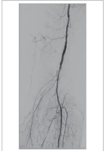
hypertension, had no pulses in the let lower limb and no posterior tibial pulse in the right lower limb. he patient was examined by a vascular surgeon, who requested aor-tography and pelvic and lower limb arteriography. Results revealed that aorta and right iliac system were normal. However, an anomalous artery in the let was shown, which originated in the internal iliac artery and extended inferi-orly towards the limb (Figure 2). he supericial femoral artery was hypoplastic (Figure 3). his anomalous artery had several wall abnormalities and was occluded distally at a point close to the transition to the popliteal artery (Figure 4). he anterior and posterior tibial arteries were occluded distally; the ibular artery was pervious, but had moderate stenosis in its proximal third (Figure 5).
he hypothesis was raised from persistent sciatic artery with atheromatous degeneration and an occluded segment. Endovascular treatment was suggested for obstructive le-sion. Using a contralateral approach and a 45-cm-long 6F introducer sheath, a 0.018 guidewire was advanced through the obstruction. Angioplasty was performed using a 5 x 20 mm balloon (Figure 6). he ibular artery was also submit-ted to angioplasty using a 3 x 120 mm balloon (Figure 7). Follow-up arteriogram showed that the artery was pervious and contrast medium low rate was good (Figure 8).
Dual antiplatelet therapy was initiated with 100 mg/day of acetyl salicylic acid and clopidogrel at 300 mg loading dose and 75 mg/day thereater. Immediately ater surgery,
Discussion
PSA is a vascular anomaly whose origin is well known. Embryos reach 9 mm at about the 6th week. In this phase, the sciatic artery, or axial artery, arises from the dorsal root of the umbilical artery and becomes the major source of blood to the primitive foot. When the embryo is 10 mm long, the femoral system starts to develop as a continuation of the external iliac artery, which expands and branches out to irrigate the thigh. When the embryo reaches 14 mm, at about 8 weeks, the lower limb has a dual blood supply sys-tem, the sciatic and the femoral ones. At about 12 weeks, the sciatic artery involutes, and the supericial femoral ar-tery develops. In adults, remnants of the sciatic arar-tery par-ticipate in the formation of gluteal arteries, popliteal artery, and the origin of anterior tibial and ibular arteries, and contribute to the terminal anastomosis of the foot1,2,7.
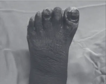
Figure 1 - Image shows trophic ulcers in foot.
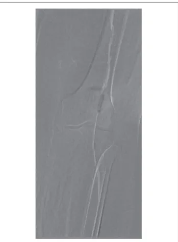
Figure 3 - Arteriogram shows hypoplastic supericial femoral artery.
Figure 4 - Arteriogram shows site of arterial occlusion.
Figure 5 - Arteriogram conirms atherosclerotic occlusion of leg arteries.
PSA has two diferent presentations:
- complete (63-79% of the cases), in which this artery is the major blood supply to the lower limb, and the supericial femoral system is hypoplastic, but rarely ab-sent. his is the presentation found in the case reported here4.
- incomplete (about 20% of the cases), in which PSA is hypoplastic and communicates via several branches with the femoral system, which, in this case, has no abnormalities8.
here is no diference in incidence between sexes, and bilateral incidence is about 12%. he anomalous artery fol-lows the trajectory of the sciatic nerve to the distal thigh. here are associations with other malformations, such as neuroibromatosis, bone hypertrophy, single kidney and other arterial and venous anomalies2. PSA is usually
symp-tomatic and associated with aneurysm formation, which is
seen in 25-58% of the cases. his high incidence of aneu-rysms is assigned to repeated micro traumas in the gluteal area and to hypoplasia of elastin ibers in the arterial wall9,10.
Another possible symptom is acute or chronic ischemia due to accelerated atherosclerotic disease and consequent thromboembolism11.
he diagnosis may be suspected if a patient presents with reduced or absent femoral pulse but palpable popliteal and distal pulses associated or not with a pulsatile gluteal mass12. he diferential diagnosis should include
lumbos-ciatalgia, arteriovenous istula, gluteal abscess and, princi-pally, gluteal artery aneurysm2.
Digital subtraction arteriography is the standard cri-terion for the diagnosis of PSA. he iliac arteries, femoral system and distal arteries should also be examined to plan treatment accurately. Other studies, such as computer-ized tomography (CT) angiography, MR angiography and Doppler ultrasound may also be useful.
here is no consensus in the literature about the best therapy for this entity, and treatments should be selected for each speciic case. Some reports describe the use of sev-eral grats, synthetic prosthesis and autologous veins, and results have been good12-14. here are also a few reports of
successful endovascular treatment using thrombolysis, em-bolization, covered stents and angioplasty10,13,15.
In the case described here, the occluded segment and the distal vascularization suggested the use of balloon an-gioplasty and, according to the results of angiographic fol-low-up that showed the artery was pervious and there was a good contrast medium low rate, we decided not to use stents. Distal angioplasty of the ibular artery was necessary to improve distal circulation and to ensure better chances of success. he immediate improvement of pain symptoms, reported by the patient immediately ater operation and three months later, conirmed the success of the treatment used.
Conclusion
PSA is a rare vascular anomaly and should be included in the diferential diagnosis of lower limb vascular diseas-es. Arteriography, the standard criterion for the diagnosis of this condition, is a useful aid in its treatment planning. Several treatment options are available and should be cho-sen individually for each speciic case. he endovascular ap-proach combined with angioplasty is a possible alternative for the treatment of this condition.
References
1. Jung AY, Lee W, Chung JW, et al. Role of computed tomographic angiography in the detection and comprehensive evaluation of persistent sciatic artery. J Vasc Surg. 2005;42:678-83.
2. Juliá J, Rimbau EM, Gómez F, Lozano P, Corominas C. Arteria ciática persistente bilateral. Rev Angiol. 1995;4:199-205.
3. Kempinas WG. O desenvolvimento do sistema vascular. In: Mafei FHA, Lastória S, Yoshida WB, Rollo HA. Doenças vasculares peri-féricas. 3a ed. Rio de Janeiro: Medsi; 2002. p. 16-7.
4. Wilson JS, Bowser AN, Miranda A, et al. Limb ischemia associated with persistent sciatic artery aneurysms-a report of 2 cases. Vasc Endovascular Surg. 2005;39:97-101.
5. Chleboun JO, Teasdale JE. A pulsatile gluteal mass due to sciatic artery aneurysm. Aust N Z J Surg. 1995;65:907-10.
6. Bez LG, Costa-Val R, Bastianetto P, et al.Persistência da artéria is-quiática: relato de caso. J Vasc Bras. 2006;5:233-6.
7. Hassan A. Symptomatic persistent sciatic artery. J Am Coll Surg. 2004;199:171-3.
8. Maldini G, Teruya TH, Kamida C, Eklof B. Combined percutane-ous endovascular and open surgical approach in the treatment of a persistent sciatic artery aneurysm presenting with acute limb-threatening ischemia--a case report and review of the literature. Vasc Endovascular Surg. 2002;36:403-8.
9. Fearing NM, Ammar AD, Hutchinson SA, Lucas ED. Endovascular stent graft repair of a persistent sciatic artery aneurysm. Ann Vasc Surg. 2005;19:438-41.
10. Mathias KD, Feldmüller M, Haarmann P, Goldbeck F. Persistent sciatic artery: bilateral percutaneous transluminal angioplasty in ischemic disease. Cardiovasc Intervent Radiol. 1993;16:377-9.
11. Ishida K, Imamaki M, Ishida A, Shimura H, Miyazaki M. A ruptured aneurysm in persistent sciatic artery: a case report. J Vasc Surg. 2005;42:556-8.
12. Nunes MA, Ribeiro RM, Aragão JA, Reis FP, Feitosa VL. Diagnóstico e tratamento de aneurisma da artéria isquiática persistente: relato de caso e revisão da literatura. J Vasc Bras. 2008;7:66-71.
13. Ito H, Okadome K, Odashiro T, et al. Persistent sciatic artery: two case reports and a review of the literature. Cardiovasc Surg. 1994;2:275-80.
14. Urayama H, Tamura M, Ohtake H, Watanabe Y. Exclusion of a sciatic artery aneurysm and an obturator bypass. J Vasc Surg. 1997;26:697-9.
15. Loh FK. Embolization of a sciatic artery aneurysm an alternative to surgery: a case report. Angiology. 1985;36:472-6.
Correspondence: Sergio Quilici Belczak Rua Sabará, 47 − Higienópolis CEP 04515-030 − São Paulo (SP), Brazil E-mail: [email protected]