ABSTRACT
C
A
SE REPOR
T
Walter Campos JúniorRoberta Murasaki Cardoso
Ronald Fidelis
Erasmo Simão da Silva
Rodrigo Ramos
A familial case of cleidocranial
dysostosis presenting upper
limb ischemia
Hospital das Clínicas, Faculdade de Medicina da Universidade
de São Paulo, São Paulo, Brazil
CONTEXT: Upper limb ischemia is not as common as lower limb ischemia but may cause severe impairment or disability if it is misdiagnosed. CASE REPORT: A case of a woman with cleido-cranial dysostosis resulting in upper right limb ischemia is presented. This uncommon condi-tion is an exceedingly rare cause of vascular compression that gives rise to thrombosis of the axillary-subclavian arteries. Only two cases have previously been reported.
KEY WORDS: Cleidocranial dysplasia. Cranio-facial dysostosis. Dysostosis. MandibuloCranio-facial dysostosis.
INTRODUCTION Acute upper limb ischemia is an uncom-mon condition, in comparison with acute lower limb ischemia. Arterial embolism, thrombosis of an atherosclerotic lesion and trauma are the principal etiological factors. Structural bone and muscle abnormalities such as cervical rib, exostosis of the first rib or clavicle, or fibrous bands may produce acute ischemia by means of mechanical damage to the axillary-subclavian arteries.
Cleidocranial dysostosis (CCD) is a congenital skeletal condition affecting mem-branous bones such as the clavicle and skull that may result in arterial compression and upper limb ischemia. The aim of this article was to describe a rare case of acute upper limb ischemia caused by cleidocranial dysostosis.
CASE REPORT A 65-year-old woman presented with a two-month history of pain in her right arm and forearm. She had also developed significant
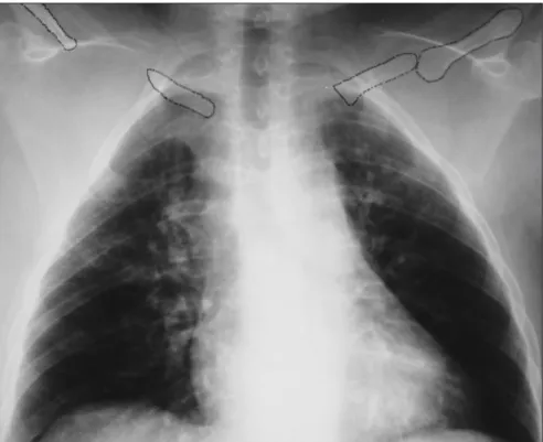
limitation of her activities but had not devel-oped any finger ulceration. Her past medical history included left mastectomy due to breast cancer. Her mother and sister presented uni-lateral clavicle agenesis. Physical examination demonstrated prognathism (Figure 1), excessive mobility of the shoulders (double-jointedness), ischemia in the right hand, and no palpable axillary, brachial and radial pulses on the right side (the left side was normal). Chest x-ray demonstrated narrowing of the thorax and both hypoplastic clavicles (Figure 2).
Color-flow duplex Doppler ultrasound scanning examination showed that the sub-clavian flow presented monophasic features. Digital angiography was performed and this displayed occlusion of the right axillary-sub-clavian arteries that spared the vertebral and internal thoracic arteries, with filling of the proximal brachial artery (Figure 3).
Due to the upper right limb limitation, angioplasty of the lesion was attempted through femoral access. It was not possible to
Figure 1. Photograph showing deformity of the upper thorax in a 65-year-old woman: appearance of drooping shoulders and elongated neck.
293
cure the lesion using hydrophilic guide wire. The procedure was halted.
Open surgery was not attempted. Follow-ing the operation, the patient evolved without severe ischemia, and clinical management was carried out. She continued to have pain, but with some improvement following a course of physiotherapy, and the symptoms remained stable.
DISCUSSION CCD is a congenital skeletal abnormal-ity with dominant autosomal inheritance, although some recessive forms have been described.1 The peculiar features of this
dis-ease result from faulty development of mem-branous bones, especially the clavicles and skull. The disease gene has been mapped to chromosome 6p21 within a region containing CBFA1, a transcription factor that activates osteoblast differentiation.1,2 Clavicular
abnor-malities give this disorder its characteristic ap-pearance of drooping shoulders, an elongated neck and an ability to adduct the shoulders anteriorly (Figure 1). The commonest defor-mity is the absence of the central clavicular segment with small bony stumps attached to the sternum and acromion3 (Figure 2). In rare
cases, the lateral portions or the entire bone is hypoplastic. Late closure of cranial sutures with Wormian bones and patent fontanels may occur, and there may also be late eruption of secondary dentition, zygomatic hypoplasia and underdevelopment of the jaw. Such pa-tients seldom present neurological symptoms due to compression, and only two other cases of vascular damage to the upper limbs have already been reported.3,4
Short,5 in 1979, was the first to report a
case of CCD causing upper limb ischemia and cerebrovascular symptoms related to this con-dition. It was of interest that the patient was a 63-year-old woman, who had both a hypoplas-tic clavicle and an ischemic right upper arm. An arteriogram showed proximal subclavian and vertebral thrombosis on the right side, and post-stenotic dilatation in the left subclavian artery, which was compressed between the first rib and a rudimentary clavicle fragment. Upon operation on both sides, it was found that the anatomy was grossly distorted and even upper right dorsal sympathectomy was impracticable. The wound was closed. On the left side, the hypoplastic proximal clavicle fragment was removed, without resection of the aneurysm.
In 1997, Qureshi et al.6 reported another
case of CCD in a 63-year-old female patient who had upper right limb ischemic pain,
result-ing in the right hand beresult-ing darker-colored and cooler than the left. Ulceration was present at the tip of the index finger and at the base of the little finger. Without palpable brachial and radial pulses, an arteriogram showed axillary occlusion beyond an abnormal clavicle, with no
Figure 2. Posteroanterior view of chest radiograph showing the position of clavicular fragments in a woman with cleidocranial dysostosis.
Figure 3. Arch aortogram of a woman presenting cleidocranial dysostosis showing occlusion of the axillary-subclavian arteries: contrast medium fills the brachial artery beyond the occlusion.
distal filling of vessels. An open-surgery repair was indicated, and, during the operation, dense scar tissue gave rise to technical difficulties in isolating and controlling the arteries. As in the earlier report,5 significant anatomical distortion
and fibrosis were present. Abnormal tissue was
294
RESUMO
Isquemia do membro superior causada por disostose cleidocraniana
CONTEXTO: A isquemia do membro superior não é tão comum quanto a isquemia do membro inferior, mas quando não diagnosticada ou diagnosticada de forma errada determina grave seqüela e até perda da extremidade.
RELATO DE CASO: O caso de uma paciente com isquemia do membro superior, devida a disostose clei-docraniana, é apresentado. Esta é uma causa extremamente rara de compressão do segmento arterial subclávio-axilar que evoluiu para trombose arterial. Somente dois casos foram previamente relatados na literatura com esta condição e nesta forma de apresentação.
PALAVRAS-CHAVE: Displasia cleidocraniana. Disostose craniofacial. Disostose. Disostose mandíbulofacial. AUTHOR INFORMATION
Walter Campos Júnior, MD. Department of Surgery, Vascular Division, Hospital das Clínicas da Faculdade de Medicina da Universidade de São Paulo
Roberta Murasaki Cardoso, MD. Department of Sur-gery, Vascular Division, Hospital das Clínicas da Faculdade de Medicina da Universidade de São Paulo
Ronald Fidelis, MD. Department of Surgery, Vascular Division, Hospital das Clínicas da Faculdade de Medicina da Universidade de São Paulo
Erasmo Simão da Silva, MD, PhD. Department of Sur-gery, Vascular Division, Hospital das Clínicas da Faculdade de Medicina da Universidade de São Paulo
Rodrigo Ramos, MD. Department of Surgery, Vascular Division, Hospital das Clínicas da Faculdade de Medicina da Universidade de São Paulo
Address for correspondence:
Erasmo Simão da Silva
Rua Martins, 96
São Paulo (SP) — Brasil — CEP 05511-000 Tel./Fax. (+55 11) 3815-1843 E-mail: [email protected]
Copyright © 2005, Associação Paulista de Medicina
1. Mundlos S. Cleidocranial dysplasia: clinical and molecular genetics. J Med Genet. 1999;36(3):177-82.
2. Cooper SC, Flaitz CM, Johnston DA, Lee B, Hecht JT. A natural history of cleidocranial dysplasia. Am J Med Genet. 2001;104(1):1-6.
3. Gupta SK, Sharma OP, Malhotra S, Gupta S. Cleido-cranial dysos-tosis – skeletal abnormalities. Australas Radiol. 1992;36(3):238-42.
4. Anspach WE, Huepel RC. Familial cleidocranial dysostosis (cleidal dysostosis). Am J Dis Child. 1939;58:786-98. 5. Short DW. A case of craniocleidal dysostosis presenting with
vascular complications. Br J Surg. 1979;66(8):596-8. 6. Qureshi KN, Lees TA, Holdsworth J. Acute upper limb
ischemia resulting from cleidocranial dysostosis. J Vasc Surg. 1997;26(5):888-90.
Sources of Funding: None
Conflicts of interest: None
Date of first submission: July 15, 2004July 15, 2004
Last received: September 13, 2005
Accepted: September 15, 2005
REFERENCES found to be bonding the clavicle fragments. A
polytetrafluoroethylene bypass was implanted. The patient was discharged with palpable distal pulse and healed hand ulcerations, while taking oral anticoagulant. Six months later, the bypass had occluded. No further intervention was at-tempted on the occlusion, despite the warfarin therapy, mainly because of the difficulties re-ported during the first operation. The ischemia did not appear to be threatening limb viability, and the patient was then treated clinically.
It is noticeable that the two previous reported cases and the present case all related
to women in their seventh decade, which may indicate high gender and age-related prevalence for the symptoms.
In spite of the scarcity of experience reported in the literature, it seems to be evident that there are significant technical difficulties in performing open vascular repairs on this kind of arterial damage. The reported cases discouraged us from trying anything more aggressive in the case of our patient, since we were not dealing with limb-threatening ischemia. Although a long arterial segment was occluded, an
endovas-cular approach was attempted in order to avoid the well-described anatomical and technical problems in the previous cases. As expected, it was not possible to cure the lesion, but the degree of ischemia did not worsen after the procedure.
CONCLUSION In spite of its rarity, cleidocranial dysplasia may lead to upper limb ischemia. A less aggres-sive approach seems to be reasonable, because of the significant anatomical distortions pres-ent in this condition.