C
ASER
EPORT| R
ELATO DEC
ASO462
Patient in chronic hemodialysis with right atrial mass:
thrombus, fungal endocarditis or atrial myxoma?
Paciente em hemodiálise crônica com massa em átrio direito:
trombo, endocardite fúngica, ou mixoma atrial?
Authors
Talita G. Salani 1
Cynthia de Moura Borges 1 Carolina S. Urbini 1 Patrícia Schincariol 1 Kélcia Rosana da Silva Quadros 1
Maria Almerinda Ribeiro--Alves 1
Rodrigo Bueno de Oliveira 1
1 Faculdade de Ciências
Médicas da Universidade Estadual de Campinas (UNICAMP), Campinas, Brazil.
Submitted on: 11/11/2015. Approved on: 11/27/2015.
Correspondence to:
Rodrigo Bueno de Oliveira. Universidade Estadual de Campinas.
Rua Tessália Vieira de Camargo, nº 126, Campinas, São Paulo, Brazil.
CEP: 13083-887
E-mail: rodrigobueno.hc@gmail. com
DOI: 10.5935/0101-2800.20160073
We present the case report of a 19-year-old patient with chronic kidney disease due to chronic glomerulonephritis, in he-modialysis (HD) by central catheter, with the incidental finding of a mass of 28x16 mm in right atrium (RA). The diagnosis of thrombus, infective endocarditis or myxo-ma were considered. Given the context of immunosuppression and difficult access vascular therapeutic practice has proved complex. Although Doppler echocardiog-raphy suggested thrombus in RA, nuclear magnetic resonance imaging (MRI) indi-cated for the diagnosis of myxoma in RA. In both conditions, the proposed surgi-cal approach was limited by intense im-munosuppression history and the risk of infectious complications. Throughout the treatment, the general state of K.M.F. re-mained satisfactory and revealed no signs or symptoms related to atrial dysfunction. The absence of fever and negative blood cultures excluded infective endocardi-tis. Prior echocardiogram report without masses in the RA decreased the chance of cardiac myxoma. The therapeutic re-sponse to anticoagulation confirmed the diagnosis of thrombosis. After 180 days of anticoagulation, there was significant reduction in mass. The patient developed asymptomatic. The diagnosis of mass in RA can be a challenge and only the evo-lution of the case was able to guide the appropriate conduit. While MRI has high sensitivity and specificity for the diagno-sis of cardiac myxoma, the interpretation of images can be subjective. Controver-sial point is the removal of the catheter in such cases, which is subject discussed throughout the report.
A
BSTRACTKeywords: catheters; endocarditis; myxo-ma; renal dialysis; thrombosis.
Apresentamos o relato de caso de uma pa-ciente de 19 anos com doença renal crô-nica devido à glomerulonefrite crôcrô-nica e em hemodiálise (HD) por cateter central, com o achado incidental de uma massa de 28x16 mm em átrio direito (AD). Foram considerados os diagnósticos de trombo, endocardite infecciosa ou mixoma. Devi-do ao contexto de imunossupressão e di-ficuldade de acesso vascular, a condução terapêutica revelou-se complexa. Apesar de Ecodopplercardiograma sugerir trombo em AD, imagens de ressonância nuclear magnética (RNM) apontaram para o diag-nóstico de mixoma em AD. Nas duas con-dições a proposta de abordagem cirúrgica esteve limitada pelo histórico de imunossu-pressão intensa e o risco de complicações infecciosas. Ao longo do tratamento, o estado geral de K.M.F. manteve-se satis-fatório e não foram observados sinais ou sintomas relacionados a disfunção atrial. A ausência de febre e hemoculturas negativas excluíram endocardite infecciosa. O relato de ecocardiograma prévio sem massas em AD tornou menor a possibilidade de mixo-ma cardíaco. A resposta terapêutica à an-ticoagulação confirmou o diagnóstico de trombo. Após 180 dias de anticoagulação, houve redução significativa da massa. A paciente evoluiu assintomática. O diagnós-tico de massa em AD pode ser um desafio e somente a evolução foi capaz de guiar a conduta apropriada. Apesar da RNM ter elevada sensibilidade e especificidade para o diagnóstico de mixoma cardíaco, a in-terpretação de imagens pode ser subjetiva. Ponto controverso é a retirada de cateter nesses casos, que é assunto discutido ao longo do relato.
R
ESUMOJ Bras Nefrol 2016;38(4):462-465 Right atrial mass in a patient on hemodialysis
463
I
NTRODUCTIONA significant portion of the individuals enrolled in hemodialysis (HD) programs without a definitive vascular access (e.g.: arteriovenous fistula) use long-term indwelling catheters. Prolonged use of indwelling catheters has been associated with severe mechanical complications and infection.1-3
A right atrial mass in an individual on hemodialysis is usually one of three possibilities: (a) a thrombus, (b) vegetation as a sign of infectious endocarditis, or (c) a heart tumor, most likely a myxoma.4 Once these
diseases call for different therapies, improper diagnosis may lead to unnecessary invasive procedures, delayed start of treatment, and increased morbidity and mortality.
This case report illustrates the diagnostic chal-lenges physicians face when finding a right atrial mass located contiguously to the HD catheter on a transthoracic echocardiogram (TTE) taken as part of the annual examination protocol in effect at the dialy-sis center. The report also aims to show the key role proper follow-up and therapy play in the production of a good clinical outcome.
C
ASE REPORTK.M.F., female, 19, had systemic vasculitis with re-nal and pulmonary involvement. Despite immuno-suppressant therapy with steroids and cyclophos-phamide, the patient progressed to renal failure. She was started on HD with a short-term catheter at the Hospital of the University of Campinas (UNICAMP) in Brazil. The patient had to undergo four catheter changes due to catheter malfunction or infection. Once the infection was eradicated, the patient had a long-term indwelling catheter implanted.
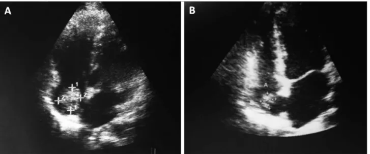
A TTE was ordered seven months after the implantation of the long-term catheter, and a mass measuring 28 x 16 mm was found in the patient’s right atrium. The mass was initially believed to be a thrombus (Figure 1A). The patient was on prednisone 10 mg/day and did not have fever or systemic symptoms, and her general condition was good. Heart auscultation was normal. The patient was not fatigued and did not have hepatomegaly, peripheral edema or signs of pulmonary embolism. A nuclear magnetic resonance (NMR) scan of the heart was ordered to verify the initial diagnostic suspicion. The scan showed a solid round non-moving pedunculated
mass on the floor of the right atrium close to the inferior vena cava, measuring 25 x 24 x 20 mm and suggestive of an atrial myxoma.
No microorganisms were detected in her blood cultures. The patient was fever-free and generally in good shape. The thoracic surgery team assessed her and the risk of removing the mass led the attending physician to choose anticoagulation with warfarin as a therapeutic test for the diagnosis of a thrombus. The long-term catheter was kept in place, as finding a new HD vascular access would not be easy. A control transesophageal echocardiogram (TEE) performed 100 days after the start of treatment (Figure 1B) showed the right atrial mass had shrunk to 12 x 10 mm.
The patient was symptom-free and was kept on warfarin for another 90 days. TEE images produced after six months of anticoagulation revealed a thick-ening of the Eustachian valve, which corresponded to remnants of the thrombus measuring 7 x 8 mm. She was offered an AVF and the long-term catheter was removed. The patient is asymptomatic and in good general condition. She had no warfarin-related ad-verse events, and was kept on anticoagulation therapy for another three months.
D
ISCUSSIONThe following diagnostic possibilities were consid-ered for the patient’s right atrial mass: thrombus, in-fectious endocarditis, and atrial myxoma. Inin-fectious endocarditis was ruled out based on the patient’s overall satisfactory progression, the absence of fever, and blood cultures negative for bacteria and fungi. Although the NMR scans suggested she had an atri-al myxoma, her previous TTE images did not show cardiac anomalies, which made a cardiac tumor a more remote possibility. However, atrial myxomas are known to develop within short periods of time due to their composition with scattered cells within a mucopolysaccharide stroma.5 The lack of
consti-tutional symptoms and her response to warfarin therapy supported the diagnosis of a right atrial thrombus.
Although low in the general population, the incidence of right atrial thrombi increases in individuals using central venous catheters.1,2,6,7 The patient described in this case
J Bras Nefrol 2016;38(4):462-465
Right atrial mass in a patient on hemodialysis
464
Figure 1. A. Transthoracic echocardiogram (TTE) showing right atrial mass measuring 28 x 16 mm consistent with thrombus. B. Control TTE showing a significant reduction on the mass to 12 x 10 mm.
embolism, endocarditis, arrhythmia, mechanical cardiac complications, and systemic embolism in individuals with a patent foramen ovale.8,9
The possible pathophysiological mechanisms involved in the formation of right atrial thrombi associated with HD catheterization include the activation of the coagulation cascade secondary to trauma to the atrial wall by the tip of the catheter. Local conditions are exacerbated by hemodynamic changes, which lead to the stagnation of blood flow in the right atrium in the area around the catheter.10,11
The location of the tip of the long-term indwelling catheter inside the right atrium - as per the guidelines of
the National Kidney Foundation: Dialysis Outcomes
Quality Initiative - has been strongly associated with
the formation of thrombi.7,12,13
TTE and TEE are the most commonly used tests to diagnose and follow the progression of right atrial masses. The specificity and sensitivity of echocardiography in the diagnosis of intracavitary masses are 86% and 95%, respectively.14,15 However,
NMR is the preferred method for the differential diagnosis of right atrial masses, as it is highly specific (100%) for the detection of heart tumors.16
In our case, the diagnoses suggested by the imaging tests differed (TTE for thrombus and NMR for myxoma), thus forcing the assisting team to make a call based on clinical findings. This shows that despite highly specific tests the interpretation of the scans still plays a significant role in achieving a diagnosis.
Treatment options range between offering anticoagulants or thrombolytic agents associated or
not to catheter removal. Anticoagulation therapy for patient with kidney disease can be challenging. Elliott et al.17found that warfarin is associated with significant
risk of bleeding in patients on HD. Additionally, the use of vitamin K antagonists such as warfarin has been associated with vascular calcifications caused by the inhibition of vitamin k-dependent proteins such as the matrix-GLA protein.18
Thrombectomy is indicated in cases suspected for infection or concomitant cardiac anomaly.19 A
study showed that individuals with autoimmune disease and recent use of immunosuppressants are assigned higher levels of surgical risk and develop greater numbers of post-op complications, including myocardial infarction, heart failure, pneumonia, sepsis, and wound dehiscence, particularly those on high-dose oral prednisone before surgery.20 This
finding supported the decision to treat our patient conservatively.
J Bras Nefrol 2016;38(4):462-465 Right atrial mass in a patient on hemodialysis
465
Surgery was recommended as the best option for patients suspected for infection, cardiac anomalies, or with contraindications to anticoagulation treatment.3
Another systematic review with 177 individuals diagnosed with right heart (atrial and ventricular) thrombi reported death rates of 28.6%, 23.8%, and 11.3% for patients offered anticoagulant therapy, embolectomy, and thrombolysis, respectively. All individuals not offered treatment died. The authors indicated that thrombolytic therapy was associated with lower death rates when compared to other treatments.9
C
ONCLUSIONDiagnosing right atrial masses can be challenging. However, patient clinical progression may be used to guide the choice of either conservative or surgical treatment, as shown in the case reported herein. There is no consensus in the literature as to the best time to remove long-term indwelling catheters or the treatment of thrombi associated with catheterization. The recommendations described in the literature suggest care should be individualized and based on the information published in prospective randomized trials.
R
EFERENCES1. Wijeyesinghe EC, Pei Y, Fenton SS, Uldall PR. Right atrial ball thrombus as a complication of subclavian catheter insertion for he-modialysis access. Int J Artif Organs 1987;10:102-4.
2. Fincher ME, Caruana RJ, Humphries A, Gross CM, Rubin JW, Bowen PA. Right atrial thrombus formation following central venous dialysis catheter placement. Am Surg 1988;54:652-4. PMID: 3190000
3. Stavroulopoulos A, Aresti V, Zounis C. Right atrial thrombi com-plicating haemodialysis catheters. A meta-analysis of reported cases and a proposal of a management algorithm. Nephrol Dial Trans-plant 2012;27:2936-44. DOI: http://dx.doi.org/10.1093/ndt/gfr739 4. Cianciulli TF, Saccheri MC, Redruello HJ, Cosarinsky LA, Cel-ano L, Trila CS, et al. Right atrial thrombus mimicking myxo-ma with pulmonary embolism in a patient with systemic lupus erythematosus and secondary antiphospholipid syndrome. Tex Heart Inst J 2008;35:454-7.
5. Pucci A, Gagliardotto P, Zanini C, Pansini S, di Summa M, Mollo F. Histopathologic and clinical characterization of car-diac myxoma: review of 53 cases from a single institution. Am Heart J 2000;140:134-8. PMID: 10874274 DOI: http://dx.doi. org/10.1067/mhj.2000.107176
6. Ross P Jr, Ehrenkranz R, Kleinman CS, Seashore JH. Thrombus associated with central venous catheters in infants and children. J Pediatr Surg 1989;24:253-6. DOI: http://dx.doi.org/10.1016/ S0022-3468(89)80006-5
7. Korones DN, Buzzard CJ, Asselin BL, Harris JP. Right atrial thrombi in children with cancer and indwelling catheters J Pediatr 1996;128:841-6. PMID: 8648545
8. Kinney EL, Wright RJ. Efficacy of treatment of patients with echo-cardiographically detected right-sided heart thrombi: a meta-anal-ysis. Am Heart J 1989;118:569-73. PMID: 2773775 DOI: http:// dx.doi.org/10.1016/0002-8703(89)90274-3
9. Rose PS, Punjabi NM, Pearse DB. Treatment of right heart thromboemboli. Chest 2002;121:806-14. PMID: 11888964 DOI:http://dx.doi.org/10.1378/chest.121.3.806
10. Fuchs S, Pollak A, Gilon D. Central venous catheter mechanical irritation of the right atrial free Wall: A cause for thrombus for-mation. Cardiology 1999;91:169-72. PMID: 10516410 DOI: http://dx.doi.org/10.1159/000006905
11. Forauer AR, Theoharis C. Histologic changes in the human vein wall adjacent to indwelling central venous catheters. J Vasc Interv Radiol 2003;14:1163-8. DOI: http://dx.doi. org/10.1097/01.RVI.0000086531.86489.4C
12. Gilon D, Schechter D, Rein AJ, Gimmon Z, Or R, Rozenman Y, et al. Right atrial thrombi are related to indwelling central venous catheter position: insights into time course and possible mechanism of formation. Am Heart J 1998;135:457-62. PMID: 9506332 DOI:http://dx.doi.org/10.1016/S0002-8703(98)70322-9
13. NKF/DOQI Clinical Practice Guidelines for Vascular. Access, 2006 Updates CPG 2.4,. [citado 19 Mar 2016]. Disponível em: http:// www2.kidney.org/professionals/KDOQI/guideline_upHD_PD_VA/ 14. Pruszczyk P, Torbicki A, Pacho R, Chlebus M, Kuch-Wocial A,
Pruszynski B, et al. Noninvasive diagnosis of suspected severe pulmonary embolism: transesophageal echocardiography vs spiral CT. Chest 1997;112:722-8. PMID: 9315806 DOI:http:// dx.doi.org/10.1378/chest.112.3.722
15. Pepi M, Evangelista A, Nihoyannopoulos P, Flachskampf FA, Athanassopoulos G, Colonna P, et al.; European Association of Echocardiography. Recommendations for echocardiography use in the diagnosis and management of cardiac sources of em-bolism: European Association of Echocardiography (EAE) (a registered branch of the ESC). Eur J Echocardiogr 2010;11:461-76. DOI:http://dx.doi.org/10.1093/ejechocard/jeq045
16. Pazos-López P, Pozo E, Siqueira ME, García-Lunar I, Cham M, Jacobi A, et al. Value of CMR for the differential diagnosis of cardiac masses. JACC Cardiovasc Imaging 2014;7:896-905. DOI: http://dx.doi.org/10.1016/j.jcmg.2014.05.009
17. Elliott MJ, Zimmerman D, Holden RM. Warfarin anticoagu-lation in hemodialysis patients: a systematic review of bleed-ing rates. Am J Kidney Dis 2007;50:433-40. PMID: 17720522 DOI: http://dx.doi.org/10.1053/j.ajkd.2007.06.017
18. Tantisattamo E, Han KH, O'Neill WC. Increased vascular cal-cification in patients receiving warfarin. Arterioscler Thromb Vasc Biol 2015;35:237-42. DOI: http://dx.doi.org/10.1161/ ATVBAHA.114.304392
19. American College of Cardiology; American Heart Association Task Force on Practice Guidelines (Writing Committee to revise the 1998 guidelines for the management of patients with valvu-lar heart disease); Society of Cardiovascuvalvu-lar Anesthesiologists; Bonow RO, Carabello BA, Chatterjee K, de Leon AC Jr, Faxon DP, Freed MD, et al. ACC/AHA 2006 guidelines for the man-agement of patients with valvular heart disease: a report of the American College of Cardiology/American Heart Association Task Force on Practice Guidelines (writing Committee to Revise the 1998 guidelines for the management of patients with val-vular heart disease) developed in collaboration with the Society of Cardiovascular Anesthesiologists endorsed by the Society for Cardiovascular Angiography and Interventions and the Society of Thoracic Surgeons. J Am Coll Cardiol. 2006;48:e1-48. 20. Papa MZ, Shiloni E, Vetto JT, Kastner DL, McDonald HD.