Authors
Thais Emanuelle Faria Botelho 1
Alamanda Kfoury Pereira 1
Patrícia Gonçalves Teixeira 1
Eura Martins Lage 1 Gabriel Costa Osanan 1
Ana Cristina Simões e Silva 1
1 Universidade Federal de
Minas Gerais.
Submitted on: 05/12/2016. Approved on: 08/08/2016.
Correspondence to: Thais Emanuelle Faria Botelho.
Universidade Federal de Minas Gerais - Faculdade de Medicina. Av. Prof. Alfredo Balena, nº
190, Sala 213, Belo Horizonte, MG, Brazil.
CEP: 30130-100
E-mail: bthais.faria@gmail. com
Uromodulin: a new biomarker of fetal renal function?
Uromodulina: um novo biomarcador de função renal fetal?
Introduction: Obstructive uropathies are main diseases affecting the fetus. Early diagnosis allows to establish the appropriate therapy to minimize the risk of damage to kidney function at birth. Biochemical markers have been used to predict the prognosis of renal function in fetuses. Uromodulin, also known by Tamm-Horsfall protein (THP) is exclu-sively produced in the kidneys and in normal conditions is the protein excret-ed in larger amounts in human urine. It plays important roles in kidneys and urinary tract. Also it participates in ion transport processes, interact with vari-ous components of the immune system and has a role in defense against urinary tract infections. Moreover, this protein was proved to be a good marker of re-nal function in adult patients with sev-eral renal diseases. Objective: To evalu-ate if uromodulin is produced and elim-inated by the kidneys during fetal life by analyzing fetal urine and amniotic fluid and to establish correlation with bio-chemical parameter of renal function already used in Fetal Medicine Center at the Clinic Hospital of UFMG (CE-MEFE/HC). Methods: Between 2013 and 2015, were selected 29 fetuses with indication of invasive tests for fetal di-agnosis in monitoring at the CEMEFE/ HC. Results: The determination of uro-modulin was possible and measurable in all samples and showed statistically significant correlation with the osmo-larity. Conclusion: There was a tenden-cy of lower levels of Uromodulin values in fetuses with severe renal impairment prenatally. Thus, high levels of this pro-tein in fetal amniotic fluid or fetal urine dosages possibly mean kidney function preserved.
A
BSTRACTKeywords: amniotic fluid;
ultrasonogra-Introdução: Uropatias obstrutivas estão entre as principais doenças que acometem o feto. O diagnóstico precoce destas doen-ças permite estabelecer a terapêutica ade-quada, visando minimizar os riscos de da-nos à função renal no nascimento. Os mar-cadores bioquímicos têm sido utilizados na predição do prognóstico da função renal em fetos. A uromodulina, também chama-da de proteína de Tamm-Horsfall (THP), é produzida exclusivamente nos rins, e em condições normais, é a proteína excretada em maior volume na urina humana. Ela desempenha importantes funções nos rins e trato urinário. Participa dos processos de transporte de íons, interage com vá-rios componentes do sistema imunológico e possui papel na defesa contra infecções do trato urinário. Além disso, se mostrou um bom biomarcador de função renal em adultos portadores de diversas doenças re-nais. Objetivos: Avaliar se a uromodulina é produzida e eliminada pelos rins durante a vida fetal através da análise de urina fe-tal e líquido amniótico, além de estabelecer correlação com o parâmetro bioquímico de função renal já utilizado no Centro de Medicina Fetal do Hospital das Clínicas da UFMG (CEMEFE/HC). Métodos: Entre 2013 e 2015, foram selecionados 29 fetos com indicação de exames invasivos para diagnóstico fetal em acompanhamento no CEMEFE/HC. Resultados: A dosagem da uromodulina foi possível e quantificá-vel em todas as amostras e mostrou cor-relação significativa com a osmolaridade.
Conclusão: A uromodulina mostrou uma tendência em apresentar valores reduzidos em fetos com grave comprometimento re-nal no pré-natal. Assim, valores elevados desta proteína em dosagens de urina fe-tal ou líquido amniótico podem significar uma função renal preservada.
R
ESUMOurogeni-I
NTRODUCTIONKidney and urinary tract malformations account for 20% of fetal anomalies detected through prenatal ultrasound examination.1 Obstructive uropathy ranks
first in the list of malformations and is the leading cause of chronic kidney disease (CKD) in children and teens.2
Obstructive uropathy is a congenital anomaly resulting from partial or complete urine flow obstruction in the urinary tract. It is initially characterized by hydronephrosis, a swelling of the kidney due to a build-up of urine, associated to other ultrasound findings depending on the type of obstruction.3 Prenatal diagnosis and early treatment
may prevent or delay the onset of chronic kidney disease.4,5
According to the Census of the Brazilian Society of Nephrology,6 approximately 100,397 individuals
had end-stage renal disease in Brazil in 2013; 90.8% of these patients were on hemodialysis and 9.2% on peritoneal dialysis. Six percent of these individuals were under the age of 18 years. A collaborative study carried out in 13 centers analyzed the records for pediatric renal transplantation procedures performed in Brazil from 2004 to 2013. The data from 1,751 transplant procedures performed in individuals under 18 years of age showed that the most common cause of transplantation was obstructive uropathy, with 31% of the cases.7
Ultrasound findings alone cannot be used to thoroughly assess fetal kidney function.8 Biochemical
tests on fetal urine bridge that gap and allow for proper patient management and timely intervention.9
The placenta maintains fluid and electrolyte balance; therefore, tests based on fetal serum do not aid in the analysis of intrauterine renal function. However, electrolyte levels in fetal urine correlate directly with renal function10 and may be assessed
through biochemical tests since week 15 of pregnancy, a time by which the fetus’ kidneys are functioning.
Some authors believe electrolyte levels can be derived from amniotic fluid, since a significant portion of it contains fetal urine.11 Biochemical tests
on urine and amniotic fluid have been performed to assess fetal renal function, but none of the tested parameters is a proven accurate marker of fetal renal function.12,13
UROMODULIN OR TAMM-HORSFALL GLYCOPROTEIN (THP)
Tamm-Horsfall glycoprotein (THP) was discovered in 1950 by Igor Tamm and Frank L. Horsfall Jr. and characterized as a protein present in the urine of humans and animals that was able to inhibit viral hemagglutination.14 In 1985, Muchmore and
Decker15 isolated a protein capable of inhibiting the
proliferation of T-cells and monocytes. Because of its source of isolation and in-vitro immune response modulation property, they called it uromodulin. Two years later, researchers studying complementary DNA found that THP and uromodulin were one and the same protein.16
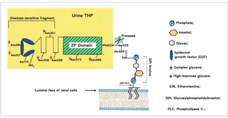
Rindler et al.17 showed that uromodulin is a
glycosylphosphatidylinositol (GPI) anchor-linked protein. The mature protein contains 616 amino acids, including 48 cysteine residues involved in 24 disulfide bridges, which are important for its conformation (Figure 1).
Eight potential sites for N-glycosylation are also present. The structure of uromodulina also contains three epidermal growth factors (EGF) and a zona pellucida-like domain. The C-terminus of the protein includes a stretch of hydrophobic amino acids that acts as a signal for transpeptidase within the endoplasmic reticulum of cells in the thick ascending limb (TAL) of the loop of Henle - where uromodulin is produced - to attach a preformed GPI anchor to the protein.
Following this addition, the anchor-linked protein is transported to the Golgi complex, where glycans are processed, then delivered to the luminal cell surface, and finally released into the urine by proteolytic cleavage.18 Ontogenetic studies indicated that the
presence of uromodulin is closely related to the development and functional maturation of the loop of Henle.19 In normal conditions, uromodulin is the
protein excreted by the kidneys in larger quantities, at a rate of approximately 50 mg/day, with levels varying as a function of a number of factors such as urine volume, nutrition, and physical exercise.20
Studies have shown a number of biological roles for uromodulin in the urinary system. Since it is produced in the cells of the TAL of the loop of Henle, uromodulin is believed to play a role in ion transport.21,22 Uromodulin has also been recognized
Figure 1. Structural model for urine uromodulin (yellow area) and GPI anchor (Source: Serafini-Cessi et al., 2003).
due to the interactions it maintains with various component of the immune system;23-25 it is also
known for helping in the defense against urinary tract infections, particularly the ones caused by Escherichia
coli.26-28 Uromodulin also acts in the prevention
against the formation of kidney stones by reducing the clustering of calcium crystals.29
Mutations on the gene encoding uromodulin (UMOD) have been associated with various autosomal dominant kidney diseases.30,31 Fifty-eight
mutations to the UMOD gene have been described in the literature.32 These conditions are assumed to
produce defective proteins that are not released in the cell membrane. In-vitro studies of kidney cells with UMOD mutations revealed that uromodulin is retained inside the cell,33,34 causing further damage to
the cells of the TAL of the loop of Henle.
According to Prajczer et al.35, urinary levels of
uromodulin were lower in individuals with CKD when compared to healthy controls and correlated positively with glomerular filtration rates (GFR).
Zhou et al.36 reported a correlation between
urine uromodulin levels and GFR in a group of patients with IgA nephropathy, with individuals showing lower levels of uromodulin as the disease progressed. These findings agree with previous studies in which uromodulin levels decreased in individuals with diseases affecting kidney function or integrity.37-40
With these evidences and the lack of studies on the production and release of uromodulin in fetal life in mind, this study aimed to verify whether uromodulin is produced during fetal life and if the protein may be used as a biomarker of renal function in fetuses with kidney malformations.
O
BJECTIVESThis study aimed to check whether uromodulin is produced and excreted by the kidneys during fetal life by analyzing samples of fetal urine and amniotic fluid collected from patients followed at the Center for Fetal Medicine of the Hospital and Clinics of the Federal University of Minas Gerais (CEMEFE/HC) and describe the correlations between uromodulin levels and osmolarity, the biochemical marker of renal function used at CEMEFE/HC.
P
ATIENTS ANDMETHODSPATIENTS
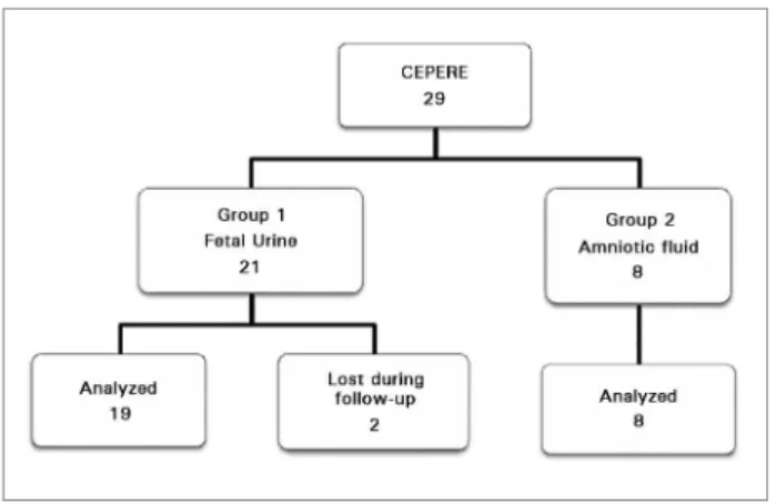
Eight fetuses (Group 2) with non-nephrourinary anomalies were prescribed genetic or infectious disease testing requiring amniotic fluid collection (Figure 2).
Figure 2. Study follow-up flowchart.
The low volumes of amniotic fluid seen in the mothers of the subjects in Group 1 warranted the collection of fetal urine. The protocol in effect for the individuals in Group 2 included amniotic fluid analysis. The patients and their fetuses were exposed to no risks other than the ones inherent to the prescribed collection procedures. At the time of the procedure, a portion of the sample was set aside to test for uromodulin levels.
ETHICS
The Research Ethics Committee of the Federal University of Minas Gerais (UFMG) approved this study and assigned it permit CAAE 35559214.1.0000.5149. The enrolled pregnant women were informed of the protocol and voluntarily joined the study after signing an informed consent term. They were followed through to labor and puerperium, according to the protocols in effect at CEMEFE, and were allowed to leave the study of they so wished.
The neonates with uropathy were followed at the Neonatal Unit and the Pediatric Nephrology Unit of the UFMG Hospital and Clinics. Only two of the 29 pregnant women initially enrolled in the study were lost during follow-up, and their data were excluded from the final analysis.
STUDY PROTOCOL
The samples of fetal urine (Group 1) and amniotic fluid (Group 2) were centrifuged at 4ºC and 300 G for 20 minutes, frozen at -80ºC, and sent for analysis
at the Interdisciplinary Medical Research Laboratory of the School of Medicine at UFMG. The biochemical parameters analyzed included osmolarity, and uro-modulin and creatinine levels.
Creatinine levels were measured to adjust uromodulin urine levels for creatinine urine clearance levels according to the recommendations for the measurement of markers in urine samples.41 Uromodulin levels were expressed
in nanograms of uromodulin per milliliter of urine or amniotic fluid (ng/ml) or in nanograms of uromodulin per microgram of urine creatinine (ng/µg). Creatinine levels were expressed in micrograms of creatinine per milliliter of urine (µg/ml).
An OSMOMAT 030 (AUTOMATIC CRYO-SCOPIC OSMOMETER) osmometer calibrated with a standard specimen of 300 mOsmol/Kg, made by Gonatec GmbH, was used in the analysis of fetal urine osmolarity. Samples with values greater than 210 mOsm/l42 were deemed anomalous, as described
in the protocol in effect at the service.
Uromodulin levels were measured from samples of fetal urine and amniotic fluid using an ELISA kit designed to perform quantitative measurements of human uromodulin. Biovendor RD191163200R Hu-man Uromodulin ELISA kits Hu-manufactured by Bio-Vendor Research and Diagnostic Products were used in this study.
A standard curve was built plotting absorbance values versus standard uromodulin values; unknown sample levels were determined based on this standard curve. Absorbance values were captured from an ELISA reader (MOLECULAR DEVICES, USA). A straight line was used to calculate the uromodulin levels in each sample.
Creatinine levels from fetal urine samples were measured with the aid of a modified Jaffee method kit made by Bioclin. The tests were run in accordance with manufacturer instructions. The method consists of a colorimetric reaction between creatinine and picric acid, producing a reddish-yellow color. An ELISA reader was used to assess the absorbance of the formed compound (MOLECULAR DEVICES, USA) at a wavelength of 510 nm.
Fetal urine osmolarity is calculated routinely at CEMEFE and analyzed jointly with ultrasound findings, amniotic fluid volumes, and aspects of renal parenchyma to aid in the prognostic assessment of fetal renal function. The results of uromodulin and creatinine biochemical tests of the two groups - fetuses with normal osmolarity and fetuses with altered osmolarity - were compared.
STATISTICAL ANALYSIS
The Shapiro-Wilk test was used to assess the distribu-tion of variables. Parametric variables were expressed in terms of their mean values and standard errors, whereas non-parametric variables were expressed in terms of their median values and interquartile ranges (25th percentile - 75th percentile).
Student’s t-test for unpaired data was applied to parametric variables; the Mann-Whitney U test was used with non-parametric variables to compare be-tween median values. Pearson’s correlation coefficient was used to test the correlations between the follow-ing variables:
Uromodulin level vs. osmolarity; Uromodulin level vs. creatinine level;
Uromodulin adjusted for creatinine vs. osmolarity. The following values were used to define the strength of the correlations:
Positive of negative 0.70 - indicative of strong correlation;
Positive or negative 0.30 to 0.70 - indicative of moderate correlation;
Zero to 0.30 - indicative of poor correlation. In statistical hypothesis testing, differences with a p < 0.05 were deemed statistically significant. The analyses were run on statistical package MINITAB version 14.13 (MINITAB, STATE COLLEGE, PA, USA).
R
ESULTSThe study enrolled 21 fetuses with gestational ages of 24.4 ± 1.2 weeks diagnosed with obstructive uropathy (Group 1) and eight fetuses with gestational ages of 26.4 4 ± 1.5 weeks free of obstructive uropathy (Group 2). No statistically significant differences were observed between the gestational ages of the individuals in groups 1 and 2.
The mothers of 16 of the 21 fetuses with obstructive uropathy (76%) had moderate to sharp decreases in amniotic fluid volume. Only five fetuses in Group 1
fluid. In contrast, all fetuses in Group 2 were exposed to normal volumes of amniotic fluid.
Five neonatal deaths occurred in Group 1 as a result of pulmonary hypoplasia. None of the neonates in Group 2 died. Posterior urethral valve (PUV), the leading cause of obstructive uropathy in the subjects in Group 1, was present in 19 of the 21 fetuses (90%).
The median level of fetal urine uromodulin in the fetuses on Group 1 was 399.4 ng/ml (interquartile range - 319.4 to 1139); the median creatinine level seen in the same urine samples was 11.35 ng/µg (interquartile range - 7.1 to 35.7).
The mean fetal urine osmolarity measured for 19 of the 21 fetuses included in Group 1 was 206.7 ± 23.4 mOsm/l. When analyzed for normal urine osmolarity (< 210 mOsm/l), ten of the 19 urine samples were normal (52.6%) and nine were anomalous (47.4%).
Figure 3 shows a comparison between urine uro-modulin levels (ng/ml) of fetuses with obstructive uropathy with urine osmolarity below 210 mOsm/l (normal) versus fetuses with altered urine osmolarity. The absolute uromodulin urine levels were signifi-cantly different between the subgroups (1152 ± 282.1 ng/ml in the fetuses with normal urine osmolarity vs. 428.1 ± 44.1 ng/ml in the group with altered urine osmolarity; p = 0.01; Figure 3).
The same comparison using uromodulin levels related to urine creatinine levels in the two subgroups (normal vs. altered osmolarity) failed to yield statistically significant differences [11.3 (interquartile range 5.7 to 23.3) versus 13.3 (interquartile range 7.9 to 25.3) ng/µg; p = 0.77; Mann Whitney U test)].
A moderate-to-significant inverse correlation was found between uromodulin levels and osmolarity (Pearson’s correlation coefficient r = -0.675; p = 0.02). The correlations between uromodulin vs. creatinine and uromodulin adjusted for creatinine vs. osmolarity were not statistically significant (data not shown).
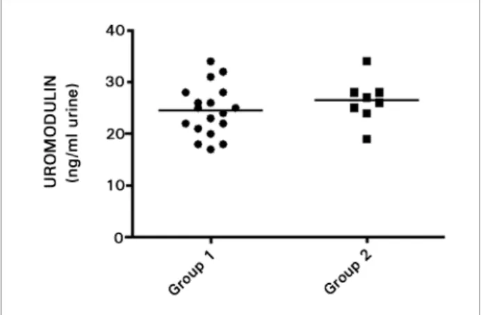
In contrast, the median level of uromodulin in amniotic fluid samples (Group 2) from fetuses free of renal involvement was 1164 (interquartile range: 528.5 to 1348) ng/ml, while the median level of uromodulin in the urine of fetuses with obstructive uropathy (Group 1) was 399.4 (interquartile range: 319.4 to 1139) ng/ml.
Figure 3. Comparison of uromodulin levels in fetuses with normal and altered osmolarity (osm).* p = 0.01, T-test for unpaired data.
in the fetuses in Group 2, the difference in relation to the levels seen in the individuals in Group 1 was not statistically significant (p = 0.23; Mann Whitney U test; Figure 4).
D
ISCUSSIONOne of the main challenges in the prenatal care of obstructive uropathy revolves around finding markers able to accurately predict fetal renal function. This information allows the medical team to establish a more suitable treatment plan with the parents designed to preserve renal function through to birth. Obstructive uropathy is known for its prevalence and treatment options. Biomarkers able to effectively predict early prenatal renal function have not been reported to date.
In this study, uromodulina levels were measured in amniotic fluid and fetal urine, indicating it is produced and excreted by fetal kidneys. Batchelder
et al.,43 in a study carried out with Rhesus monkey
fetuses, found uromodulin expression in cells of the TAL of the loop of Henle starting on the second trimester of pregnancy.
THP or uromodulin has been attributed numerous roles in renal physiology and protection against urinary tract infection, in addition to associations with genetic renal diseases. Adult individuals diagnosed with conditions leading to kidney involvement were shown to have minute uromodulin levels.
Uromodulin has also been used as a marker of acute kidney injury (AKI) in neonates. Individuals with AKI had lower uromodulin levels than injury-free neonates.44
Figure 4. Comparison of urine uromodulin levels of fetuses with obstructive uropathy (group 1) and uromodulin levels in amniotic fluid of fetuses without obstructive uropathy (group 2). p = 0.23, Mann Whitney U test.
With such information in mind, the authors of this study compared uromodulin levels in fetuses with renal involvement (Group 1) and fetuses without renal involvement (Group 2). In Group 2, uromodulin levels in amniotic fluid were higher than the levels seen in the individuals in Group 1, thus supporting the findings in the literature on adult and neonate health in which individuals with probable loss of renal function have lower uromodulin levels.
Thus far low uromodulin levels have almost unanimously been considered the consequence of damage to cells of the TAL of the loop of Henle and correlated with decreased renal function.
Uromodulin levels measured from fetal urine were sorted into subgroups with normal or altered osmolarity. A statistically significant inverse correlation was found between these markers. Uromodulin was shown to be similar to osmolarity in diagnosing fetal renal function.
The results reported in this study showed that uromodulin levels were lower in the fetuses with severe prenatal renal involvement, indicating possible cell injury and compromised excretion. Uromodulin might possibly become a chemical biomarker of fetal renal function and contribute to the early diagnosis and better management of fetuses with severe obstructive uropathy. Uromodulin levels may be measured from fetal urine and amniotic fluid.
C
ONCLUSIONSUromodulin levels may be derived and quantified from fetal urine or amniotic fluid samples with the aid of ELISA kits for human uromodulin. All tested samples were read and measured quantitatively based on calculated uromodulin absorbance.
A statistically significant inverse correlation was found between uromodulin levels and osmolarity.
A trend toward lower uromodulin levels in urine was observed in fetuses with prenatal renal involvement. Thus, high uromodulin levels in fetal urine or amniotic fluid may mean preserved renal function.
R
EFERENCES1. Elder JS. Antenatal hydronephrosis. Fetal and neonatal manage-ment. Pediatr Clin North Am 1997;44:1299-321. PMID: 9326963 2. Soares CM, Diniz JS, Lima EM, Oliveira GR, Canhestro MR,
Colosimo EA, et al. Predictive factors of progression to chro-nic kidney disease stage 5 in a predialysis interdisciplinary pro-gramme. Nephrol Dial Transplant 2009;24:848-55. DOI:http:// dx.doi.org/10.1093/ndt/gfn547
3. Callen PW. Ultrassonografia em Ginecologia e Obstetrícia, 5 ed. Rio de Janeiro: Saunders-Elsevier; 2009.
4. Pereira AK, Oliveira EA, Leite HV, Cabral ACV. Correlação entre o diagnóstico morfológico pré e pós-natal das nefrou-ropatias fetais. Rev Bras Ginel Obstet 2000;22:365-71. DOI: http://dx.doi.org/10.1590/S0100-72032000000600007 5. Melo BF, Aguiar MB, Bouzada MC, Aguiar RL, Pereira AK,
Pai-xão GM, et al. Early risk factors for neonatal mortality in CAKUT: analysis of 524 affected newborns. Pediatr Nephrol 2012;27:965-72. DOI: http://dx.doi.org/10.1007/s00467-012-2107-y
6. Sociedade Brasileira de Nefrologia. Censo de diálise SBN 2013. [Cited 2016 Oct 7]. Disponível em: http://arquivos.sbn.org.br/ pdf/censo_2013-14-05.pdf
7. Garcia C, Pestana JM, Martins S, Nogueira P, Barros V, Rohde R, et al. Collaborative Brazilian Pediatric Renal Transplant Regis-try (CoBrazPed-RTx): A Report From 2004 to 2013. Transplant Proc 2015;47:950-3. doi: 0.1016/j.transproceed.2015.03.020. DOI:http://dx.doi.org/10.1016/j.transproceed.2015.03.020 8. Grupe WE. The dilemma of intrauterine diagnosis of congenital
renal disease. Pediatr Clin North Am 1987;34:629-38. PMID: 3295720 DOI:http://dx.doi.org/10.1016/S0031-3955(16)36259-9 9. Muller F, Dommergues M, Bussières L, Lortat-Jacob S, Loirat
C, Oury JF, et al. Development of human renal function: refe-rence intervals for 10 biochemical markers in fetal urine. Clin Chem 1996;42:1855-60. PMID: 8906088
10. Ekblom P. Embryology and Prenatal development. In: Holliday MA, Barratt TM, Avner ED, eds. Pediatric Nephrology. 3rd ed. Baltimore: Williams & Wilkins; 1994. p. 2-20.
11. Grannum PA, Copel JA. Invasive fetal procedures. Radiol Clin North Am 1990;28:217-26.
12. Oliveira FRD. Líquido amniótico: Perfil Bioquímico do desen-volvimento Renal Fetal [dissertação]. Porto Alegre: Universida-de FeUniversida-deral do Rio GranUniversida-de do Sul; 2001.
13. Morris RK, Quinlan-Jones E, Kilby MD, Khan KS. Systematic view of accuracy of fetal urine analysis to predict poor postnatal re-nal function in cases of congenital urinary tract obstruction. Prenat Diagn 2007;27:900-11. DOI: http://dx.doi.org/10.1002/pd.1810 14. Tamm I, Horsfall FL Jr. Characterization and separation
of an inhibitor of viral hemagglutination present in urine. Proc Soc Exp Biol Med 1950;74:106-8. DOI: http://dx.doi. org/10.3181/00379727-74-17825
15. Muchmore AV, Decker JM. Uromodulin: A unique 85-kilo-dalton immunosuppressive glycoprotein isolated from urine of pregnant women. Science 1985;229:479-81. PMID: 2409603 DOI: http://dx.doi.org/10.1126/science.2409603
16. Pennica D, Kohr WJ, Kuang WJ, Glaister D, Aggarwal BB, Chen EY, et al. Identification of human uromodulin as the Tamm--Horsfall urinary glycoprotein. Science 1987;236:83-8. PMID: 3453112 DOI: http://dx.doi.org/10.1126/science.3453112 17. Rindler MJ, Naik SS, Li N, Hoops TC, Peraldi MN.
Uro-modulin (Tamm-Horsfall glycoprotein/uromucoid) is a phosphatidylinositol-linked membrane protein. J Biol Chem 1990;265:20784-9.
18. Serafini-Cessi F, Malagolini N, Cavallone D. Tamm-Horsfall glycoprotein: biology and clinical relevance. Am J Kidney Dis 2003;42:658-76. PMID: 14520616 DOI: http://dx.doi. org/10.1016/S0272-6386(03)00829-1
19. Hoyer JR, Resnick JS, Michael AF, Vernier RL. Ontogeny of Tamm-Horsfall urinary glycoprotein. Lab Invest 1974;30:757-61. PMID:4209495
20. Kobayashi K, Fukuoka S. Conditions for solubilization of Tamm-Horsfall protein/uromodulin in human urine and es-tablishment of a sensitive and accurate enzyme-linked im-munosorbent assay (ELISA) method. Arch Biochem Biophys 2001;388:113-20. PMID: 11361126 DOI:http://dx.doi. org/10.1006/abbi.2000.2265
21. Ying WZ, Sanders PW. Dietary salt regulates expression of Tamm-Horsfall glycoprotein in rats. Kidney Int 1998;54:1150-6. PMID:9767530 DOI: http://dx.doi.org/10.1046/j.1523-1755.1998.00117.x
22. Mutig K, Kahl T, Saritas T, Godes M, Persson P, Bates J, et al.
Activation of the bumetanide-sensitive Na+,K+,2Cl− Cotrans -porter (NKCC2) is facilitated by Tamm-Horsfall protein in a chloride-sensitive manner. J Biol Chem 2011;286:30200-10. DOI:http://dx.doi.org/10.1074/jbc.M111.222968
23. Cavallone D, Malagolini N, Serafini-Cessi F. Binding of human neutrophils to cell-surface anchored Tamm-Horsfall glycopro-tein in tubulointerstitial nephritis. Kidney Int 1999;55:1787-99. DOI: http://dx.doi.org/10.1046/j.1523-1755.191999;55:1787-99.00439.x 24. Rhodes DC. Binding of Tamm-Horsfall protein to complement
1q measured by ELISA and resonant mirror biosensor techni-ques under various ionic-strength conditions. Immunol Cell Biol 2000;78:474-82. PMID: 11050529 DOI: http://dx.doi. org/10.1111/j.1440-1711.2000.t01-3-.x
25. Säemann MD, Weichhart T, Zeyda M, Staffler G, Schunn M, Stuhlmeier KM, et al. Tamm-Horsfall glycoprotein links innate immune cell activation with adaptive immunity via a Toll-like receptor-4-dependent mechanism. J Clin Invest 2005;115:468-75. DOI:http://dx.doi.org/10.1172/JCI200522720
26. Pak J, Pu Y, Zhang ZT, Hasty DL, Wu XR. Tamm- Horsfall protein binds to type 1 fimbriated Escherichia coli and pre-vents E coli from binding to uroplakin Ia and Ib receptors. J Biol Chem 2001;276:9924-30. PMID: 11134021 DOI: http:// dx.doi.org/10.1074/jbc.M008610200
27. Mo L, Zhu XH, Huang HY, Shapiro E, Hasty DL, Wu XR. Ablation of the Tamm-Horsfall protein gene increases suscepti-bility of mice to bladder colonization by type 1-fimbriated Es-cherichia coli. Am J Physiol Renal Physiol 2004;286:F795-802. PMID: 14665435
28. Raffi HS, Bates JM Jr, Laszik Z et al. Tamm-Horsfall protein acts as a general host-defense factor against bacterial cystitis. Am J Nephrol. 2005 Nov-Dec;25(6):570-8. PMID: 16244464 DOI: http://dx.doi.org/10.1159/000088990
29. Mo L, Huang HY, Zhu XH, Hasty DL, Wu XR. Tamm-Hor-sfall protein is a critical renal defense factor protecting against calcium oxalate crystal formation. Ki dney Int 2004;66:1159-66. DOI: http://dx.doi.org/10.1111/j.1523-1755.2004.00867.x 30. Hart TC, Gorry MC, Hart OS, Woodard AS, Shihabi Z,
31. Rampoldi L, Caridi G, Santon D, Boaretto F, Bernascone I, Lamorte G, et al. Allelism of MCKD, FJHN and GCKD cau-sed by impairment of uromodulin export dynamics. Hum Mol Genet 2003;12:3369-84. DOI: http://dx.doi.org/10.1093/hmg/ ddg353
32. Vyletal P, Bleyer AJ, Kmoch S. Uromodulin biology and patho-physiology - an update. Kidney Blood Press Res 2010;33:456-475. doi: 10.1159/000321013. DOI: http://dx.doi. org/10.1159/000321013
33. Choi SW, Ryu OH, Choi SJ, Song IS, Bleyer AJ, Hart TC. Mu-tant Tamm-Horsfall glycoprotein accumulation in endoplasmic reticulum induces apoptosis reversed by colchicine and so-dium 4-phenylbutyrate. J Am Soc Nephrol 2005;16:3006-14 DOI:http://dx.doi.org/10.1681/ASN.2005050461
34. Vylet'al P, Kublová M, Kalbácová M, Hodanová K, Bareso-vá V, Stibůrková B, et al. Alterations of uromodulin biolo-gy: a common denominator of the genetically heterogeneous FJHN/MCKD syndrome. Kidney Int 2006;70:1155-69. PMID: 16883323 DOI:http://dx.doi.org/10.1038/sj.ki.5001728 35. Prajczer S, Heidenreich U, Pfaller W, Kotanko P, Lhotta K,
Jen-nings P. Evidence for a role of uromodulin in chronic kidney disease progression. Nephrol Dial Transplant 2010;25:1896-903. DOI: http://dx.doi.org/10.1093/ndt/gfp748
36. Zhou J, Chen Y, Liu Y, Shi S, Wang S, Li X, et al. Urinary uro-modulin excretion predicts progression of chronic kidney disea-se resulting from IgA nephropathy. PLoS One 2013;8:e71023. doi: 10.1371/journal.pone.0071023. DOI: http://dx.doi. org/10.1371/journal.pone.0071023
37. Lynn KL, Marshall RD. Excretion of Tamm-Horsfall glyco-protein in renal disease. Clin Nephrol 1984;22:253-7. PMID: 6518675
38. Thornley C, Dawnay A, Cattell WR. Human Tamm-Horsfall glycoprotein: urinary and plasma levels in normal subjects and patients with renal disease determined by a fully validated ra-dioimmunoassay. Clin Sci (Lond) 1985;68:529-35.
39. Torffvit O, Jørgensen PE, Kamper AL, Holstein-Rathlou NH, Leyssac PP, Poulsen SS, et al. Urinary excretion of TammHor-sfall protein and epidermal growth factor in chronic nephro-pathy. Nephron 1998;79:167-72. PMID: 9647496 DOI: http:// dx.doi.org/10.1159/000045020
40. Chakraborty J, Below AA, Solaiman D. Tamm-Horsfall pro-tein in patients with kidney damage and diabetes. Urol Res 2004;32:79-83. DOI: http://dx.doi.org/10.1007/s00240-003-0374-6
41. Vasconcelos MA, Bouzada MC, Silveira KD, Moura LR, Santos FF, Oliveira JM, et al. Urinary levels of TGF β-1 and of cytoki-nes in patients with prenatally detected nephrouropathies. Pediatr Nephrol 2011;26:739-47. doi: 10.1007/s00467-011-1802-4. DOI:http://dx.doi.org/10.1007/s00467-011-1802-4 42. Crombleholme TM, Harrison MR, Golbus MS, Longaker MT,
Langer JC, Callen PW, et al. Fetal intervention in obstructive uropathy: prognostic indicators and efficacy of intervention. Am J Obstet Gynecol 1990;162:1239-44 PMID: 2187354 DOI:http://dx.doi.org/10.1016/0002-9378(90)90026-4 43. Batchelder CA, Keyser JL, Lee CCI, Taranta AF.
Characteriza-tion of growth, glomerular number, and tubular proteins in the developing rhesus monkey kidney. The Anat Rec (Hoboken) 2013;296:1747-57. DOI: http://dx.doi.org/10.1002/ar.22756 44. Askenazi DJ, Koralkar R, Hundley HE, Montesanti A, Parwar