Heterotopic intraabdominal ossification: report
of a case and review of the literature
Ossificação mesentérica heterotópica: relato de caso e revisão da literatura
Daniel Abensur Athanazio1; Andre Luiz Lopes de Carvalho2; Natália Oliveira e Silva3; Paulo Roberto Fontes Athanazio4.
There are 28 unequivocal reports of heterotopic mesenteric ossiication (HMO) in the medical literature. Most cases are poorly deined lesions in intra-abdominal structures that cause intestinal obstruction. A small well-delineated solid mass was reported in only one patient with no previous history of trauma. We report herein the case of a 67 year-old female patient with calciied mass in the left adnexal region. The awareness of HMO may avoid an erroneous diagnosis of extraskeletal osteosarcoma. This case differs from most cases of HMO as it is the third one reported in females and does not present a diffuse involvement, which leads to obstructive symptoms.
resumo
abstract
key words
unitermos Myositis ossificans
Soft tissue neoplasms
Ossification heterotopic
Miosite ossificante
Neoplasias de tecidos moles
Ossificação heterotópica
1. Ph.D.; deputy professor of the Federal University of Bahia (UFB). 2. Doctor, doctor surgeon of Hospital Espanhol.
3. Student of medicine of the Bahiana School of Medicine and Public Health (EBMSP). 4. Master, deputy professor of the UFB.
Primeira submissão em 08/04/09
Última submissão em 20/04/09
Aceito para publicação em 20/04/09
Publicado em 20/04/09
Introduction
Heterotopic ossiication (HO) is a metaplasic process that usually follows trauma and infections. The most familiar synonym of this process in somatic soft tissues is myositis ossiicans. In 1999, Wilson et al. suggested the designation heterotopic mesenteric ossiication (HMO) for the ive cases of intraabdominal lesions reported by them(11), in addition to two
other previous reports(7, 12). These lesions share with classical
soft tissue myositis ossiicans the characteristics of frequent association with trauma, rapid but predictable course, an mesenchymal proliferation with zoning phenomenon (ordely pattern of bone formartion)(11). Importantly, myositis ossiicans
is a misnomer since this process is not conined to muscles and usually exhibit little inlammation(13).
In English literature, there are 28 reports of HMO (“intraabdominal myositis ossificans” [IMO]) meeting the criteria proposed by Wilson et al.(11). Most of these
cases present a diffuse reaction or poorly deined lesions in intraabdominal structures (18 of 19 cases), usually involving the mesentery (19 of 28 cases), and induce a high frequency of intestinal obstruction (14 of 20 cases). Small well-delineated solid masses are rare and indeed reported in only one case with no previous history of trauma(11).
We reported herein a rare case of HMO/IMO presenting an isolated pelvic mass in a female patient with long term history of pelvic surgeries (cesarean operations).
Case report
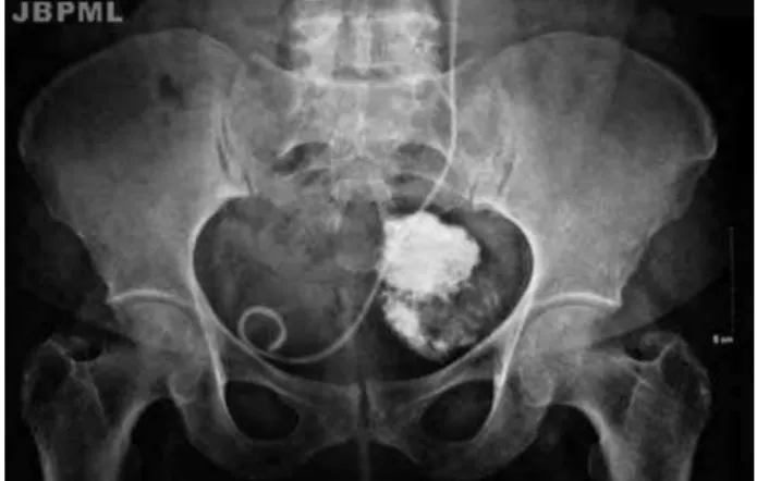
A 67 year old female patient sought the surgery service because of an incidental inding of calciied mass in the left adnexal region. She had a past history of urolithiasis diagnosed two years before when an abdominal radiograph suggested the presence of calciied leiomyomas. By that time, a double-J ureteral catheter was placed in the left side. After a year and a half, she started complaining of dysuria, urinary incontinence and nocturia. A pelvis radiograph performed six months before the current admission was also indicative of calciied leiomyomas (Figure 1). A magnetic resonance (MR)was performed two months before admission, revealing a heterogeneous mass in left adnexal region suggestive of an ovarian tumor. The tumor had a bilobated appearance with a densely calciied posterior lobe and an anterior lobe with lower density (Figure 2). Serum tests of tumor markers one month before admission
indicated non elevated levels of CA-125, human chorionic gonadotropin, α-fetoprotein, carcinoembryonic antigen and lactate dehydrogenase.
The patient had a long term history of systemic arterial hypertension and dyslipemia. Her obstetric history includes 10 pregnancies and ten deliveries (with two cesarean operations). The last surgical obstetric delivery occurred 35
Figure 1 – Radiograph of the pelvis. A calcified mass with ovoid shape in left pelvis
years ago. She also reported a previous surgical procedure for uterine leiomyoma removal 10 years ago.
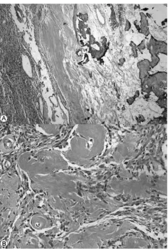
At surgery, the tumor was unrelated to left ovary and uterine tube which exhibited normal appearance. The tumor emerged from the retro-rectal adipose tissue and broad ligament. At macroscopy, it was a 9 x 4.5 x 4.5 cm well delineated tumor with bilobated appearance. The external surface had a yellowish appearance. At cut surface, one lobe had a 2 cm cavity with yellowish serous content. The cut surface had a soft grayish glistening center while the periphery appeared grayish-white, irm and densely calciied. Microscopically, the tumor was mainly composed by hypercellular population of immature stromal cells. The classic zonation phenomenon described in myositis ossiicans was easily observed with orderly progression of osteoid formation to mature bone from the center to tumor periphery (Figure 3). Stromal cells including osteoblasts lacked cytologic features of malignancy. Chronic inlammation was minimal. Foreing body or granulomas were not observed.
The patient reported relief of the urinary symptoms three months after surgery. No recurrence was detected during a 12-month period of follow-up.
Discussion
Awareness on the existence of HMO/IMO is important to avoid an erroneous diagnosis of extraskeletal osteosarcoma, which is its most important morphologic mimic(11, 13).
Typical zonation of stromal cell proliferation, immature osteoid and mature bone formation, and lack of cytologic atypia are important clues for the identiication of HMO/ IMO(9). In addition, the rare well differentiated extraskeletal
osteosarcoma will most probably exhibit a reverse zonation effect: immature woven bone or osteoid in the tumor periphery while mature osseous trabeculae are centrally located(4).
Alertness to HMO/IMO increased since 1999 when Wilson et al. reported ive similar cases and reviewed previous reports. The authors found only two similar cases in previous reports(7, 12) and suggested that this
entity should not be confounded with previous reported of well-documented ossificafion of laparotomy scars or heterotopic bone formation in metastatic colonic carcinoma(11). Since them, 21 new cases have been
reported in English literature.
HO is a nonspecific term used to describe bone formation in tissues that normally do not ossify. It occurs in soft tissue locations associated with trauma, burns, prolonged immobilization, prior surgery, and neoplasia. HO is distinct from dystrophic calciication (DC), which refers to the deposition of amorphous calcium salts in the absence of osteoblastic activity. The origin of HO is not known, although it has been postulated to be due to osteoblastic metaplasia of multipotent mesenchymal cells in response to trauma, or occasionally by traumatic or surgical implantation of bone or periosteum into the soft tissues(8).
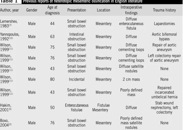
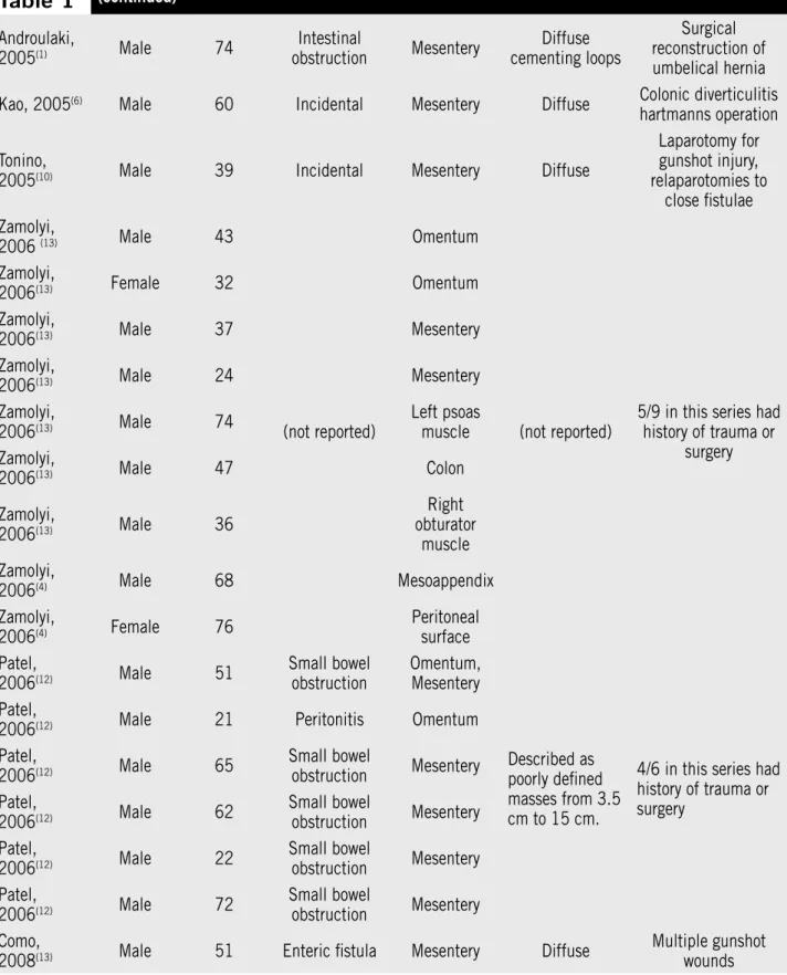
Table 1 summarizes the available information of all cases of HMO/IMO in English literature. The mean ± SD age at the diagnosis is 53.8 ± 18.7 years. The process is strongly associated with male gender with 26 reports to date from men and only two, similar to our case, occurring in women. These features suggest an even higher male predominance when compared to the soft tissue counterpart (which is 3:2) and a concentration of cases at an older age group (mean age of soft tissue myositis ossiicans is 32 years)(9). Figure 3 – Microscopy appearance. (A) Classic zonation phenomenon described in
Most cases of IMO present a small bowel obstruction and only three among 28 cases were incidental indings, mirroring the present case. A history of trauma or previous surgery was positive in 18 of 27 cases (67% of all). Only one case is described as a well deined lesion such as in the present case, and this case had no known history of trauma(11). In the present case, the long period between
pelvic surgeries and the incidental detection of the lesion due to imaging tests may have been responsible for the organization of this reactive process that seems to be diffuse soon after triggering traumatic events. Unfortunately, we could not locate the previous material from the reported myomectomy 10 years ago to verify if the same process could be already present at that moment.
It is common to ind hemorrhage or gelatinous material in the center of soft tissue myositis ossiicans, and some cases may indeed present cystic changes with yellowish clear luid paralleling the observation of the present case(4).
In contrast to a single report that described elevated serum levels of CA-125 associated with a HMO/IMO tumor with similar dimensions (9 x 3.5 cm)(2), we did not observe
abnormal levels of that and other tumor markers.
In conclusion, we report a rare case of HMO/IMO involving pelvis and leading to differential diagnosis with leiomyomas and ovarian tumors. Awareness on this reactive process will prevent pathologist to misdiagnosis such lesions as sarcomatous process, such as extraskeletal osteosarcoma. This case is clearly different from most cases of HMO/IMO since is the third reported in a female patient and lacks the diffuse mesenteric involvement leading to obstructive symptoms. In addition, past trauma/surgical events are too distant to be obviously implicated in tumor emergence. Alternatively, such an insidious course could lead to the organization of this reactive process into a silent isolated pelvic mass.
Acknowledgements
This work was approved by ethics committee of Hospital Espanhol and the patient gave written consent for publication of this case report (September 22, 2008 – Register Number 026/2008).
Table 1
Previous reports of heterotopic mesenteric ossification in English literature
Author, year Gender Age at
diagnosis Presentation Location
Intraoperative
findings Trauma history
Lemershev,
1983(7) Male 44
Small bowel obstruction Mesentery Diffuse enterocutaneous fistula Laparotomies Yannopoulos,
1992(12) Male 63
Intestinal
obstruction Mesentery Diffuse
Aortic bifemoral bypass Wilson,
1999(11) Male 75
Small bowel
obstruction Mesentery
Diffuse cementing loops
Repair of aortic aneurysm Wilson,
1999(11) Male 76
Small bowel
obstruction Mesentery
Diffuse cementing loops
Left colectomy repair of aortic aneurysm Wilson,
1999(11) Male 43
Small bowel
obstruction Mesentery
Diffuse satellite
nodules None Wilson,
1999(11) Male 80 Incidental Mesentery 2 cm mass None
Wilson,
1999(11) Male 43
Small bowel obstruction Mesentery Poorly defined mass Repaired incarcerated umbelical hernia Hakim,
2001(5) Male 50
Enterocutaneous fistulae Fistulae Mesentery Diffuse Stab wound nephrectomy, left colectomy Bovo,
2004(2) Male 76
Table 1
(continued)
Androulaki,
2005(1) Male 74
Intestinal
obstruction Mesentery
Diffuse cementing loops
Surgical reconstruction of umbelical hernia
Kao, 2005(6) Male 60 Incidental Mesentery Diffuse Colonic diverticulitis
hartmanns operation
Tonino,
2005(10) Male 39 Incidental Mesentery Diffuse
Laparotomy for gunshot injury, relaparotomies to
close fistulae Zamolyi,
2006 (13) Male 43
(not reported)
Omentum
(not reported)
5/9 in this series had history of trauma or
surgery Zamolyi,
2006(13) Female 32 Omentum
Zamolyi,
2006(13) Male 37 Mesentery
Zamolyi,
2006(13) Male 24 Mesentery
Zamolyi,
2006(13) Male 74
Left psoas muscle Zamolyi,
2006(13) Male 47 Colon
Zamolyi,
2006(13) Male 36
Right obturator
muscle Zamolyi,
2006(4) Male 68 Mesoappendix
Zamolyi,
2006(4) Female 76
Peritoneal surface Patel,
2006(12) Male 51
Small bowel obstruction
Omentum, Mesentery
Described as poorly defined masses from 3.5 cm to 15 cm.
4/6 in this series had history of trauma or surgery
Patel,
2006(12) Male 21 Peritonitis Omentum
Patel,
2006(12) Male 65
Small bowel
obstruction Mesentery Patel,
2006(12) Male 62
Small bowel
obstruction Mesentery Patel,
2006(12) Male 22
Small bowel
obstruction Mesentery Patel,
2006(12) Male 72
Small bowel
obstruction Mesentery Como,
2008(13) Male 51 Enteric fistula Mesentery Diffuse
1. ANDROULAKI, A. et al. Heterotopic mesenteric ossification: a rare reactive process. J Gastroenterol Hepatol, v. 20, n. 4, p. 664-6, 2005.
2. BOVO, G. et al. Heterotopic mesenteric ossification (“intraabdominal myositis ossificans): a case report. Int J Surg Pathol, v. 12, n. 4, p. 407-9, 2004.
3. COMO, J. J. et al. Extensive heterotopic mesenteric ossification after penetrating abdominal trauma. J Trauma, v. 65, n. 6, p. 1567, 2008.
4. FOREST, M. Myositis Ossificans. In: FOREST, M.; TOMENO, B.; VANEL, D. (eds.). Orthopedic surgical pathology. Edinburgh: Churchill Livingstone, 1998. p. 663-70. 5. HAKIM, M.et al. Heterotopic mesenteric ossification. AJR
Am J Roentgenol, v. 176, n. 1, p. 260-1, 2001. 6. KAO, H. W. et al. Imaging features of heterotopic mesenteric
ossification: a case report and literature review. Chinese Journal of Radiology, v. 30, p. 55-8, 2005.
7. LEMESHEV, Y. et al. Heterotopic bone formation associated with intestinal obstruction and small bowel resection.
Ala J Med Sci, v. 20, n. 3, p. 314-7, 1983.
Mailing Address
Daniel Athanazio
Universidade Federal da Bahia / Depto. de Biointeração Av. Reitor Miguel Calmon, s/n – Campus do Canela CEP 40110-100 – Salvador-BA
Tel: (71) 3245-8602 Fax: (71) 3240-4194 e-mail: [email protected]
References
8. PATEL, R. M. et al. Heterotopic mesenteric ossification: a distinctive pseudosarcoma commonly associated with intestinal obstruction. Am J Surg Pathol, v. 30, n. 1, p. 119-22, 2006.
9. ROSENBERG, A. E. Myositis ossificans and fibroosseus pseudotumour of digits. In: Fletcher, C. D. M.;
UNNI, K. K.; MERTENS, F. (eds.) Tumours of
Soft tissue and bone. Lyon: IARCPress, 2002. p. 52-4.
10. TONINO, B. A. et al. Heterotropic mesenteric ossification: a case report (2004:10b). Eur Radiol, n. 15, v. 1, p. 195-7, 2005.
11. WILSON, J. D. et al. Heterotopic mesenteric ossification (intraabdominal myositis ossificans): report of five cases.
Am J Surg Pathol, n. 23, v. 12, p. 1464-70, 1999. 12. YANNOPOULOS, K. et al. Mesenteritis ossificans. Am J
Gastroenterol, n. 87, v. 2, p. 230-3, 1992.