Effect of alloxan-induced diabetes
mellitus
and ethanol on
pregnancy outcome in mice
Efeito do diabetes
mellitus
induzido por aloxana e etanol na gestação de camundongos
Luiz Cesar Peres1; Camila Nunes de Morais Ribeiro2; Cristiane Minot Gutierrez3; Milton Cesar Foss4
Introduction and objectives: To investigate the effects of ethanol, diabetes mellitus and the combination
of both on mouse fetuses. Methods: We used 24 female Swiss mice, dividing them into four groups
of 6 each: control (C), ethanol (E), diabetes (D) (blood glucose > 200 mg/dL) and diabetes + ethanol (DE). Diabetes was induced by alloxan (40 mg/kg) on day 7 of pregnancy. Groups E and DE received 4 g/kg of 25% v/v ethanol intraperitoneally, whereas groups C and D received saline. On day 18, all fetuses were harvested. Results: In group DE the following anomalies were found: exencephaly, situs inversus totalis, situs inversus partialis, eyelid skin tag and one animal from group E had pulmonary artery hypoplasia. Ethanol administration partially reverted diabetes-fetal resorption caused by diabetes, yet it induced late fetal death. Both diabetes and ethanol reduced placental diameter and increased its weight. Ethanol had more effect on fetal length in males than in females, however, such bias was not found for diabetes. Ethanol prevented diabetes-induced tail shortening in both genders. Conclusions: These results show that, although ethanol might improve energy metabolism in early gestation, it causes cell damage that leads to cardiovascular, limb and neural tube defects, late fetal death and reduced placental size.
abstract
key words
Diabetes mellitus
Ethanol
Neural tube defects
Fetus mice
resumo
Introdução e objetivos: Investigar o efeito do etanol, do diabetes mellitus (DM) e da associação de ambos sobre os fetos de camundongo. Métodos: Foram utilizadas 24 fêmeas de camundongos Swiss divididas em quatro grupos de seis animais cada: controle (C); etanol (E); diabetes (D) (glicemia > 200 mg/dl), e diabetes + etanol (DE). O diabetes foi induzido pela aloxana (40 mg/kg) no dia 7 da gestação. Os animais dos grupos E e DE receberam 4 g/kg de solução a 25% v/v de etanol intraperitoneal (IP), enquanto os animais dos grupos C e D receberam salina. No dia 18, todos os fetos foram coletados. Resultados: Foram encontradas as seguintes anomalias no grupo DE: exencefalia, situs inversus totalis, situs inversus partialis e apêndice cutâneo palpebral. Um animal do grupo E apresentou hipoplasia da artéria pulmonar. A administração de etanol reverteu parcialmente a reabsorção fetal induzida pelo diabetes, porém aumentou a morte fetal tardia. Ambos, diabetes e etanol, reduziram o diâmetro placentário e aumentaram o seu peso. O etanol teve mais efeito no comprimento de fetos machos, contudo isso não ocorreu com o diabetes. O etanol preveniu a redução da cauda induzida pelo diabetes em ambos os sexos. Conclusão: Esses resultados indicam que, embora o etanol possa melhorar o metabolismo energético no início da gestação, ele causa lesão celular que leva a defeitos cardiovasculares, dos membros e do tubo neural, além de morte fetal tardia e redução do tamanho da placenta.
unitermos
Diabetes
Defeitos do tubo neural
Feto
Camundongo
Primeira submissão em 09/09/09
Última submissão em 09/09/09
Aceito para publicação em 02/12/09
Publicado em 20/10/09
1. Livre-docente; histopatologista pediátrico pelo Department of Histopathology, Shefield Children’s Hospital, Shefield, UK. 2. Doutora em Patologia Experimental; professora doutora e coordenadora do curso de Biomedicina da
Universidade Tuiuti do Paraná (UTP).
3. Doutora em Patologia Experimental; pós-doutoranda do Departamento de Ginecologia e Obstetrícia da Faculdade de Medicina de Ribeirão Preto da Universidade de São Paulo (FMRP/USP). 4. Professor titular de Clínica Médica (Endocrinologia) do Departamento de Clínica Médica da FMRP/USP.
Introduction
Major congenital anomalies are found in 2% to 3% of liveborn infants. The prevalence of such anomalies has been increasing in parallel with the decline in infant mortality rates, which is related to the number of diseases that are currently preventable. Among the congenital anomalies classiied as isolated defects, those affecting the central nervous system are second only to cardiovascular anomalies(30).
Periconceptional folic acid supplementation has been shown to reduce signiicantly the irst occurrence and recurrence of neural tube defects (NTDs) in humans, pointing to its role in the pathogenesis of this condition. However, in experimental animals, a variety of environmental agents have been implicated, including ethanol(10, 28), inhibition
of cholesterol synthesis(6, 10), retinoic acid(45), vitamin A(23),
hyperglycemia(12, 43), hypoxia(2, 13), ionizing radiation/
hyperthermia(30) and nicotine(18, 24).
It is well recognized that babies born to diabetic mothers are 3 to 4 times more prone to present congenital anomalies than is the general population(21, 34). Such anomalies have
been correlated with ineficient blood glucose control(34, 43) which may induce diabetic embryopathy, which
encompasses NTDs and other system defects(12, 16).
Ethanol, which is widely consumed worldwide, has been implicated in the etiology of NTDs and of heart and craniofacial defects(27, 40). According to 1997 data
provided by the United States Centers for Disease Control and Prevention(3), approximately 50% of all women of
childbearing age drink alcoholic beverages. Additionally, 60% of pregnant women may not realize they are pregnant until the fourth week of gestation(13, 28).
Animal models of ethanol-induced congenital anomalies have provided invaluable data regarding critical developmental periods and pathogenesis(37). Administering
a dose of ethanol corresponding to the limit of human tolerance to C57B/6J mice during gastrulation has been shown to result in craniofacial defects, including midfacial hypoplasia and prosencephalic anomalies(38).
Recently, Padmanabhan and Shaiullah(28) demonstrated
an increase in resorptions, intrauterine growth restriction and congenital anomalies in the litters of TO mice treated with streptozotocin-induced diabetes mellitus receiving a single dose of ethanol on gestational day 7 or 8.
The combination of experimental maternal diabetes mellitus and ethanol has received little attention. Therefore,
it is important to determine how these two common environmental teratogenic factors interact. This approach is relevant since diabetes mellitus is quite common(15, 28)
and ethanol is widely consumed by young women of reproductive age(5, 28).
Material and methods
This study was approved by the University of São Paulo Committee on Ethics in Animal Research (Process no 010/2004).
A total of 24 young nulliparous female Swiss mice weighing 40 to 50 g were used. The animals were housed in appropriate individual plastic cages in a 22oC, 12-h
light/12-h dark environment and given ad libitum access to tap water and commercial mouse chow throughout the experiment.
Alloxan (40 mg/kg) was injected into the dorsal vein of the tail of 12 randomly-selected mice. The remaining 12 mice received similar injections of saline. The diabetic mice were treated with subcutaneous injection of 0.25 IU of insulin (10 ml of Humulin® and 100 U/ml of neutral
protamine hagedorn (NPH) human insulin; Eli Lilly, Indianapolis, IN, USA) until mating. Beginning at four days after the induction of diabetes, ive females were caged together with 1 male for two hours daily, from 9:00 to 11:00 am. Day 0 of pregnancy was determined by the presence of a vaginal plug. Pregnant mice were divided into four groups of 6 mice each: control (C); ethanol (E); diabetes (D) and diabetes plus ethanol (DE). Diabetic mice were considered those which presented blood glucose of over 200 mg/dl.
On day 7 of pregnancy, the mice in groups E and DE received an intraperitoneal injection of 25% ethanol (4 g/kg) in saline (v/v) and those in groups C and D received the same volume of saline.
Blood glucose from the tail was measured after a fasting time of 12 hours one day before treatment with alloxan or saline, three days after treatment and on the day of sacriice using a MediSense Optium blood glucose meter (Abbott Laboratories, Abbott Park, IL, USA) and MediSense Optium blood glucose test strips (MediSense UK Ltd, Cambridge, England, UK).
were recorded: gender; body weight, crown-rump length and tail length. Placentas were also weighed and its diameter measured. Fetal weight and measurements were considered only for live fetuses. The number of resorptions (representing early fetal death), late fetal deaths (considered those fetuses that have completed their development and were in a process of degeneration), normal live fetuses and fetuses with NTD, as well as the total number of all anomalies, were recorded. Implants were deined as the sum of all resorptions, late fetal deaths and live fetuses with or without any anomaly. After ixation in buffered formalin, all fetuses were submitted to autopsy according to Sterz and Lehmann in situ sectioning method(35) using a
stereomicroscope coupled to a digital camera.
All data were statistically analyzed using the GraphPad Prism 4.0 program for analysis of variance (ANOVA), the Kruskal-Wallis test, the Mann Whitney test and the Cytel StatXact® 7 program for the Fisher-Freeman-Halton test.
Values of p < 0.05 were considered statistically signiicant.
Results
There was no difference in blood glucose level among the groups before treatment. After alloxan administration, however, blood sugar levels characteristic of diabetes were observed in groups D and DE (Figure 1).
Blood glucose level
Before treatment 3 days after treatment At sacrifice
460
400
360
300
260
C E D DE C E D DE C E D DE
200
mg/dL
160
100
60
0
Figure 1 – Blood glucose level in mg/dl in the different groups and in different moments
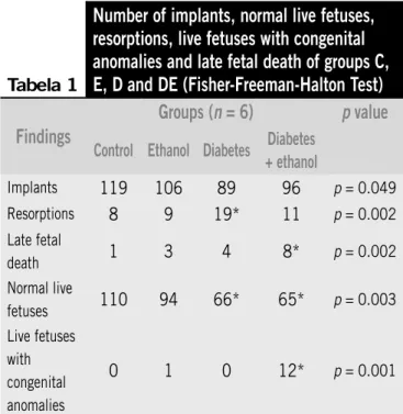
There was no difference among the groups in terms of the number of implants. However, although the number of normal live fetuses did not differ between groups C (110) and E (94), there was a difference between these two groups and groups D and DE (Table 1).
Tabela 1
Number of implants, normal live fetuses,
resorptions, live fetuses with congenital
anomalies and late fetal death of groups C,
E, D and DE (Fisher-Freeman-Halton Test)
Findings
Groups (
n
= 6)
p
value
Control Ethanol Diabetes Diabetes + ethanol
Implants 119 106 89 96 p = 0.049
Resorptions 8 9 19* 11 p = 0.002
Late fetal
death 1 3 4 8* p = 0.002
Normal live
fetuses 110 94 66* 65* p = 0.003
Live fetuses with congenital anomalies
0 1 0 12* p = 0.001
*Statistically different from control.
Resorption, the result of early fetal death, was signiicantly higher in group D animals, whereas late fetal death was higher in group DE ones (Table 1).
There were less normal live fetuses in groups D and DE and congenital anomalies were conined to groups DE and E, which received ethanol (Table 1). The congenital anomalies found were exencephaly (nine fetuses from group DE) (Figure 2A, Figure 2B is a control fetus), cutaneous tag over the eyelid (1 fetus from group DE) (Figures 3A and 3B), situs inversus totalis (Figures 4A and 4B) and situs inversus partialis (Figures 5A and 5B) (1fetus each from group DE) and pulmonary artery hypoplasia (one fetus from group E) (Figures 6A and 6B).
Figure 6 – Photograph of the autopsy of a group E fetus. Note the severe hypoplasia of the pulmonary artery (PA) compared to the aorta in A. Histologic section at the level of the base of the heart depicts the hypoplastic pulmonary artery (PA) close to the normal sized aorta (Ao) in B (hematoxylin and eosin, 50x original magniication)
RA: right atrium, LA: left atrium.
Figure 3 – A cutaneous appendage on the right eyelid is seen in one fetus of group DE (A and B)
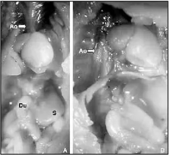
Figure 4 – Macroscopic pictures of the other anomalies found in a group DE fetus with situs inversus totalis. Note in A the apex of the heart turned to the right. After trimming off the lungs and displacement of the heart to the left, one can clearly see the aorta running along the right side of the vertebral column (arrow) in B. The tube running above the column is the esophagus (asterisk)
A gender-based difference was found in terms of fetal length. Male crown-rump length was reduced in animals from groups E, D and DE, whereas female fetal crown-rump length
was decreased only in groups D and DE. Male fetuses of group DE presented less crown-rump reduction than female ones and the most severely affected group of all was group D (Table 2).
Tabela 2
Median fetal crown-rump length of Swiss mouse fetuses recovered on gestational
day 18 from dams which received saline (control), ethanol 4 mg/kg (ethanol),
alloxan-induced diabetes + saline (diabetes) and alloxan-induced diabetes + ethanol
4 mg/kg on gestational days 7, 8 and 9 (One way ANOVA, Kruskal-Wallis Test)
Control
Ethanol
Diabetes
Diabetes + ethanol
n
n
n
n
Crown-rump length
(male + female) (cm) 2.5 110 2.4 94 2 74 2.2 74
Male crown-rump
length (cm) 2.5 56 2.4 49 2 38 2.2 29
Female crown-rump
length (cm) 2.5 54 2.4 45 2 36 2.1 45
Dunns multiple comparison test for fetal crown-rump length
Male + female
Male
Female
Control vs. ethanol p < 0.05 p < 0.001 p > 0.05
Control vs. diabetes p < 0.001 p < 0.001 p < 0.001
Control vs. diabetes ethanol p > 0.05 p < 0.001 p < 0.001
Ethanol vs. diabetes p < 0.001 p < 0.001 p < 0.001
Ethanol vs. diabetes ethanol p > 0.05 p > 0.05 p < 0.01
Diabetes vs. diabetes ethanol p < 0.001 p > 0.05 p > 0.05
Fetal body weight was equally decreased in groups E, D and DE for both sexes and this inding correlated with IUGR (Table 3).
Tail length reduction was observed in both male and female fetuses from groups D and DE, although less intensely in the latter (Table 4).
Placental diameter was reduced in groups E, D and DE whereas placental weight was increased in groups E and DE (Table 5).
Discussion
The reproductive outcome is affected by different conditions that can be endogenous or exogenous. Maternal diabetes is a well known metabolic endogenous factor associated with poor pregnancy outcome, inducing
congenital anomalies. Ethanol, on the other hand, is a common exogenous offender, recognized by its many deleterious actions on the developing fetus. When associated, diabetes and ethanol are expected do induce more harmful effects due to their synergistic action.
In the present study, Swiss mice became diabetic after alloxan administration before mating but were supplemented with insulin until they became pregnant. This approach was effective since we avoided any inluence of alloxan on the fetuses at the same time fertility was kept unaltered with insulin treatment, which is relected by the absent difference regarding implant number among all groups.
Tabela 3
Median fetal body weight and percentage of intrauterine growth restriction
(IUGR) of Swiss mouse fetuses recovered on gestational day 18 from dams
which received saline (control), ethanol 4 mg/kg (ethanol), alloxan-induced
diabetes + saline (diabetes) and alloxan-induced diabetes + ethanol 4 mg/kg on
gestational days 7, 8 and 9 (One way ANOVA, Kruskal-Wallis Test)
Control
Ethanol
Diabetes
Diabetes + ethanol
n
n
n
n
Body weight (male +
female) (g) 1.55 110 1.31 94 0.99 74 1.2 60
IUGR (male + female) -2
SD (%) 1.82 110 13.8 94 68.9 74 43.3 60
Male weight (g) 1.59 56 1.27 49 1.08 38 1.22 29
IUGR (male) -2 SD (%) 1.75 56 40.8 49 74.5 38 68.9 29
Female crown-rump
length (cm) 1.47 54 1.37 45 0.96 36 1.15 46
IUGR (female) -2 SD (%) 1.85 54 8.8 45 61.1 36 37.7 46
SD: standard deviation.
Dunns multiple comparison test for fetal crown-rump length
Male + female
Male
Female
Control vs. ethanol p < 0.001 p < 0.001 p < 0.01
Control vs diabetes p < 0.001 p < 0.001 p < 0.001
Control vs. diabetes ethanol p < 0.001 p < 0.001 p < 0.001
Ethanol vs. diabetes p < 0.001 p < 0.001 p < 0.001
Ethanol vs. diabetes ethanol p < 0.001 p < 0.001 p < 0.01
Diabetes vs. diabetes ethanol p > 0.05 p > 0.05 p > 0.05
increased late fetal death in group DE. It may be speculated that ethanol provides enhanced caloric availability in early gestation and thus prevents early fetal death. This may also be the explanation why IUGR, fetal body weight, crown-rump length and tail length were less severely affected in group DE than in group D.
The mechanism involved in IUGR seen with both diabetes and ethanol is the production of reactive oxygen species and free radicals(28), which damage tissues,
inducing apoptosis. It is postulated that the oxidative stress, possibly more intense with ethanol administration, may induce late fetal death and congenital anomalies. In fact, this was observed in the present study, since congenital anomalies were only observed in groups which received ethanol, either alone, group E or associated with diabetes, group DE, and late fetal death was higher in the latter. Additionally, cases of NTD were found only in
the diabetic animals receiving ethanol, indicating that the combination of both factors is essential. This inding can be interpreted in the light of epidemiological studies which have found an increased risk for major systemic anomalies in human maternal diabetes without an overall incidence of anomalies, suggesting that diabetes is not a primary teratogen but acts by promoting an initial insult and potentializing the detrimental effects of other teratogens(22). The reverse has also been proposed, i.e.,
that ethanol potentiates the effects of diabetes or that anomalies are attributable to the interaction between ethanol and diabetes-induced metabolic disorders(19).
Apoptosis and decreased migration and differentiation of neural crest cells(4) may be implicated in NTD. Therefore,
Tabela 4
Median fetal tail length of Swiss mouse fetuses recovered on gestational day 18
from dams which received saline (control), ethanol 4 mg/kg (ethanol),
alloxan-induced diabetes + saline (diabetes) and alloxan-alloxan-induced diabetes + ethanol
4 mg/kg on gestational days 7, 8 and 9 (One way ANOVA, Kruskal-Wallis Test)
Control
Ethanol
Diabetes
Diabetes + ethanol
n
n
n
n
Tail length (male +
female) (cm) 1.3 110 1.3 94 1.2 74 1.3 74
Male tail length (cm) 1.3 56 1.3 49 1.15 38 1.3 29
Female tail length
(cm) 1.3 54 1.3 45 1.2 36 1.2 45
Dunns multiple comparison test for fetal tail length
Male + female
Male
Female
Control vs. ethanol p > 0.05 p > 0.05 p > 0.05
Control vs. diabetes p < 0.001 p < 0.001 p < 0.001
Control vs. diabetes ethanol p > 0.05 p > 0.05 p > 0.05
Ethanol vs. diabetes p < 0.001 p < 0.001 p < 0.001
Ethanol vs. diabetes ethanol p > 0.05 p > 0.05 p > 0.05
Diabetes vs. diabetes ethanol p < 0.001 p < 0.001 p < 0.05
Tabela 5
Median placental weight and median placental diameter of Swiss mouse fetuses recovered
on gestational day 18 from dams which received saline (control), ethanol 4.0 mg/kg
(ethanol), alloxan-induced diabetes + saline (diabetes) and alloxan-induced diabetes +
ethanol 4 mg/kg on gestational days 7, 8 and 9 (One way ANOVA, Kruskal-Wallis Test)
Control
Ethanol
Diabetes
Diabetes + ethanol
n
n
n
n
Placental
weight (g) 0.107 110 0.12 94 0.11 74 0.111 74
Placental
diameter (cm) 0.8 110 0.7 94 0.5 74 0.6 74
Dunns multiple comparison test for placental weight and diameter
Placental weight
Placental diameter
Control vs. ethanol p < 0.001 p < 0.001
Control vs. diabetes p > 0.05 p < 0.001
Control vs. diabetes ethanol p < 0.01 p < 0.001
Ethanol vs. diabetes p > 0.05 p < 0.001
Ethanol vs. diabetes ethanol p > 0.05 p > 0.05
Swiss mouse seems to be less affected by diabetes and/or ethanol than do other strains, such as TO(28) and C57BL/6J(39).
The latter is particularly prone to the teratogenic effects of ethanol, resulting in the facial dysmorphisms seen in the fetal alcohol syndrome (FAS)(39), which were not found in
the swiss mouse fetuses. However, it is not possible to assure that in the diabetic group not receiving ethanol there were no NTDs or other major defects.
One of the fetuses of group E presented pulmonary artery hypoplasia. This inding may be interpreted in the light of the studies that present congenital heart disease as part of FAS, what has already been demonstrated in animal models as well(5).
Situs inversus, totalis or partialis, a congenital defect characterized by the mirror image of viscera, is found in humans(32, 41) and animals(8, 42). This defect can be
accompanied by other anomalies(20, 32) and is 84 times
more common in infants born to diabetic women(14). Situs
inversus is rare in mouse strains, including C57Bl/6J(25).
Recently, a transgenic mouse strain (inv/inv mice) was reported, in which almost 100% of the homozygous individuals were characterized by situs inversus totalis as a consequence of insertional mutagenesis of the inversin gene(44). Therefore, the cases occurring in our study were
probably related to diabetes although it is not possible to exclude any participation of the ethanol since it was found in group DE. In the same group, we found a case of a cutaneous appendage on the eyelid. No such defect has been reported previously, although there is evidence that ethanol interferes with ocular development, inducing anophthalmia, microphthalmia, hypo- and hypertelorism and cyclopia(33, 38).
IUGR was observed in all experimental groups, with increasing frequency in groups E, DE and D. The results of clinical studies have demonstrated that the development of embryos of diabetic mothers is impaired, and that growth restriction is a risk factor for congenital anomalies(29). Lin et al.(19) showed that rat infants born
to diabetic dams receiving or not receiving ethanol weighed less than did those born to dams receiving ethanol or receiving no treatment. Although ours was a mouse model, this is in agreement with our indings. Those authors stated that fetal growth is accelerated after gestational day 11 in the rat, and that using a single dose prior to that moment, as was done in the present study, would therefore have a less intense effect at the end of gestation. In addition, Padmanabhan and Shaiullah(28)
showed that the IUGR induced by ethanol injection on
gestational day 8 in a mouse model of diabetes was less intense than that caused by ethanol injection on day 7, indicating that the effect is gestational stage-dependent.
In group E, fetal weight of male and female fetuses was lower than that seen in group C. This inding is in accordance with those previously described(11, 36).
Diabetes also induced IUGR that was more pronounced than that induced by ethanol.
We found that diabetes impaired tail growth, although the same was not found for ethanol. Once again, ethanol was found to block further reductions in tail length in male and female infant mice born to diabetic dams, possibly because of the higher calorie provided by ethanol.
Although the combination of ethanol and diabetes induced less IUGR and tail growth, it was the cause of the NTDs and other major anomalies seen. Therefore, the net result is decidedly negative.
Contrary to diabetes, which affects both genders in a similar way, ethanol affects male fetuses crown-rump length more intensely than female ones.
The inluence of ethanol alone or in association with diabetes on the placenta was more marked, resulting in a heavier placenta. The correlation between IUGR and loss of placental size might constitute a cause and effect relationship or might be the net result of the deleterious effects that both diabetes and ethanol have on the fetuses and placentas simultaneously. In humans, the placenta is usually larger and heavier in diabetic women, possibly related to the chronically reduced blood low(17) caused
by diabetes-induced systemic hypertension, which in turn might be linked to the diabetic embryopathy(31).
Although we have not tested this hypothesis, it is possible that an abnormal uterine environment is the cause of the placental size reduction seen in our animals.
The perinatal deaths observed in the present study indicate placental insuficiency, since the apparent cause was asphyxia, as it typically is in humans(9). In a rat model
of preeclampsia, diabetes-induced oxidative stress was the cause of the placental insuficiency(26). Akay and
Koçkaya(1) showed that ibrin deposition, inlammation,
ibroblast proliferation and synthesis of the extracellular matrix in the placentas of rats exposed to ethanol, that could explain the weight increase in groups E and DE placentas, together with the diabetes-related impairment of the inlux of nutrients from the maternal circulation(7),
explain at least in part the larger number of perinatal deaths observed in the diabetic groups, all of which presented reduction of placental size.
In summary, the present study shows that, although a single high dose of ethanol administered to Swiss mice with alloxan-induced diabetes mellitus on gestational day 7 has some favorable effects in terms of less IUGR and tail
reduction, it induces congenital anomalies, cardiovascular defects and NTDs in particular, reduction of the size and increase in the weight of the placenta and late fetal death. The synergism of the two teratogenic factors is a relevant issue and, despite interspecies differences, can serve as a warning of the risk that this combination poses to human population.
References
1. AKAY, M. T.; KOÇKAYA, E. A. The effects of alcohol on rat
placenta. Cell Biochem Funct, v. 23, p. 435-45, 2005.
2. BLOCH, J. R. antenatal events causing neonatal bain injury
in premature infants. JOGNN, v. 34, p. 358-66, 2005.
3. CENTERS FOR DISEASE CONTROL AND PREVENTION. Alcohol consumption among pregnant and
childbearing-aged women – United States, 1991 and 1995. MMWR,
v. 46, p. 345-50, 1997.
4. CHEN, S. Y.; SULIK, K. K. Iron-mediated free radical injury in
ethanol-exposed mouse neural crest cells. J Pharmacol
Exp Ther, v. 294, p. 134-40, 2000.
5. CHUDLEY, A. E. et al. Fetal alcohol spectrum disorder:
Canadian guidelines for diagnosis. CMAJ, v. 172, p.
S1-S20, 2005.
6. EDISON, R.; MUENKE, M. The interplay of genetic and environmental factors in craniofacial morphogenesis:
holoprosencephaly and the role of cholesterol. Congenit
Anom (Kyoto), v. 43, p. 1-21, 2003.
7. ERIKSSON, U. J.; JANSSON, L. Diabetes in pregnancy: decreased placental blood flow and disturbed fetal
development in the rat. Pediatr Res, v. 18, p. 735-8, 1984.
8. EVANS, H. E. Cyclopia, situs inversus and widely patent
ductus arteriosus in a new-born pig, Sus scrofa. Anat
Histol Embryol, v. 16, p. 221-6, 1987.
9. EVERS, I. M. et al. Placental pathology in women with type
1 diabetes and in a control group with normal and
large-for-gestational-age infants. Placenta, v. 24, p.
819-25, 2003.
10. GUIZZETTI, M.; COSTA, L. G. Disruption of cholesterol homeostasis in the developing brain as a potential mechanism contributing to the developmental
neurotoxicity of ethanol: a hypothesis. Med Hypot, v.
64, p. 563-7, 2005.
11. HANSON, J. W.; JONES K. L.; SMITH, D. W. Fetal alcohol
syndrome, experience with 41 patients. JAMA, v. 235,
p. 1458-60, 1976.
12. HERRERA, J. N.; HUIDOBRO, M. G.; OVALLE, L. C. Malformaciones congénitas en hijos de madres con
diabetes gestacional. Rev Méd Chile, v. 133, p.
547-54, 2005.
13. JOHNSTON, M. C.; BRONSKY, P. T. Prenatal craniofacial development: new insights on normal and abnormal
mechanisms. Crit Rev Oral Biol Med, v. 6, p. 368-422, 1995.
14. JOVANOVIC-PETERSON, L.; PETERSON, C. M. Abnormal Metabolism and the risk for birth defects with
emphasis on diabetes. Ann NY Acad Sci, v. 678, p.
228-43, 1993.
15. KALTER, H. Case reports of malformations associated with
maternal diabetes: history and critique. Clin Genet, v.
43, p. 174-9, 1993.
16. KANWAR, Y. S. et al. Hyperglycemia: its imminent effects
on mammalian nephrogenesis. Pediatr Nephrol, v. 20,
p. 858-66, 2005.
17. LANG, U. et al. Uterine blood low a determinant of fetal
growth. Eur J Obst Gynecol Reprod Biol, v. 110, suppl.
1, p. S55-S61, 2003.
18. LE FOLL, B.; GOLDBERG, S. R. Ethanol does not affect
discriminative-stimulus effects of nicotine in rats. Eur
J Pharmacol, v. 519, p. 96-102, 2005.
19. LIN, Y.; LEE, M.; LEICHTER, J. Interactive effects of alcohol
and diabetes during pregnancy on the rat fetus. Terat,
Carcinog and Mutag, v. 15, p. 147-53, 1995.
20. LOHSE, C. L.; SELCER, R. R.; SUTER, P. F.
Hepatoencephalopathy associated with situs inversus
of abdominal organs and vascular anomalies in a dog.
J Am Vet Med Assoc, v. 168, p. 681-8, 1976.
21. MARTÍNEZ-FRÍAS, M. L. et al. Epidemiological analysis of
outcomes of pregnancy in gestational diabetic mothers.
Am J Med Gen, v. 78, p. 140-5, 1998.
22. MCCARTER, R. J.; KESSLER, H.; COMSTOCK, G. W. Is
Diabetes mellitus a teratogen or a co-teratogen? Am J
Epidemiol, v. 125, p. 195-205, 1987.
23. MENDONÇA, E. D.; GUTIERREZ, C. M.; PERES, L. C. Brain tissue fragments in the amniotic luid of rats with
neural tube defect. Pathology, v. 37, p. 152-6, 2005.
24. Miller Jr, R. R. et al. Ethanol and nicotine induced
membrane changes in embryonic and neonatal chick
brains. Comp Biochem Physiol, part C, v. 130, p.
163-78, 2005.
25. MORISHIMA, M. et al. Inluence of genetic and maternal
diabetes in the pathogenesis of visceroatrial heterotaxy
in mice. Teratology, v. 54, p. 183-90, 1996.
26. NASH, P.; OLOVSSON, M.; ERIKSSON, U. J. Placental dysfunction in Suramin-treated rats: impact of maternal
diabetes and effects of antioxidative treatment. J Soc
27. NODEN, D. M. Fetal alcohol syndrome. BioAP475: Mechanisms Underlying Mammalian Developmental Defects, p. 1-12, 2002.
28. PADMANABHAN, R.; SHAFIULLAH, M. Effect of maternal diabetes and ethanol interactions on embryo
development in the mouse. Mol Cell Biochem, v. 261,
p. 43-56, 2004.
29. PEDERSEN, J. F.; MOLSTED-PEDERSON, L. Early fetal growth delay detected by ultrasound marks increased risk of congenital malformation in diabetic pregnancy.
Br Med J, v. 283, p. 269-71, 1981.
30. PERES, L. C. Review of pediatric autopsies performed at a
University Hospital in Ribeirão Preto, Brazil. Arch Pathol
Lab Med, v. 130, p. 62-8, 2006.
31. PUNAREEWATTANA, K. et al. Reduced birth defects
caused by maternal immune simulation in diabetic ICR mice: lack of correlation with placental gene expression.
Immunol Invest, v. 34, p. 71-89, 2005.
32. RAINES, K. H.; ARMSTRONG, B. E. Aortic atresia with
visceral situs inversus with mirror-image dextrocardia.
Pediatr Cardiol, v. 10, p. 232-5, 1989.
33. ROGERS, J. M. et al. Methanol exposure during
gastrulation causes holoprosencephaly, facial dysgenesis, and cervical vertebral malformations in
C57Bl/6J mice. Birth Defects Res (Part B), v. 71, p.
80-8, 2004.
34. SHEFFIELD, J. et al. Diabetes mellitus and infants
malformations. Obstet & Gynecol, v. 100, p. 925-30, 2002.
35. STERZ, H.; LEHMANN, H. A critical comparison of the freehand razor-blade dissection method according to
Wilson with an in situ sectioning method for rat fetuses.
Teratog Carcinog Mutagen, v. 5, p. 347-54, 1985.
36. STREISSGUT, A. P. et al. Teratogenic effects of alcohol
in human and laboratory animals. Science, v. 209, p.
353-61, 1980.
37. SULIK, K. K.; COOK, C. S.; WEBSTER, W. S. Teratogens and craniofacial malformations: relationships to cell
death. Development, v.103, suppl., p. 213-32, 1988.
38. SULIK, K. K.; LAUDER, J. M.; DEHART, D. B. brain malformations in prenatal mice following acute maternal
ethanol administration. Int J Dev Neurosci, v. 2, p.
203-14, 1984.
39. SULIK, K. K. Genesis of alcohol-induced craniofacial
dysmorphism. Exp Biol Med, v. 230, p. 366-75, 2005.
40. SZABO, K. T. Congenital malformations in laboratory and farm animals. Academic Press Inc., San Diego, California, USA, 1989.
41. VIJAYAKUMAR V.; BRANDT, T. Prolonged survival with
isolated levocardia and situs inversus. Cleve Clin J Med,
v. 58, p. 243-7, 1991.
42. VOGEL, O. Situs inversus in the cat. Berl Munch Tierarztl
Wochenschr, v. 92, p. 48-9, 1979.
43. WALKINSHAW, S. Pregnancy in women with pre-existing
diabetes: Management issues. Sem Fetal Neonat Med,
v. 10, p. 307-15, 2005.
44. YOKOYAMA, T. et al. Reversal of left-right asymmetry: a situs
inversus mutation. Science, v. 260, p. 679-82, 1993.
45. YU, J. et al. Effects of retinoic acid on the neural
crest-controlled organs of fetal rats. Pediatr Surg Int, v. 19,
p. 355-8, 2003.
Mailing adress Luiz Cesar Peres