ABSTRACT
Objective: To describe respiratory function in a series of patients with late-onset Pompe disease after the deinitive diagnosis and before enzyme replacement therapy. Methods: This was a cross-sectional study involving patients with a deinitive molecular diagnosis of late-onset Pompe disease. The data analyzed included age at symptom onset; age at deinitive diagnosis; type of initial symptoms; time from symptom onset to diagnosis; FVC in the sitting and supine positions; six-minute walk distance; and locomotor ability. Analyses were carried out using frequencies, medians, minimum values, and maximum values. Results: Six patients were included in the study. The median age at symptom onset was 15 years (range, 13-50 years), and the median age at diagnosis was 39.5 years (range, 10-64 years). The median time from symptom onset to diagnosis was 8 years (range, 0-45 years). In all cases, the initial manifestation of the disease had been motor weakness. The median FVC in percentage of the predicted value (FVC%) in the sitting and supine positions was 71.0% (range, 22.9-104.6%) and 58.0% (range, 10.9-106.9%), respectively. The median ΔFVC% was 24.5% (range, −4.59 to 52.40%).The median six-minute walk distance was 391.7 m (range, 97-702 m). Conclusions: In this case series, the time from symptom onset to diagnosis was long. Although respiratory signs or symptoms were not the initial manifestations of the disease, 66.7% of the patients showed reduced FVC% in the sitting and supine positions at diagnosis.
Keywords: Glycogen storage disease type II; Respiratory function tests; Respiratory muscles/pathology.
Respiratory manifestations in late-onset
Pompe disease: a case series conducted in
Brazil
Bruna de Souza Sixel1,2, Luanda Dias da Silva3, Nicolette Celani Cavalcanti4,
Glória Maria Cardoso de Andrade Penque5, Sandra Lisboa3,
Dafne Dain Gandelman Horovitz6, Juan Clinton Llerena Jr6
Correspondence to:
Bruna de Souza Sixel. Avenida Rui Barbosa, 716, Flamengo, CEP 22250-020, Rio de Janeiro, RJ, Brasil. Tel.: 55 21 2554-1768. E-mail: [email protected]
Financial support: None. INTRODUCTION
Pompe disease (PD), also known as glycogen storage disease type II, is an autosomal recessive hereditary disease caused by mutations in the gene encoding acid alpha-glucosidase, an enzyme that is responsible for the degradation of glycogen, especially at the muscle level.
Data on the incidence of PD are inaccurate, because of the rarity, underdiagnosis, and ethnic distribution of the disease. Data from the United States estimate that its overall incidence is approximately 1:40,000.(1) More
recent studies that have been conducted in Taiwan and Austria and are based on neonatal screening programs have found higher incidences of approximately 1:28,000. (2,3) In
Latin America, only 88 patients had been reported to have the disease by 2012.(4) Data from Brazil are unavailable.
PD is characterized by lysosomal accumulation of gly-cogen, especially in skeletal and cardiac striated muscles, beginning when acid alpha-glucosidase activity falls below the critical level of 30%. It is classiied as classic infantile PD, with symptom onset occurring before the irst year of life, accounting for approximately 28% of all cases; and as late-onset PD, when symptoms appear after that period, including children, youths, and adults. Disease progression in late-onset PD is slower than that in infantile PD, but it is quite variable. Clinical manifestations and disease severity vary according to age at symptom onset, rate of progression, and extent of organ involvement.(5-7)
1. Programa de Pós-Graduação em Pesquisa Aplicada à Saúde da Criança e da Mulher, Instituto Nacional de Saúde da Mulher, Criança e Adolescente Fernandes Figueira, Fundação Oswaldo
Cruz, Rio de Janeiro (RJ) Brasil. 2. Setor de Fisioterapia Respiratória,
Instituto Nacional de Saúde da Mulher, Criança e Adolescente Fernandes
Figueira, Fundação Oswaldo Cruz, Rio de Janeiro (RJ) Brasil. 3. Setor de Prova de Função Pulmonar,
Instituto Nacional de Saúde da Mulher, Criança e Adolescente Fernandes
Figueira, Fundação Oswaldo Cruz, Rio de Janeiro (RJ) Brasil. 4. Setor de Fisioterapia Motora, Instituto
Nacional de Saúde da Mulher, Criança e Adolescente Fernandes Figueira,
Fundação Oswaldo Cruz, Rio de Janeiro (RJ) Brasil.
5. Ambulatório de Doenças Musculares e
Neuroisiologia, Instituto de Neurologia Deolindo Couto, Universidade Federal do Rio de Janeiro, Rio de Janeiro (RJ) Brasil.
6. Centro de Genética Médica, Instituto Nacional de Saúde da Mulher, Criança e Adolescente Fernandes Figueira,
Fundação Oswaldo Cruz, Rio de Janeiro (RJ) Brasil.
Submitted: 20 December 2015. Accepted: 12 September 2016.
Until recently, treatment of PD was considered to be only palliative. In 2006, the commercial use of enzyme replacement therapy (ERT) with recombinant human alpha-glucosidase (alglucosidase alpha; Myozyme®, Genzyme, Cambridge, MA, USA) was approved in the USA and Europe, and, in 2007, it was also approved in Brazil. The treatment seems to improve respiratory and locomotor functions, as well as survival, in both forms of the disease.(8-10)
Muscle weakness is the major symptom in late-onset PD. The paravertebral and proximal lower limb muscles are usually the irst to be affected, making it dificult to perform activities of daily living and favoring postural changes.(5) The respiratory consequences of muscle
weakness result in restrictive lung disease, with a reduction in vital capacity accompanied by a reduction in FEV1. Initially, breathing is compromised only during sleep, but later on, hypoventilation will occur during the day as well. There is impairment in the cough mechanism and airway clearance, leading to recurrent respiratory infections. Respiratory dysfunction will occur in approximately 75% of patients.(11) Without
treatment, FVC is expected to decrease by 1.0% to 4.6% annually.(5,12,13) Respiratory failure is the major
cause of death.(14-16)
The predominance of diaphragmatic weakness over weakness of other respiratory muscles seems to be a characteristic of PD.(17,18) Therefore, the use of methods
capable of assessing the activity of the diaphragm alone can be useful in describing and monitoring the disease. Measurement of transdiaphragmatic pressure is the gold standard for the diagnosis of diaphragmatic dysfunction; however, other simpler methods, such as measurement of FVC in the supine position and difference between sitting and supine FVC have been described and recommended for the clinical follow-up of PD.(16,19-21)
At present, little is known about pulmonary manifes-tations in patients with PD in Brazil, which contributes to the management dificulty in this population. The objective of the present study was to characterize the proile of patients with PD and to describe respiratory function in a series of such patients followed at a referral center for rare diseases in Brazil, after the deinitive diagnosis and before ERT.
METHODS
This was a cross-sectional study involving data obtained from the medical records of patients with a deinitive diagnosis of late-onset PD and followed at the Center for Medical Genetics of the Fernandes Figueira National Institute for Women’s, Children’s, and Adolescents’ Health, Fundação Oswaldo Cruz (Fiocruz, Oswaldo Cruz Foundation), located in the city of Rio de Janeiro, Brazil, between 2010 and 2015. Data on clinical history and respiratory function for the period after the diagnosis and prior to the initiation of ERT were analyzed. The exclusion criterion was a lack of data on respiratory function at diagnosis. The study
was approved by the local research ethics committee, as part of the International Pompe Disease Registry.
Patient’s characteristics and clinical history included data on gender, type of pathogenic mutation, age at symptom onset, type of initial symptoms (motor or respiratory), age at deinitive diagnosis, and time from symptom onset to deinitive molecular diagnosis. The major initial motor symptoms that are commonly reported and described for PD and that were sought from the medical records included lower limb proximal muscle weakness and/or upper limb proximal muscle weakness; dificulty running, climbing stairs, or walking; frequent falls; trunk muscle weakness; and scoliosis. Respiratory symptoms included orthopnea, dyspnea after exercise, dyspnea at rest, and sleep-disordered breathing.(22) The sample was further characterized
on the basis of locomotor function as assessed by the Walton and Gardner-Medwin (WGM) scale(23) and the
six-minute walk distance (6MWD). The WGM scale characterizes locomotor ability and has a score ranging from 0 to 10, with 0 indicating that the patient performs all activities normally and 10 indicating that the patient is completely bedridden. The 6MWD was recorded in meters and as a percentage of the predicted value, using equations from Iwama et al.(24) and Priesnitz et
al.,(25) for each age group.
The respiratory function variables of interest included FVC, as measured in the sitting and supine positions, and FEV1, both of which are expressed as a percentage of the predicted value (FVC% and FEV1%); as well as FEV1/FVC ratio (in %)(26); difference between sitting and
supine FVC (∆FVC%), as calculated using the equation [(sitting FVC – supine FVC)/sitting FVC] × 100; use of (invasive or noninvasive) mechanical ventilatory support; and presence of an artiicial airway. Volumes were measured with a MasterScope® spirometer (Jaeger, Hoenberg, Germany), in accordance with the criteria established by the American Thoracic Society,(27) and
FVC values ≥ 80% of predicted were considered normal for the sitting position.
Data were analyzed using descriptive statistics via IBM SPSS Statistics for Windows, version 20.0 (IBM Corp., Armonk, NY, USA). Nominal variables are presented as frequency, and numerical variables are presented as median and range (minimum to maximum).
RESULTS
During the study period, we identiied seven patients with late-onset PD, all of whom were followed at the institution. Only one patient was excluded because he had no spirometry results for the period in question. The individual results are described in Tables 1 and 2. Of the included patients, four (66.7%) were male. All patients were compound heterozygous for the mutation found. The intronic mutation c-32-13T>G, which is known as potentially mild, was present in 100% of the cases, and the nonsense mutation c.2560C>T, which is known as very severe,(28) was present in three
The median age at symptom onset was 15 years (range, 13-50 years), and muscle weakness was found as the initial symptom in all patients, except in patient 2, who was asymptomatic at diagnosis. Frequent falls and dificulty climbing stairs, running, or performing vigorous exercise were reported. The median age at diagnosis was 39.5 years (range, 10-63 years), and diagnosis was made in two adolescents and four adults in accordance with the criteria established by the World Health Organization. The median time from irst symptoms to conirmation of the diagnosis of PD was 8 years (range, 0-45 years), ranging from 0 to 2 years for the adolescents and from 4 to 45 years for the adults. All patients were able to walk. The minimum WGM scale score achieved was zero and the maximum WGM scale score achieved was 6, which indicates walking only with assistance. The median 6MWD was 391.7 m (range, 97-702 m and 19-110% of the predicted value for age).
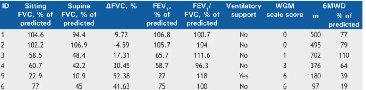
Taking into account FVC in the sitting position, four patients (66.7%) showed respiratory system impairment at diagnosis, with FVC% < 80% of predicted and normal FEV1/FVC ratio, characterizing restrictive lung disease, as is expected for neuromuscular diseases. Only one patient already used noninvasive mechanical ventilatory support intermittently, being the one who showed the lowest FVC% in the sitting and supine positions (22.9% and 10.9%, respectively) and the highest ∆FVC% (52.38%). None of the patients used invasive ventilatory support or had been tracheostomized. The median FVC% in the sitting position was 71% (range, 22.9-104.6%), the median FVC% in the supine position was 58% (range, 10.9-106.9%), the median ∆FVC%
was 24.5% (range, −4.59% to 52.4%), the median FEV1% was 70.35% (range, 27.0-106.8%), and the median FEV1/FVC ratio (in %) was 102.4% (range, 96.3-118.0%). Stratiication by age group showed that only two adolescents had spirometry results within the normal range.
DISCUSSION
The clinical history characteristics of our patients with PD were similar to those found in the literature. The type of initial symptoms was predominantly motor, the median age at diagnosis was 39.5 years, and the delay between symptom onset and diagnosis was 8 years. Data from Byrne et al.,(16) obtained through
analysis of the PD patient registry administered by the Genzyme Corporation, revealed a predominance of motor symptoms, a median age at diagnosis of 37.1 years, and a delay in diagnosis of 4 years. The delay in diagnosis was slightly greater in the analysis carried out by Kishnani et al.(22) The median age at
symptom onset was lower in our group of patients (15.0 years vs. 28.8 years).
The rarity of PD, the variability of its clinical pres-entation, its overlap of signs and symptoms with other neuromuscular diseases, and limited access to the health care system often result in a very long time to diagnosis. The delay in diagnosis seems to be greater in older subjects, which indicates improved knowledge of the disease today.(16,22) Taking into account that
patients who are younger and less severely affected respond more favorably to administration of ERT,(13)
the importance of early diagnosis and early treatment initiation is evident.
Table 1. General description of the patients in the present case series.a
ID Gender Birth, year
AGA mutation Age at symptom
onset, years
Type of initial symptoms
Age at molecular diagnosis,
years
Delay in diagnosis,
years Allele 1 Allele 2
1 M 1992 c.-32-13T>G c.2560C>T 15 Motor 17 2
2 F 2000 c.-32-13T>G c.2560C>T - Asymptomatic 10
-3 M 1988 c.-32-13T>G c.2646+2T>A 13 Motor 25 10
4* M 1958 c.-32-13T>G c.1912G>T 50 Motor 54 4
5** M 1954 c.-32-13T>G c.1912G>T 13 Motor 58 45
6 F 1951 c.2560C>T, c.-32-13T>G 40 Motor 63 23
ID: identiication; F: female; and M: male. *Patients 1 and 2 and patients 4 and 5 were siblings.
Table 2. Functional description of the patients in the present case series.
ID Sitting FVC, % of
predicted
Supine FVC, % of
predicted
∆FVC, % FEV1, % of predicted
FEV1/ FVC, % of
predicted
Ventilatory support
WGM scale score
6MWD
m % of
predicted
1 104.6 94.4 9.72 106.8 100.7 No 0 500 77
2 102.2 106.9 -4.59 105.7 104 No 0 495 79
3 58.5 48.4 17.31 65.7 111.6 No 1 702 110
4 60.7 42.2 30.45 58.7 96.3 No 3 376 64
5 22.9 10.9 52.38 27 118 Yes 6 180 39
6 77 45 41.63 75 100 No 6 97 19
More than 500 mutations have currently been identi-ied, and the expected effects range from very severe to non-pathogenic.(28) The mutation most commonly
observed in our group of patients is also the one most commonly reported by other authors.(16,29) However,
the phenotypic behavior is not explained exclusively by the genotype found, especially in late-onset disease. Phenotypic differences are present even in members of the same family, including siblings.(29) Patients 1
and 2 and patients 4 and 5, respectively, were siblings with the disease. In both cases, differences were observed in presentation and severity. However, the diagnosis of the younger siblings was facilitated by their family history, enabling a better functional condition at diagnosis. Records show that 32% of patients with late-onset PD had a sibling with a diagnosis of PD(16);
therefore, we believe that family screening may be useful in identifying asymptomatic patients and may contribute to a better prognosis.
Monitoring of respiratory function in patients with PD is imperative.(6,7,20) In 2013, Ambrosino et al.(21)
described basic management of respiratory dysfunction in PD, including periodic evaluations every 3-12 months, depending on the rate of disease progression; moni-toring of respiratory signs and symptoms; spirometry in the sitting and supine positions; measurement of MIP; measurement of peak cough low; blood gas analysis; and, in some cases, polysomnography and swallowing studies. Consensus statements and guidelines for the management of PD also have similar recommendations. (6,7,20,30)
The pathophysiology of chronic respiratory failure in neuromuscular diseases includes not only respiratory muscle weakness but also changes in chest wall compliance, central respiratory control, and swal-lowing, which, in turn, are responsible for Ineffective cough, alveolar hypoventilation, chest deformities, sleep apnea, atelectasis, airway hyperreactivity, and recurrent pneumonia.(31) Unlike other neuromuscular
diseases, in which loss of walking ability precedes ventilatory failure,(5,17) in PD, respiratory symptoms
may manifest early, being the initial symptom in 8.5% of cases.(4) Despite our small sample size, the
results for locomotor ability and the 6MWD results, when compared against the spirometry results, seem to corroborate the hypothesis that impairment of the respiratory and locomotor systems is heterogeneous. (32)
The patient with the greatest 6MWD already showed reduced FVC% in the sitting and supine positions and reduced ∆FVC%, whereas the patient with the shortest 6MWD did not have the most severe lung disease.
In our study, none of the patients followed had respiratory symptoms as the irst manifestation. However, at diagnosis, we observed signs of respiratory system impairment, as identiied by reduced FVC% in the sitting position (FVC < 80% of predicted), in 66.7% of them. Despite the absence of respiratory symptoms as the initial manifestation of the disease and the delay between symptom onset and the irst spirometry, we cannot rule out the existence of some
degree of respiratory impairment in the very early stages of PD, but without ignoring that age and duration of symptomatic disease also seem to contribute to a worsening of functional indings. It is possible that mild respiratory symptoms were present but went unnoticed because they overlapped with motor symptoms that were more prominent. Questioning and standardized description of the signs and symptoms found, especially at the onset of the disease, may facilitate knowledge and follow-up of patients.
Measures of respiratory muscle strength such as MIP and MEP may be highly relevant to identifying the onset of respiratory muscle impairment, given that changes in them may precede volume reduction as identiied by vital capacity. Unfortunately, in our group of patients, we found no such data in the medical records of one of the patients, and two were unable to perform acceptable and reproducible maneuvers. Therefore, MIP and MEP measures could not be included in the analysis, and this represents a limitation of the study.
Diaphragmatic weakness is a dysfunction that is char-acteristic of PD,(17,18) being considered the major cause
of disordered breathing during sleep and respiratory failure.(33) Prigent et al.,(34) using magnetic stimulation
of the phrenic nerve, and Wens et al.,(18) using magnetic
resonance imaging, conirmed the predominance of diaphragmatic weakness over weakness of thoracic respiratory muscles in PD. The most accurate method for assessing diaphragmatic function is to measure transdiaphragmatic pressure during maximal respiratory effort or during spontaneous breathing or use bilateral magnetic stimulation of the phrenic nerves. These methods have the disadvantage of being invasive and not being well accepted by patients, especially when they need to be repeated several times, resulting in them rarely being indicated in clinical practice.(35) A
simpler way to assess diaphragmatic weakness is to measure sitting and supine FVC% and calculate their difference, which correlates strongly with variation in cranio-caudal diameter as observed by magnetic resonance imaging.(18,36) Normal subjects may show
a reduction from sitting to supine FVC% as high as 10%. (37) Reductions > 25% characterize diaphragmatic
weakness, with a sensitivity of 79% and a speciicity of 90%.(19) In our sample, 66.7% of the patients
showed ∆FVC% > 10%, and 50% showed ∆FVC% > 25%. This assessment is recommended for the diagnosis and follow-up of patients with PD because it is a potential marker of the severity of the respiratory dysfunction. A > 10% reduction strengthens the diagnosis of PD.(30) Other methods have also been
described for diaphragmatic assessment, including luoroscopy, ultrasonography, electromyography, and optoelectronic plethysmography(38); however, they are
and this contributes to a more pronounced deicit in the motor function of the diaphragm.(17,39,40)
As respiratory muscle weakness progresses, the use of noninvasive ventilatory support is indicated, helping to control nocturnal hypoventilation and sleep apnea syndrome, as well as acute and chronic respiratory failure.(14,21) Only one patient in our case series used
this resource. Speciic indications regarding when to start using ventilatory support in PD have not been described. The use of recommendations for neuromus-cular diseases in general contributes to this process. Respiratory system involvement was present in 66.7% of our patient sample, and diaphragmatic
dysfunction as characterized by ∆FVC% > 25% was present in 50% of our series at diagnosis, suggesting that even if it is not the initial manifestation, respiratory system involvement may occur early in a signiicant number of cases. Further studies are needed for a better understanding of this involvement, especially of diaphragmatic dysfunction. The sign and symptom proile used by Llerena et al.(7) and Kishnani et al.,(22) the
recommendations included in the International Pompe Disease Registry, and the respiratory management proposed by Ambrosino et al.(21) may be of great
importance in the approach to patients with suspected PD or already diagnosed with PD.
REFERENCES
1. Martiniuk F, Chen A, Mack A, Arvanitopoulos E, Chen Y, Rom WN, et al. Carrier frequency for glycogen storage disease type II in New York and estimates of affected individuals born with the disease. Am J Med Genet. 1998;79(1):69-72. https://doi.org/10.1002/(SICI)1096-8628(19980827)79:1<69::AID-AJMG16>3.0.CO;2-K
2. Chiang SC, Hwu WL, Lee NC, Hsu LW, Chien YH. Algorithm for Pompe disease newborn screening: results from the Taiwan screening program. Mol Genet Metab. 2012;106(3):281-6. https:// doi.org/10.1016/j.ymgme.2012.04.013
3. Mechtler TP, Stary S, Metz TF, De Jesús VR, Greber-Platzer S, Pollak A, et al. Neonatal screening for lysosomal storage disorders: feasibility and incidence from a nationwide study in Austria. Lancet. 2012;379(9813):335-41. https://doi.org/10.1016/S0140-6736(11)61266-X
4. Kishnani PS, Amartino HM, Lindberg C, Miller TM, Wilson A, Keutzer J; et al. Methods of diagnosis of patients with Pompe disease: Data from the Pompe Registry. Mol Genet Metab. 2014;113(1-2):84-91. https://doi.org/10.1016/j.ymgme.2014.07.014
5. van der Beek NA, de Vries JM, Hagemans ML, Hop WC, Kroos MA, Wokke JH, et al. Clinical features and predictors for disease natural progression in adults with Pompe disease: a Nationwide prospective observational study. Orphanet J Rare Dis. 2012;7:88. https://doi. org/10.1186/1750-1172-7-88
6. Kishnani PS, Steiner RD, Bali D, Berger K, Byrne BJ, Case LE, Crowley JF, et al. Pompe disease diagnosis and management guideline. Genet Med. 2006;8(5):267-88. https://doi.org/10.1097/01. gim.0000218152.87434.f3
7. Llerena JC Jr, Horovitz DM, Marie SK, Porta G, Giugliani R, Rojas MV, et al. The Brazilian consensus on the management of Pompe disease. J Pediatr. 2009;155(4 Suppl):S47-56. https://doi. org/10.1016/j.jpeds.2009.07.006
8. van der Ploeg AT, Clemens PR, Corzo D, Escolar DM, Florence J, Groeneveld GJ, et al. A randomized study of alglucosidase alfa in late-onset Pompe’s disease. N Engl J Med. 2010;362(15):1396-1406. https://doi.org/10.1056/NEJMoa0909859
9. Kishnani PS, Corzo D, Leslie ND, Gruskin D, Van der Ploeg A, Clancy JP, et al. Early treatment with alglucosidase alpha prolongs long-term survival of infants with Pompe disease. Pediatr Res. 2009;66(3):329-35. https://doi.org/10.1203/PDR.0b013e3181b24e94
10. Güngör D, Kruijshaar ME, Plug I, D’Agostino RB, Hagemans ML, van Doorn PA, et al. Impact of enzyme replacement therapy on survival in adults with Pompe disease: results from a prospective international observational study. Orphanet J Rare Dis. 2013;8:49. https://doi. org/10.1186/1750-1172-8-49
11. van der Beek NA, van Capelle CI, van der Velden-van Etten KI, Hop WC, van den Berg B, Reuser AJ, et al. Rate of progression and predictive factors for pulmonary outcome in children and adults with Pompe disease. Mol Genet Metab. 2011;104(1-2):129-36. https://doi. org/10.1016/j.ymgme.2011.06.012
12. Wokke JH, Escolar DM, Pestronk A, Jaffe KM, Carter GT, van den Berg LH, et al. Clinical features of late-onset Pompe disease: a prospective cohort study. Muscle Nerve. 2008;38(4):1236-45. https:// doi.org/10.1002/mus.21025
13. de Vries JM, van der Beek NA, Hop WC, Karstens FP, Wokke JH, de Visser M, et al. Effect of enzyme therapy and prognostic factors in 69 adults with Pompe disease: an open-label single-center study.
Orphanet J Rare Dis. 2012;7:73. https://doi.org/10.1186/1750-1172-7-73
14. Mellies U, Lofaso F. Pompe disease: a neuromuscular disease with respiratory muscle involvement. Respir Med. 2009;103(4):477-84. https://doi.org/10.1016/j.rmed.2008.12.009
15. Güngör D, de Vries JM, Hop WC, Reuser AJ, van Doorn PA, van der Ploeg AT, et al. Survival and associated factors in 268 adults with Pompe disease prior to treatment with enzyme replacement therapy. Orphanet J Rare Dis. 2011;6:34. https://doi.org/10.1186/1750-1172-6-34
16. Byrne BJ, Kishnani PS, Case LE, Merlini L, Müller-Felber W, Prasad S, et al. Pompe disease: design, methodology, and early indings from Pompe Registry. Mol Genet Metab. 2011;103(1):1-11. https:// doi.org/10.1016/j.ymgme.2011.02.004
17. Fuller DD, ElMallah MK, Smith BK, Corti M, Lawson LA, Falk DJ, et al. The respiratory neuromuscular system in Pompe disease. Respir Physiol Neurobiol. 2013;189(2):241-9. https://doi.org/10.1016/j. resp.2013.06.007
18. Wens SC, Ciet P, Perez-Rovira A, Logie K, Salamon E, Wielopolski P, et al. Lung MRI and impairment of diaphragmatic function in Pompe disease. BMC Pulm Med. 2015;15:54. https://doi.org/10.1186/ s12890-015-0058-3
19. Fromageot C, Lofaso F, Annane D, Falaize L, Lejaille M, Clair B, et al. Supine fall in lung volumes in the assessment of diaphragmatic weakness in neuromuscular disorders. Arch Phys Med Rehabil. 2001;82(1):123-8. https://doi.org/10.1053/apmr.2001.18053
20. Cupler EJ, Berger KI, Leshner RT, Wolfe GI, Han JJ, Barohn RJ, et al. Consensus treatment recommendations for late-onset Pompe disease. Muscle Nerve. 2012;45(3):319-33. https://doi.org/10.1002/ mus.22329
21. Ambrosino N, Confalonieri M, Crescimanno G, Vienello A, Vitacca M. The role of respiratory management of Pompe disease. Respir Med. 2013;107(8):1124-32. https://doi.org/10.1016/j.rmed.2013.03.004
22. Kishnani PS, Amartino HM, Lindberg C, Miller TM, Wilson A, Keutzer J. Timing of diagnosis of patients with Pompe Disease: data from the Pompe Registry. Am J Med Genet A. 2013;161A(10):2431-43. https://doi.org/10.1002/ajmg.a.36110
23. Gardner-Medwin D, Walton JN. The clinical examination of voluntary muscles. In: Walton JN, editor. Disorders of Voluntary Muscles, 3rd ed., Edinburgh: Churchill Livingstone; 1974, p. 517-60.
24. Iwama AM, Andrade GN, Shima P, Tanni SE, Godoy I, Dourado VZ. The six-minute walk test and body weight-walk distance product in health Brazilian subjects. Braz J Med Biol Res. 2009;42(11):1080-5. https://doi.org/10.1590/S0100-879X2009005000032
25. Priesnitz CV, Rodrigues GH, Stumpf Cda S, Viapiana G, Cabral CP, Stein RT, et al. Reference value for the 6-min walk test in health children aged 6-12 years. Pediatr Pulmonol. 2009;44(12):1174-9. https://doi.org/10.1002/ppul.21062
26. Knudson RJ, Lebowitz MD, Holberg CJ, Burrows B. Changes in the normal maximal expiratory low-volume curve with growth and aging. Am Rev Respir Dis. 1983;127(6):725-34.
27. Miller MR, Hankinson J, Brusasco V, Burgos F, Casaburi R, Coates A, et al. Standardization of spirometry. Eur Respir J. 2005;26(2):319-38. https://doi.org/10.1183/09031936.05.00034805
c2015 [cited 2015 Dec 12]. Available from: www.pompecenter.nl
29. Kross M, Hoogeveen-Westerveld M, van der Ploeg A, Reuser AJ. The genotype-phenotype correlation in Pompe disease. Am J Med Genet C Semin Med Genet. 2012;160C(1):59-68. https://doi. org/10.1002/ajmg.c.31318
30. American Association of Neuromuscular & Electrodiagnostic Medicine. Diagnostic criteria for late-onset (childhood and adult) Pompe disease. Muscle Nerve. 2009;40(1):149-60. https://doi. org/10.1002/mus.21393
31. Khatwa UA, Dy FJ. Pulmonary Manifestations of Neuromuscular Diseases. Indian J Pediatr. 2015;82(9):841-51. https://doi. org/10.1007/s12098-015-1814-3
32. Pellegrini N, Laforet P, Orlikowski D, Pellegrini M, Caillaud C, Eymard B, et al. Respiratory insuficiency and limb muscle weakness in adults with Pompe’s disease. Eur Respir J. 2005;26(6):1024-31. https://doi. org/10.1183/09031936.05.00020005
33. Mellies U, Ragette R, Schwake C, Baethmann M, Voit T, Teschler H. Sleep-disordered breathing and respiratory failure in acid maltase deiciency. Neurology. 2001;57(7):1290-5. https://doi.org/10.1212/ WNL.57.7.1290
34. Prigent H, Orlikowski D, Laforêt P, Letilly N, Falaize L, Pellegrini N, et al. Supine volume drop and diaphragmatic function in adults with Pompe disease. Eur Respir J. 2012;39(6):1545-6. https://doi. org/10.1183/09031936.00169011
35. Meric H, Falaize L, Pradon D, Orlikowski D, Prigent H, Lofaso F. 3D analysis of the chest wall motion for monitoring late-onset Pompe disease patients. Neuromuscul Disord. 2016;26(2):146-52. https:// doi.org/10.1016/j.nmd.2015.11.003
36. Gaeta M, Musumeci O, Mondello S, Ruggeri P, Montagnese F, Cucinotta M, et al. Clinical and pathophysiological clues of respiratory dysfunction in late-onset Pompe disease: New insights from a comparative study by MRI and respiratory function assessment. Neuromuscul Disord. 2015;25(11):852-8. https://doi.org/10.1016/j. nmd.2015.09.003
37. Allen SM, Hunt B, Green M. Fall in vital capacity with posture. Br J Dis Chest. 1985;79(3):267-71. https://doi.org/10.1016/0007-0971(85)90047-6
38. Boon AJ, O’Gorman C. Ultrasound in the Assessment of Respiration. J Clin Neurophysiol. 2016;33(2):112-9. https://doi.org/10.1097/ WNP.0000000000000240
39. DeRuisseau LR, Fuller DD, Qiu K, DeRuisseau KC, Donnelly WH Jr, Mah C, et al. Neural deicits contribute to respiratory insuficiency in Pompe disease. Proc Natl Acad Sci U S A. 2009;106(23):9419-24. https://doi.org/10.1073/pnas.0902534106