Non-paraneoplastic
Lambert-Eaton myasthenic syndrome
A brief review of 10 cases
Paulo J. Lorenzoni, Rosana H. Scola, Cláudia S. Kamoi Kay, Sérgio F. Parolin, Lineu C. Werneck
ABSTRACT
Lambert-Eaton myasthenic syndrome (LEMS) is an immune-mediated disorder of the presynaptic neuromuscular transmission, which more frequently occurs as the remote effect of a neoplasm, in the paraneoplastic form (P-LEMS), or in a non-paraneoplastic form (NP-LEMS); but few studies describe the clinical features of NP-LEMS. We analyzed the clinical manifestations, laboratory findings, electrophysiological studies, and treatment responses in ten Brazilian patients suffering from NP-LEMS. The mean age was 41.5 years. More often neurological findings were hyporeflexia or areflexia with a post-exercise improvement. Treatment response occurred with pyridostigmine, guanidine, prednisone, azathioprine, and cyclosporine; but not response was observed after intravenous immunoglobulin and plasma exchange. Age at onset, clinical manifestations, and electrophysiological abnormalities can help more in the diagnosis than serum antibodies; the symptomatic treatment with pyridostigmine was effective; and the immunosuppressive treatment with prednisone, azathioprine, or cyclosporine was more beneficial than plasma exchange or intravenous immunoglobulin treatment.
Key words: Lambert-Eaton myasthenic syndrome, myasthenic syndrome, P/Q-type voltage-gated calcium channel antibody, repetitive nerve stimulation, electrophysiological study, treatment.
Síndrome miastênica de Lambert-Eaton não paraneoplásica: uma breve revisão de dez casos
RESUMO
A síndrome miastênica de Lambert-Eaton (LEMS) é uma desordem imunomediada da transmissão neuromuscular pré-sinaptica, que mais frequentemente ocorre como efeito à distância de uma neoplasia, na forma paraneoplásica (P-LEMS), ou na forma não paraneoplásica (LEMS); porém poucos estudos têm descrito as características da NP-LEMS. Nós analisamos as manifestações clínicas, laboratoriais, eletrofisiológicas, e resposta ao tratamento em dez pacientes brasileiros com NP-LEMS. A idade média foi de 41,5 anos. A manifestação neurológica mais freqüente foi hiporeflexia ou arreflexia com melhora após o exercício. A resposta ao tratamento ocorreu com piridostigmina, guanidina, prednisona, azatioprina, e ciclosporina; mas não com imunoglobulina intravenosa e plasmaférese. A idade de início, manifestações clínicas e eletrofisiológicas ajudaram mais no diagnóstico do que os anticorpos séricos; o tratamento sintomático com piridostigmina foi efetivo; e o tratamento imunossupressor com prednisona, azatioprina, ou ciclosporina beneficiou mais do que a plasmaférese ou a imunoglobulina intravenosa.
Palavras-chave: síndrome miastênica de Lambert-Eaton, síndrome miastênica, anticorpo anti-canal de cálcio voltagem dependente, estimulação nervosa repetitiva, estudo eletrofisiológico, tratamento.
Correspondence
Rosana Herminia Scola
Serviço de Doenças Neuromusculares Hospital de Clínicas da UFPR Rua General Carneiro 181 / 3º andar 80060-900 Curitiba PR - Brasil E-mail: [email protected]
Received 14 December 2009 Received in final form 15 May 2010
Lambert-Eaton myasthenic syndrome (LEMS) is an immune-mediated disorder of the neuromuscular trans-mission in which serum autoantibodies against the P/Q-type voltage-gated calcium channels (VGCCs) at the pre-synaptic nerve terminal lead to a decrease in the presyn-aptic release of acetycholine1-3. hese antibodies impair the acetylcholine release on the neuromuscular and on the autonomic neuroefective junctions and cause the on-set of the symptoms3.
LEMS can frequently occur as remote efect of a neo-plasm, in the paraneoplastic form (P-LEMS), usually in as-sociation with small cell lung cancer (SCLC), or in the non-paraneoplastic form (NP-LEMS)1,2,4-8. A review of 50 cases of LEMS demonstrated about a 60% risk of SCLC, which was diagnosed in most of these cases within 2 years of onset of the myasthenic syndrome9. hen, the diagnosis of NP-LEMS requires a long-term follow up because the neurolog-ical symptoms can precede a diagnosis of a neoplasm1,5,8,9.
P-LEMS and NP-LEMS have diferent characteris-tics, but few studies describe the clinical features of NP-LEMS after a long-term follow up4,8. he objective of this study was to analyze the clinical manifestations, labora-tory indings, electrophysiological studies and treatment responses in a series of Brazilian patients sufering from NP-LEMS.
METHOD
We retrospectively analyzed the LEMS patients seen in our hospital from 1976 to 2008, and we studied ten pa-tients with a diagnosis of NP-LEMS. Our hospital is a ref-erence neuromuscular center with a special interest and an expertise in disorders of the neuromuscular junction. he LEMS diagnosis was based on typical clinical fea-tures, characteristic electrophysiological changes, and an absence of cancer in a follow up at of least four years af-ter the LEMS diagnosis2,10,11. Relevant data, including the
age, gender, clinical evaluation, course of the disease, se-rum anti-P/Q-type VGCC antibody, sese-rum anti-acetyl-choline receptor (AChR) antibody, electrophysiological study, and treatment response were recorded.
he LEMS electrophysiological measurement criteria comprised a reduced compound muscle action potential (CMAP) amplitude with a CMAP amplitude increment of more than 100% after a brief maximal voluntary contrac-tion or high frequency (20 Hz) repetitive nerve stimula-tion (RNS) according to standard procedures1,10,11.
he treatment response was classiied as total, par-tial, or absent according to the objective improvement in the neurological indings and autonomic symptoms dur-ing the drug therapy.
All studies were done following patient consent.
RESULTS
he sample consisted of ten patients (six female and four male), age 26 to 60 years (mean 41.5 years), showing predominance of young adult patients in this series (eight patients with age less than 45 years). he time of the fol-low-up varied between 4 and 13 years, with a mean time of 8 years (Table 1).
NP-LEMS was associated with another autoimmune disease (thyroiditis with hypothyroidism) in two cases (Table 1).
he neurological indings that were found more fre-quently in our group before treatment were hyporelex-ia or arelexhyporelex-ia (10/10) with a post-exercise improvement (6/6). he other neurological indings before treatment are described in Table 1. he more frequent autonom-ic dysfunction symptoms before treatment were dry mouth (6/10), constipation (2/10), a reduction in the li-bido (2/10), and blurred vision (2/10) (Table 1).
Serological analysis of the anti-P/Q-type VGCC an-tibody was performed in six patients and demonstrated
Table 1. Clinical manifestations, laboratory indings, eletrophysiological studies and treatment responses of our NP-LEMS patients.
Case 1 2 3 4 5 6 7 8 9 10
Gender M M M F F F F F M F
Age at onset (years) 60 38 39 26 42 48 34 45 39 45
Follow up time (years) 7 4 12 4 8 4 13 9 8 10
Associated autoimmune disorder
Hypothyroidism – – – – + – – + – –
Neurologic indings
Diplopia + + + – – + – – + –
Ophthalmoplegia + + – – – – – – + –
Ptosis + + + + + – – + + –
Dysphagia + + + + + – + – – +
Facial weakness – + – + + – – – + –
Proximal upper limb weakness + – + + + + + – – +
Distal upper limb weakness – – – – – + – – + –
Proximal lower limb weakness + + + + + + + – – +
Distal lower limb weakness – – – – – + – + + –
Hyporelexia or arelexia + + + + + + + + + +
Increased relex post-exercise ND ND + ND + + + + + ND
Muscular pain – – – – + + + – – –
Cerebellar ataxia – + – – – – – + + –
Respiratory failure – + – – – – – – – –
Autonomic dysfunction
Dry mouth – + – – – + + + + +
Constipation – – + – + – – – – –
Libido reduction – – – – – + + – – –
Blurred vision – – – – + – – – – +
Serum antibodies
Anti-P/Q-type VGCC antibody ND ND ND ND – – – + + +
Anti-AChR antibody ND ND ND ND – – – ND – –
Electrophysiological test
Low CMAP amplitude + + + + + + + + + +
Incremental CMAP post–exercise + + + + + + + + + +
CMAP decrement at low-rate RNS + + + + – + + + + +
CMAP increment at high-rate RNS ND + + + + + + + + +
Treatment response Symptomatic therapy
Pyridostigmin + + + + + ± + + + +
Guanidine ND ND ND ND ND ND ND ND ND +
Immunosuppressive therapy
Prednisone + + + ± ± ND ± ± ± +
Azathioprine ND ND + ND * ND ± ± ± ±
Cyclosporin ND ND ND ND + ND + + + ND
Intravenous immunoglobulin ND ND ND ND ND ND – – – –
Plasma exchange ND ND ND ND ND ND – ND – ND
the antibody presence in three patients. he anti-AChR antibodies were tested in ive patients and were nega-tive (Table 1).
he most common abnormalities found in the elec-trophysiological studies were a low CMAP amplitude (10/10), an incremental CMAP amplitude post-exercise (10/10), a CMAP amplitude decrease with a low-rate RNS (9/10), and a CMAP amplitude increase with a high-rate RNS (9/9) (Table 1 and Figure).
A treatment response was total in patients who used pyridostigmine (9/10), guanidine (1/1), prednisone (4/9), azathioprine (1/5), and cyclosporine (4/4). Partial treat-ment response was observed with pyridostigmine (1/10), prednisone (5/9) and azathioprine (4/5). No treatment re-sponse was reported after intravenous immunoglobulin (4/4) and plasma exchange (2/2) (Table 1).
DISCUSSION
NP-LEMS has been rarely described in Brazil, includ-ing our previous reports (cases 1, 6, 7, 8, and 9), in
con-trast to other places3,12,13. NP-LEMS usually occurs in 30 to 50% of LEMS patients6,8. No gender difference was found in the NP-LEMS patients, as in our series (Table 2), while the P-LEMS patients show a male predominance6,8. he mean age at onset of LEMS in the NP-LEMS patients is lower than in the P-LEMS patients7-9. In NP-LEMS, on-set can be from childhood to old age2,9,14.
In NP-LEMS, a mean interval between the onset of symptoms and a diagnosis of LEMS is longer than in P-LEMS cases7-9,14. he probability of an underlying SCLC, at presentation of the myasthenic syndrome, range from 50 to 60%, which falls sharply after 2 years and becomes less than a few per cent after 4 years from onset7-9.
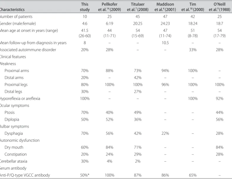
LEMS almost invariably starts with a proximal weak-ness (Table 2), especially of the legs7,14. Hyporelexia is also a common sign (Table 2), which shows a functional im-provement after a physical efort by the facilitation phe-nomenon that occurs by Ca+2 accumulations on the ner-vous terminal and, therefore, acetylcholine release on the synaptic gap3. Ocular symptoms are regularly seen in pa-Table 2. Characteristics of our NP-LEMS patients compared to previous published cohorts.
Characteristics studyThis et al.Pellkofer 14 (2009) et al.Titulaer 7 (2008) et al.Maddison 4 (2001) et al.Tim 18 (2000) et al.O’Neill 9 (1988)
Number of patients 10 25 45 47 42 25
Gender (male:female) 4:6 6:19 20:25 24:23 18:24 18:7
Mean age at onset in years (range) 41.5 (26-60)
44 (11-71)
54 (15-69)
47 (11-74)
51 (8-78)
54 (17-79)
Mean follow-up from diagnosis in years 8 – – 10.5 – –
Associated autoimmune disorder 20% 28% – – 33% 28%
Clinical features Weakness
Proximal arms 70% 88% 73% 94% 100% –
Distal arms 20% – 42% – – –
Proximal legs 80% 100% 100% 96% 100% 100%
Distal legs 30% – 27% – – –
Hyporelexia or arelexia 100% – – – 100% 92%
Ocular symptoms
Ptosis 70% 40% 49% – – 44%
Diplopia 50% 52% 36% – – 56%
Bulbar symptoms
Dysphagia 70% 56% 42% 22% – 28%
Autonomic dysfunction
Dry mouth 60% 84% 71% – – 84%
Constipation 20% 24% 29% – – 28%
Cerebellar ataxia 30% 4% 2% – – –
Serum antibody
Anti-P/Q-type VGCC antibody 50%* 100% 87% 86% 65% –
tients with LEMS, but the severity is rather mild, especial-ly compared to the severity in myasthenia gravis7. Cere-bellar ataxia is found more in P-LEMS than in NP-LEMS, but our patients had a high incidence of this symptom (Table 2), which sometimes dominated the clinical pic-ture so that the presence of LEMS was overlooked7,12. An-ti-VGCC antibodies also underlie the autonomic symp-toms in LEMS patients, especially dry mouth (Table 2), which occurs in between 71% and 84% of cases4,7,9,14.
Despite all the symptoms that appear during the course of disease, no neurologic indings distinguished between the P-LEMS and the NP-LEMS patients, but weakness of the distal muscles in the legs (Table 2), weight loss, and respiratory failure appeared signiicant-ly less frequentsigniicant-ly in NP-LEMS6-8. In addition, NP-LEMS showed a slower progressive course of disease than the P-LEMS patients7.
Associated autoimmune disorders can occur usually after an interval of six months in between 28% and 33% of NLEMS patients (Table 2), but only occur in 6% of P-LEMS patients8,14. he high frequency of associated auto-immune disorders, together with the high proportion of women and the relative younger age at onset, suggests a similar etiology as other non-paraneoplastic autoimmune diseases in NP-LEMS patients8.
he antibodies to the P/Q-type VGCC can be detected in over 90% of the LEMS patients2,4,7. he antibodies are speciic for LEMS, but the site of the antigenic stimulus in NP-LEMS is unknown2. Our patients had a low frequency of the anti-P/Q-type VGCC antibody (Table 2); therefore, the LEMS patients without the anti-P/Q-type VGCC an-tibody more frequently have NP-LEMS than P-LEMS6. he classic electrophysiological abnormalities (a low CMAP amplitude, a CMAP decrease at low rate of stim-ulation, and a CMAP increase above 100% after a brief maximal voluntary contraction or high-frequency RNS) are present in almost all of the NP-LEMS patients, similar to P-LEMS, although this is not seen in all muscles, and it may be necessary to examine several muscles to dem-onstrate this feature1-3,10,13.
The NP-LEMS treatment consists of symptomatic treatment of the acetylcholine deiciency as well as an immunosuppressive treatment3,15-17. Some patients show symptom improvement by use of cholinesterase inhibi-tors such as pyridostigmine, but this drug is less efec-tive in LEMS than in myasthenia gravis3,15,17,18. Other pa-tients require drugs such as guanidine and 3,4-diamopyridine, which increase the pre-synaptic calcium in-lux and the acetylcholine release improving LEMS symp-toms15-17. Moreover, an additional therapeutic efect can be obtained if guanidine or 3,4-diaminopyridine are com-bined with pyridostigmine15-18. he unresponsive patients to symptomatic treatment may respond to
immunosup-pressive treatment with prednisone alone or combined with azathioprine, and, most recently, cyclosporine15-17. Although the evidence of beneit for the immunosuppres-sive treatment of LEMS is limited to a series of case re-ports, it is reasonable to adopt treatment procedures by analogy with myasthenia gravis15-17. In LEMS patients with respiratory failure or bulbar dysfunction, plasma exchange and intravenous immunoglobulin are useful in bridging the gap until other immunosuppressive therapy takes ef-fect15. In NP-LEMS, where weakness is severe, plasma ex-change or intravenous immunoglobulin treatment confers a short-term beneit, but this treatment did not alter the disease course in our patients, while corticosteroids and immunosuppressive drugs improved their symptoms15-17.
Although about half of the NP-LEMS patients achieved sustained clinical remission, most of them re-quired substantial and continuing doses of immunosup-pressive medication4. A review of 47 cases of NP-LEMS demonstrated that the only predictor of long-term out-come (clinical remission or independent ambulation) was initial clinical score (comprising muscle strength mea-surements by Medical Research Council scale) in limb muscles4. he electrophysiological study in NP-LEMS can start to improve after one year of treatment in some pa-tients, but no direct relation was seen between the NP-LEMS treatment, the anP/Q-type VGCC antibody ti-ter and the CMAP amplitude4,17. Immunological and elec-trophysiological measurements were useful, however, for monitoring disease progression and response to treat-ment in individual patients4.
In this study, the age at onset, clinical manifestations, and electrophysiological abnormalities helped more in the LEMS diagnosis than the serum antibodies; the symptom-atic treatment with pyridostigmine was efective; and an immunosuppressive treatment with prednisone, azathio-prine, or cyclosporine were more beneicial than plasma exchange or intravenous immunoglobulin treatment.
ACKNOWLEDGEMENTS – We are grateful to Dr. Vanda A. Lennon/ Mayo Clinic College of Medicine (cases 6 and 7) and Dr. Bethan Lang/ University of Oxford (cases 8 and 9) for measuring the serum anti-P/Q-type VGCC antibody; and Dr. Helio A. G. Teive/UFPR and Dr. Pedro A. Kowacs/UFPR for their collaboration (case 8).
REFERENCES
1. Levin KH. Paraneoplastic neuromuscular syndromes. Neurol Clin 1997;15: 597-614.
2. Newsom-Davis J. Neuromuscular junction channelopathies: a brief overview. Acta Neurol Belg 2005;105:181-186.
3. Scola RH, Iwamoto FM, Ramos CS, et al. Lambert-Eaton myasthenic syn-drome: report of two cases. Arq Neuropsiquiatr 1998;56:457-464. 4. Maddison P, Lang B, Mills K, Newsom-Davis J. Long term outcome in
Lam-bert-Eaton myasthenic syndrome without lung cancer. J Neurol Neurosurg Psychiatry 2001;70:212-217.
6. Nakao YK, Motomura M, Fukudome T, et al. Seronegative Lambert-Eaton myasthenic syndrome: study of 110 japanese patients. Neurology 2002;59: 1773-1775.
7. Titulaer MJ, Wirtz PW, Kuks JBM, et al. The Lambert-Eaton myasthenic syn-drome 1988-2008: a clinical picture in 97 patients. J Neuroimmunol 2008;201-202:153-158.
8. Wirtz PW, Smallegange TM, Wintzen AR, Verschuuren JJ. Diferences in clin-ical features between the Lambert-Eaton myasthenic syndrome with and without cancer: an analysis of 227 published cases. Clin Neurol Neurosurg 2002;104:359-363.
9. O’Neill JH, Murray NMF, Newsom-Davis J. The Lambert-Eaton myasthenic syndrome: a review of 50 cases. Brain 1988;111:577-596.
10. AAEM Quality Assurance Committee. Literature review of the usefulness of repetitive nerve stimulation and single iber EMG in the electrodiagnostic evaluation of patients with suspected myasthenia gravis or Lambert-Eaton myasthenic syndrome. Muscle Nerve 2001;24:1239-1247.
11. Amato AA, Dumitru D. Neuromuscular junction disorders. In: Dumitru D, Am-ato AA, Zwarts MJ (Eds). Electrodiagnostic medicine, 2nd Edition. Philadelphia,
Hanley & Belfus 2002:1149-1153.
12. Lorenzoni PJ, Scola RH, Lang B, et al. Cerebellar ataxia in non-paraneoplastic Lambert-Eaton myasthenic syndrome. J Neurol Sci 2008;270:194-196. 13. Werneck LC, Bittencourt PCT, Nóvak EM. Myasthenia gravis with
electro-graphic indings of myasthenic syndrome: a case report. Arq Neuropsiqui-atr 1985;43:198-205.
14. Pellkofer HL, Armbruster L, Linke R, Schumm F, Voltz R. Managing non-para-neoplastic Lambert-Eaton myasthenic syndrome: clinical characteristics in 25 German patients. J Neuroimmunol 2009;217:90-94.
15. Weimer MB, Wong J. Lambert-Eaton myasthenic syndrome. Curr Treat Opt Neurol 2009;11:77-84.
16. Skeie GO, Apostolski S, Evoli A, et al. Guidelines for the treatment of au-toimmune neuromuscular transmission disorders. Eur J Neurol 2006;13: 691-699.
17. Verschuuren JJGM, Wirtz PW, Titulaer MJ, Willems LNA, Gerven JV. Available treatment options for the management of Lambert-Eaton myasthenic syn-drome. Expert Opin Pharmacother 2006;7:1323-1336.