572
1. Cardiovascular surgeon.
2. PhD; Cardiovascular surgeon. Clinic Director of Heart Institute of Distrito Federal, Fundação Zerbini, Brasília.
Work carried out at Heart Institute of Distrito Federal, Fundação Zerbini, Brasília, DF, Brazil.
Correpondence address:
Dr. Fernando A. Atik. Instituto do Coração do Distrito Federal,
Ricardo B. CORSO1,Fernando A. ATIK1,Cristiano N. FABER1,Luiz Fernando CANEO2
Rev Bras Cir Cardiovasc 2008; 23(4): 572-574 CASE REPORT
RBCCV 44205-1035
Involução de artérias colaterais aneurismáticas após a correção da coarctação da aorta
Involution of aneurysmal collateral arteries after
correction of aortic coarctation
Abstract
A 47 year-old man with coarctation of the aorta had undergone extra-anatomic bypass through a left thoracotomy. He presented 10 years later with uncontrolled hypertension due to residual coarctation, graft obstruction and multiple aneurysmal subclavian to aortic collateral arteries. He was submitted to an ascending aorta to descending aorta bypass through a median sternotomy with the aid of standard cardiopulmonary bypass. His postoperative recovery was unremarkable, and there was complete involution of all aneurysmal collateral arteries after the operation.
Descriptors: Aorta, coarctation/surgery. Heart defects, congenital. Collateral circulation.
Resumo
Paciente de 47 anos de idade, com diagnóstico de coarctação da aorta, foi submetido ao implante de tubo extra-anatômico por toracotomia esquerda. Dez anos após o procedimento, o paciente retorna com hipertensão arterial de difícil controle relacionada a coarctação da aorta residual, obstrução do tubo e múltiplos aneurismas de artérias colaterais entre a artéria subclávia e a aorta. O paciente foi submetido então a correção extra-anatômica entre a aorta ascendente e a descendente por esternotomia mediana, com auxílio de circulação extracorpórea convencional. Sua recuperação pós-operatória foi boa, e houve involução completa de todas as artérias colaterais aneurismáticas após a operação.
Descritores: Coartação aórtica/cirurgia. Cardiopatias congênitas. Circulação colateral.
Fundação Zerbini Estrada Parque Contorno do Bosque s/n, 1 andar, sala 13 – Brasília, DF – Brasil – CEP 70658-700.
E-mail: [email protected]
Article received on April 22nd, 2008 Article accepted on July, 28th, 2008 INTRODUCTION
Development of aneurysmal thoracic collateral arteries has been rarely reported in the literature [1]. We aim to present a case of an adult patient with coarctation of the aorta with a previous distal aortic arch to descending aorta bypass that was referred for surgery due to residual coarctation and development of multiple aneurysmal subclavian to aortic collateral arteries.
CASE REPORT
573 CORSO, RB ET AL - Involution of aneurysmal collateral arteries
after correction of aortic coarctation
Rev Bras Cir Cardiovasc 2008; 23(4): 572-574
Although the anatomic repair was initially contemplated as a primary option, the presence of a previous thoracotomy, with known aneurysmal collateral arteries and possibly dense adhesions changed our plan. Under general anesthesia, a median sternotomy was constructed, and a 26-millimeter ascending aorta to descending aortic knitted double velour graft (Boston Scientific, Natick, MA) was performed with the aid of standard cardiopulmonary bypass. His postoperative recovery was unremarkable, being discharged home on the eighth day in good condition.
Control chest computed tomography six weeks later showed a patent graft and complete involution of all aneurysmal collateral arteries [Figure 2]. A year later, he is currently asymptomatic and in normotensive state.
DISCUSSION
Development of collaterals in intercostals arteries and internal thoracic arteries is a common phenomenon in untreated coarctation of the aorta [2]. These arteries connect the subclavian artery or costocervical trunk to the descending aorta. There is a direct correlation with patient age at presentation, morphology of the coarctation and transverse arch and concomitant cardiac defects. However, the occurrence of aneurysmal collateral arteries in supra-aortic position is rare and it may have relation to the previous surgical approach. They probably represent hypertrophied preexisting normal vessels of an embryologic origin [3].
Indication for surgery in the present case was related to the risk of aneurysm expansion with extrinsic compression of adjacent structures, arterial rupture and pseudoaneurysm formation, in addition to deleterious effects of untreated coarctation.
There was a debate whether the anatomic repair in addition to collateral arteries ligation or embolization would provide better long-term outcomes. Although we generally favor the anatomic repair, this particular problem seemed to be better managed with an extra-anatomic bypass through a median sternotomy, avoiding manipulation of a densely adherent and well-vascularized area. Those factors are often responsible for extensive lung manipulation, airway injury and parenchyma hematomas. Aortic repair is usually demanding, which increases the risk of bleeding and paraplegia. Therefore, deep hypothermic circulatory arrest may be advocated [4]. In this setting, extra-anatomic bypass is a reasonable option in order to lessen surgical risk, eventually permitting simultaneous treatment of cardiac problems and to promote a sustained long-term distal aortic perfusion [5].
Percutaneous intervention is another treatment option and it has been particularly indicated in recurrent coarctation [6]. However, as opposed to children, adult patients often present with fragile, atherosclerotic and
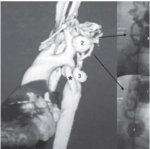
Fig. 1 - Preoperative chest computed tomography with three-dimensional reconstruction (left image), showing obstructed extra-anatomic bypass (*) and multiple aneurysmal thoracic collateral arteries (1,2,3) corresponding to their angiographic appearance on the right-sided images
obstructed extra-anatomic bypass through a juxta ductal coarctation of the aorta, and multiple left subclavian artery to descending aorta aneurysmal collateral arteries (maximum diameter 45 millimeters).
574
REFERENCES
1. Lee AD, Shyamkumar NK, Nayak S, Agarwal S, Perakath B. Collateral artery aneurysm: a unique presentation of thoracic outlet syndrome. Eur J Vasc Endovasc Surg. 2005;29(6):611-2.
2. Mathew R, Simon G, Joseph M. Collateral circulation in coarctation of aorta in infancy and childhood. Arch Dis Child. 1972;47(256):950-3.
3. Kirks DR, Currarino G, Chen JT. Mediastinal collateral arteries: important vessels in coarctation of the aorta. AJR Am J Roentgenol. 1986;146(4):757-62.
4. Lange R, Thielmann M, Schmidt KG, Bauernschmitt R, Jakob H, Hasper B, et al. Spinal cord protection using hypothermic cardiocirculatory arrest in extended repair of recoarctation and persistent hypoplastic aortic arch. Eur J Cardiothorac Surg. 1997;11(4):697-702.
5. Oliveira SA, Lisboa LA, Dallan LA, Abreu Filho CA, Rochitte CE, Souza JM. Extraanatomic aortic bypass for repair of aortic arch coarctation via sternotomy: midterm clinical and magnetic resonance imaging results. Ann Thorac Surg. 2003;76(6):1962-6.
6. Preventza O, Wheatley GH 3rd, Williams J, Hughes K, Ramaiah VG, Rodriguez-Lopez JA, et al. Endovascular approaches for complex forms of recurrent aortic coarctation. J Endovasc Ther. 2006;13(3):400-5.
CORSO, RB ET AL - Involution of aneurysmal collateral arteries after correction of aortic coarctation
Rev Bras Cir Cardiovasc 2008; 23(4): 572-574
calcified aortas, which certainly limits procedure success and poses patients at additional risk.